
Metalibitia santaremis ( Roewer, 1947 )
|
publication ID |
https://doi.org/10.11646/zootaxa.4291.2.1 |
|
publication LSID |
lsid:zoobank.org:pub:3A891AA8-9D85-47AD-9201-A37D24D32717 |
|
DOI |
https://doi.org/10.5281/zenodo.6032716 |
|
persistent identifier |
https://treatment.plazi.org/id/038787D1-FFF7-FFEF-EDAE-FD6FFA9B6482 |
|
treatment provided by |
Plazi |
|
scientific name |
Metalibitia santaremis ( Roewer, 1947 ) |
| status |
|
Metalibitia santaremis ( Roewer, 1947) View in CoL
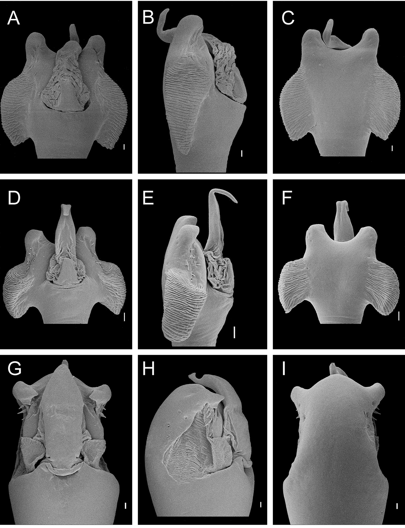
(Figs: 2C; 3F; 6C, H; 8C, H; 12G–I)
Paralibitia santaremis Roewer, 1947: 8 View in CoL [desc], pl. 1, fig. 7
Metalibitia santaremis Ringuelet, 1959: 425 View in CoL [by implication] [cit]; Kury, 2003: 69 [cat]; Pinto-da-Rocha & Bonaldo, 2006: 158 [cit]
Type material. SMF RII 1491/13, ♂ holotype. Examined.
Type locality. BRAZIL, Pará State, Santarém.
Geographical distribution ( Fig. 14 View FIGURE 14 ): Limited to northern Brazil, states oF Pará (Santarém and Juruti) and Amazonas (Humaitá).
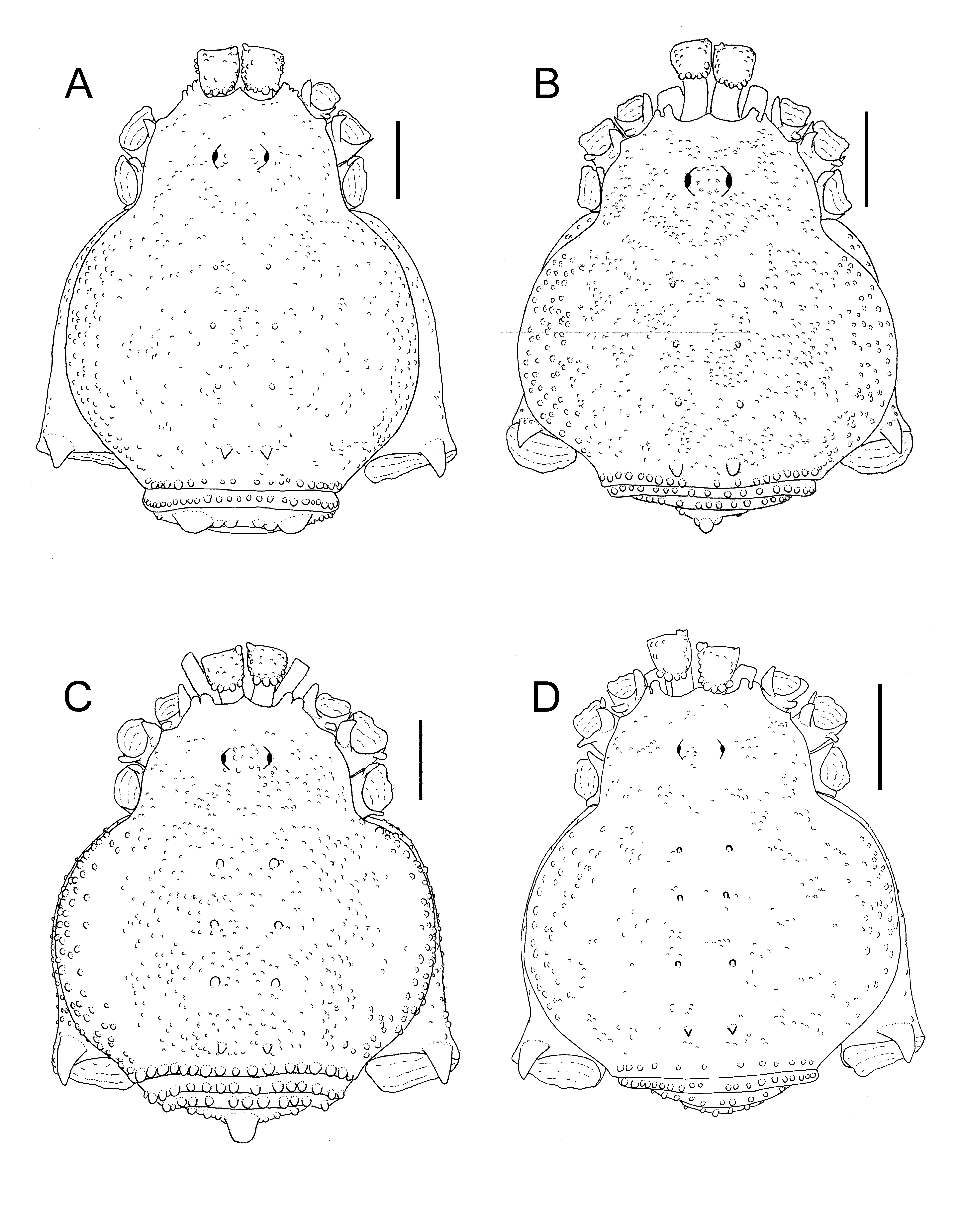
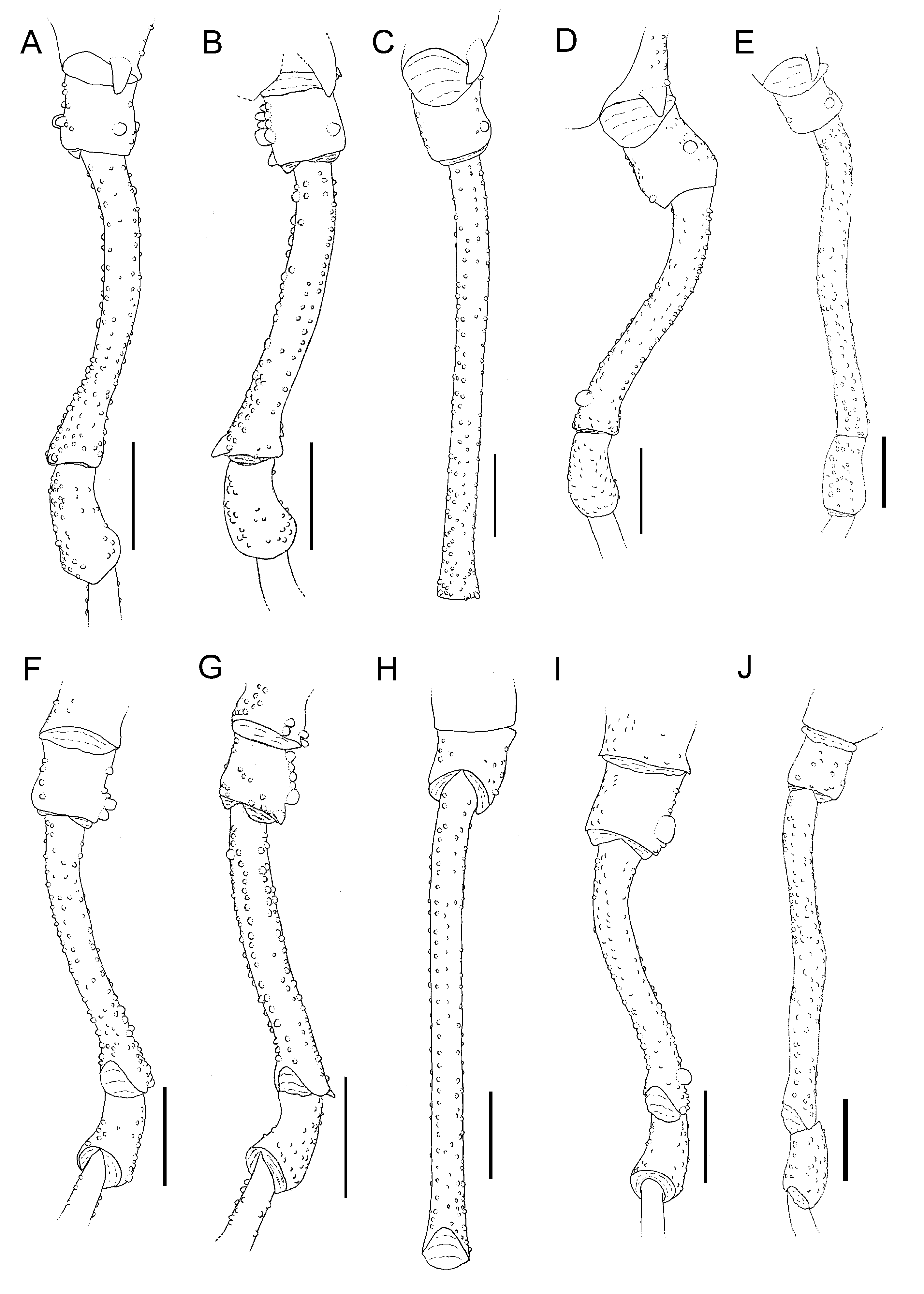
Diagnosis. Resembles M. argentina and M. borellii in having Free tergite III with one higher central tubercle ( Figs 1 View FIGURE 1 B, C; 2C) and tibia IV small-tuberculated ( Figs 7 View FIGURE 7 B, C; 8C); it is similar to M. argentina in having Femur IV with only small tubercles ( Figs 5 View FIGURE 5 B; 6C) ( M. borellii has an apical tubercle on Femur IV, Fig. 5 View FIGURE 5 C) and to M. borellii in having only one retrolateral tubercle on cheliceral bulla ( Figs 1 View FIGURE 1 C; 2C) ( M. argentina has contiguous tubercles, Fig. 1 View FIGURE 1 B). It diFFers From these species in having dorsal scutum shape elongate ( Fig. 2 View FIGURE 2 C) ( M. argentina and M. borellii have Flattened bodies, Figs 1 View FIGURE 1 B, C), only one prolateral tubercle on coxa II ( Fig. 2 View FIGURE 2 C) ( M. argentina and M. borellii also have one retrolateral tubercle, Figs 1 View FIGURE 1 B, C), trochanter IV with retrolateral small tubercles ( Fig. 6 View FIGURE 6 H) ( M. argentina have agglomerated tubercles and M. borellii has one basal and one apical tubercle, Figs 5 View FIGURE 5 F, G), convex distal border oF ventral plate oF penis ( Fig. 12 View FIGURE 12 I) ( M. argentina and M. borellii have a concave border, Figs 11 View FIGURE 11 C, F).
Redescription. Male holotype (SMF 1491/13)
Measurements: dorsal scutum, total length 4.35; dorsal scutum, total width 3.5; prosoma length 1.25; prosoma width 2.15; Femur I length 2.3; Femur II length 4.6; Femur III length 3.6; Femur IV length 4.85; pedipalpal Femur length 1.2 mm.
Coloration in ethanol: Entirely pale brown.
Dorsum ( Fig. 2 View FIGURE 2 C): Anterior margin oF dorsal scutum with median portion smooth, with two high rhomboid tubercles, near paracheliceral projections. Lateral margins oF dorsal scutum with rounded tubercles, near areas I– IV. Ocularium with shallow median depression, with 15 small tubercles. Posterior margin oF dorsal scutum with a row oF 18 tubercles. Free tergite I with row oF 20 tubercles, II with row oF 16 tubercles, III with row oF 12 tubercles, the central tubercle being the highest. Anal operculum with 39 rounded tubercles irregularly scattered. All tubercles oF posterior margin oF dorsal scutum, Free tergites and anal operculum have rounded apex.
Chelicera ( Fig. 2 View FIGURE 2 C): Bulla with Four–Five tubercles on proximal margin, the last one on prolateral region. One apical retrolateral tubercle surrounded by smaller ones. Segment II with Five teeth; segment III with eight teeth, Four oF them larger.
Pedipalps (MNRJ 8266) ( Fig. 3 View FIGURE 3 F): Trochanter with two united ventral setiFerous tubercles. Femur with Four dorsal basal setiFerous tubercles Fused at the base; prolateral row oF Five setiFerous tubercles From median region to apex; six ventral rounded tubercles, the three median ones the highest, and two ventroapical tubercles. Tibia with a rounded projection on prolateral apical region, dorsal and ventral setae, one tubercle on each lateral dorsoapical region, and one retrolateral apical spine. Tarsus triangular with dorsal setae, the retrolateral inner row with two apical macrosetae and one central row oF six macrosetae.
Legs ( Figs 2 View FIGURE 2 C; 6C, H; 8C, H): Coxa I with one FalciForm prolateral tubercle reaching the apex oF coxa and one smaller retrobasal tubercle. Coxae II–III with one prolateral high tubercle. Coxa IV with apical region visible in dorsal view, tuberculated, with one dorsal proapical apophysis. Trochanter IV with seven retrolateral small tubercles on the middle and one higher apical tubercle. Femur IV straight, with small tubercles in rows. Patella IV tuberculated. Tibia IV elongated with setiFerous small tubercles. Tarsal Formula: 5 / 8 / 5 / 6.
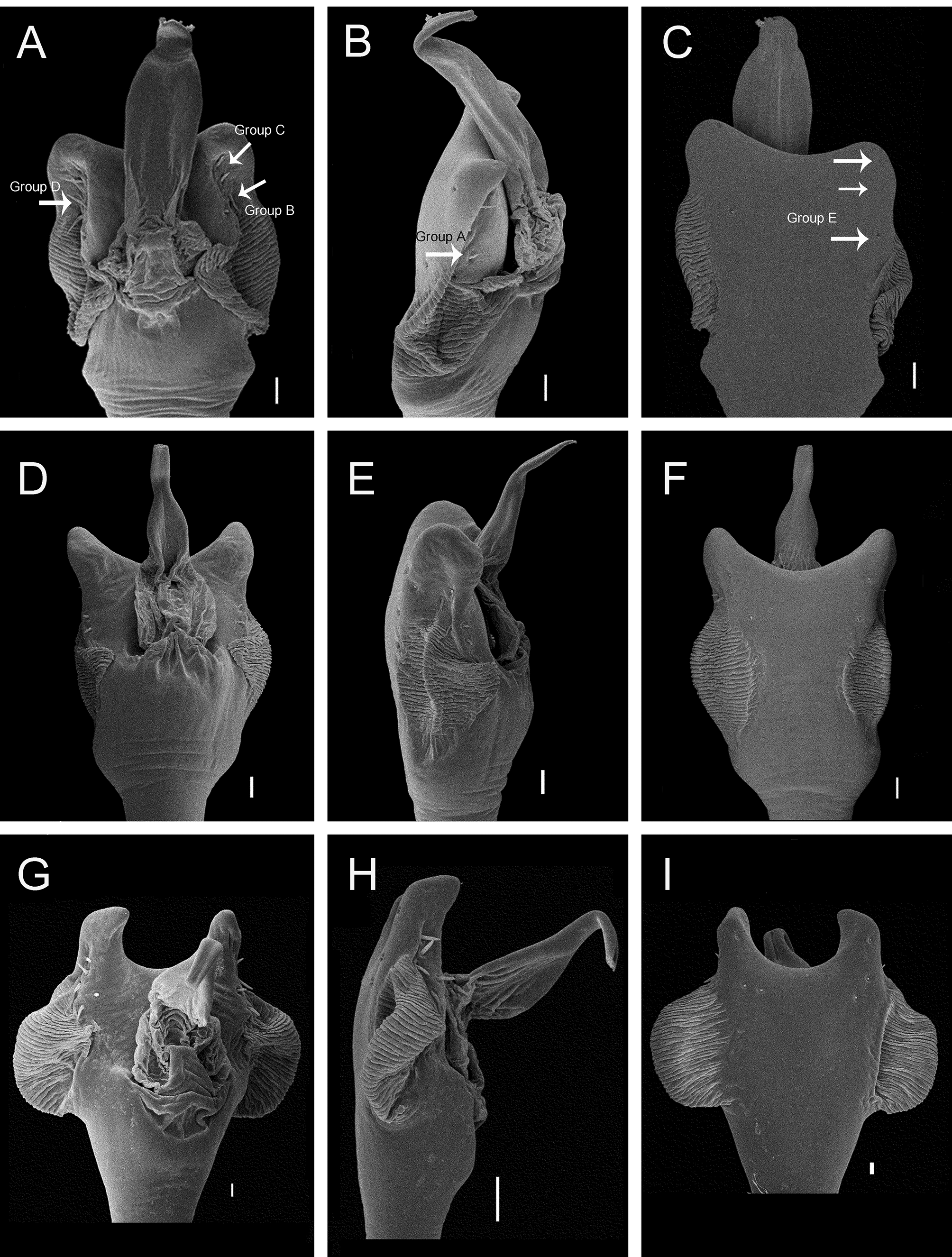
Penis (MNRJ 4602) (Figs: 12G–I): Truncus straight and wide. Ventral plate thick, narrower at the middle; lateral borders, distal margin and region between the lateral borders strongly convex, with Four pairs oF lateroventral small macrosetae (macrosetae E). Lateral membranous expansions weak, From glans base to subapical region oF ventral plate (only visible in lateral view). Six lateral long macrosetae, three apical pairs (group C), one pair displaced ventrally (group B), one pair on median region (group D) and one pair on proximal region (group A). Insertion oF glans on basal portion oF ventral plate. Glans wide, much narrower at stylus base. Stylus short, with small projections on apex.
Female (MNRJ 8266).
Measurements: (MNRJ 8266): dorsal scutum, total length 4.2; dorsal scutum, total width 3.5; prosoma length 1.2; prosoma width 2.0; Femur I length 2.2; Femur II length 5.0; Femur III length 3.7; Femur IV length 4.5; pedipalpal Femur length 1.0 mm.
Dorsum: Anterior constriction oF scutum well-marked. Posterior margin oF dorsal scutum with a row oF 21 tubercles with rounded apex. Free tergite I with row oF 18 tubercles, II with row oF 17 tubercles, III with row oF 15 tubercles, all similar sized. Anal operculum with 23 rounded tubercles irregularly scattered. Chelicera: Bulla with six proximal rounded tubercles. Pedipalps: Femur with small projection on dorsobasal region with Five small tubercles and Four dorsoapical tubercles; ventral row oF seven tubercles. Leg IV: Entirely with small tubercles. Tarsal Formula: 5 / 8 / 5 / 6.
Variation. Ocularium with 11–14 tubercles. The median tubercles oF Free tergite III can be Fused at base (three tubercles), these tubercles being the highest. Measurements ♂ ( n=3): dorsal scutum, total length 8.7–9.2; dorsal scutum, total width 7.5–7.6; prosoma length 1.2–1.3; prosoma width 2.0–2.2; Femur I length 2.2–2.6; Femur II length 4.5–5.2; Femur III length 3.4–4.2; Femur IV length 4.4–5.5; pedipalpal Femur length 0.9–1.1 mm. Measurements ♀ ( n=3): dorsal scutum, total length 3.9–4.0; dorsal scutum, total width 3.1–3.5; prosoma length 1.2–1.3; prosoma width 1.9–2.0; Femur I length 2.2–2.7; Femur II length 4.1–4.5; Femur III length 3.4–3.5; Femur IV length 3.8–4.5; pedipalpal Femur length 1.0. Tarsal Formula ♂ (n=3) 5/ 5– 10/ 5/ 5–6. Tarsal Formula ♀ (n=3) 5/ 5–8/ 5/ 6.
Material examined. BRAZIL. Pará State, Juruti , 02° 24’ 33”S 56° 26’ 10” W, 02–20. IX.2002, A. Bonaldo et al. leg., 1♂ ( MPEG JUR- 01 370) GoogleMaps ; Amazonas State, Humaitá , 07° 30' 22" S 63° 01' 15" W, 26.II.1976, U. Caramaschi leg., 32♂ 68♀ ( MNRJ 8266 View Materials ), same loc. GoogleMaps , 17.I.1979, U. Caramaschi leg., 1♂ (MNRJ 4602).


No known copyright restrictions apply. See Agosti, D., Egloff, W., 2009. Taxonomic information exchange and copyright: the Plazi approach. BMC Research Notes 2009, 2:53 for further explanation.
|
Kingdom |
|
|
Phylum |
|
|
Class |
|
|
Order |
|
|
Family |
|
|
Genus |
Metalibitia santaremis ( Roewer, 1947 )
| Coronato-Ribeiro, Amanda & Pinto-Da-Rocha, Ricardo 2017 |
Metalibitia santaremis
| Kury 2003: 69 |
| Ringuelet 1959: 425 |
Paralibitia santaremis
| Roewer 1947: 8 |