
Pleioplana okusi, Bulnes & Kalkan & Karhan, 2009
|
publication ID |
https://doi.org/10.1080/00222930903094662 |
|
publication LSID |
lsid:zoobank.org:pub:F97CD81B-A397-48F8-8A4A-1E1627425D91 |
|
persistent identifier |
https://treatment.plazi.org/id/3D9EE08A-7374-4DE3-8C3B-DA10A69DC24B |
|
taxon LSID |
lsid:zoobank.org:act:3D9EE08A-7374-4DE3-8C3B-DA10A69DC24B |
|
treatment provided by |
Felipe |
|
scientific name |
Pleioplana okusi |
| status |
sp. nov. |
Pleioplana okusi View in CoL sp. nov.
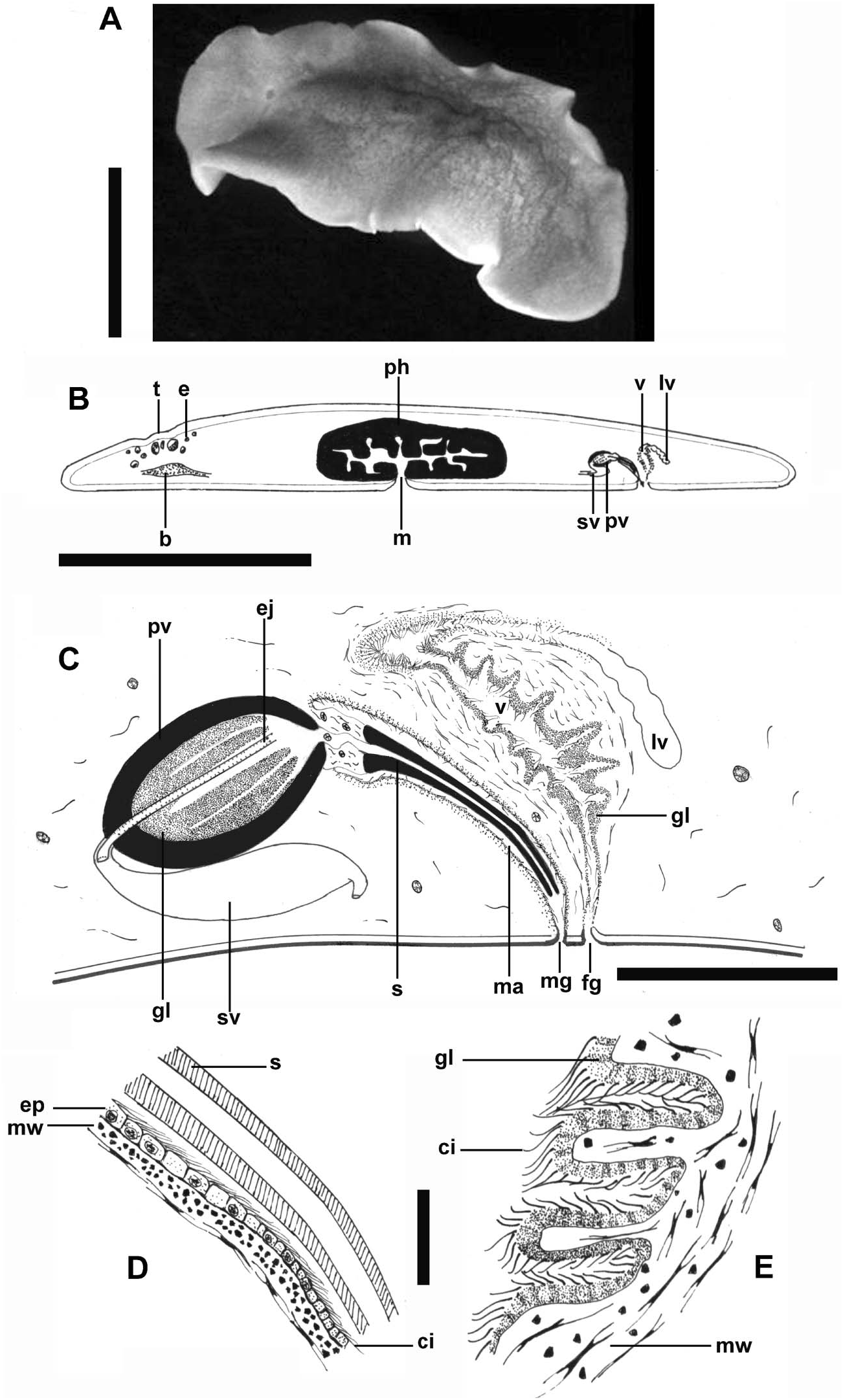
( Figure 3A–E View Figure 3 )
Type material
Holotype 9.0 µm sagittally sectioned specimen, mounted on nine slides. Altinkum , Bosphorus Strait, Turkey, 41°11′21.22′′ N, 29°04′57.10′′ E, on hard-bottom mussel bed of M. galloprovincialis (Lamarck, 1819) , collected by E. Kalkan, 2 November 2004. BMNH 2007.9 .26.8. GoogleMaps
Additional material examined
Five specimens from Altinkum, Bosphorus Strait, collected from hard-bottom mussel bed of M. galloprovincialis (Lamarck, 1819) by E. Kalkan, 2 November 2004.
Etymology
The specific epithet is a patronymic honouring the memory of Prof. Erdoǧan Okuş, who dedicated much of his brief life to the progress of marine science in Turkey.
Description
Fixed specimen about 6 mm in length by 3.3 mm in width. Body oval, more or less elongated, translucent, with few smooth marginal undulations ( Figure 3A View Figure 3 ). Body with brownish ground colour, speckled with tiny black or brownish-black spots densely packed on mid-line of body, decreasing towards the margins. Body margin free of spots; ventral surface colourless. Small tentacular knobs present. Tentacular eyes present, cerebral eye spots in two scattered clusters. Pharynx ruffled and short, one-quarter of the body length, centrally arranged at the mid-body; the mouth opens in the middle of the pharyngeal cavity ( Figure 3B View Figure 3 ).
Ventral body wall 82 µm high, consisting of cellular epidermis 82 µm high, with scattered rhabdites. The epidermis is underlain by a circular muscle layer followed by longitudinal muscular fibres, a well-developed layer of circular muscles and an inner layer of longitudinal muscles mixed with diagonal muscle fibres. Transverse muscle fibres are numerous and well differentiated. Granular pigmentation distributed in small patches between the inner muscle layers. Dorsal body wall about 50 µm high. Cellular epidermis completely ciliated, with intraepithelial nuclei and densely packed with dermal rhabdites. Cilia covered by a dense film of mucous secretion, which persists after fixation and staining. Ventral epidermis underlain by a circular muscle layer, followed by a layer of longitudinal fibres. Clusters of granular pigmentation regularly distributed between the muscle layer and the body parenchyma.
Testis follicles situated ventrally and ovaries extending from dorsal to ventral between intestinal branches.
The male copulatory apparatus consists of a true seminal vesicle, an interpolated prostatic vesicle, and a distal penis stylet, the latter housed in a narrow atrium that communicates with a male gonopore ( Figure 3C View Figure 3 ). The vasa deferentia enter the posterior wall of the seminal vesicle. The muscular seminal vesicle is elongated, located ventrally to the prostatic vesicle. Prostatic vesicle of Atomata-type, slightly elongated, with uniform muscular wall ( Figure 3D View Figure 3 ). The folded glandular lining of the prostatic vesicle forms six long tubular chambers filling its entire lumen. Inner lining of the prostatic vesicle non-ciliated. The ejaculatory duct projects into the prostatic vesicle. The distal section of the prostatic duct extends into the male atrium. The prostatic duct starts as a small muscular duct but distally develops to form a long (390 µm), slightly curved, sclerotized stylet that projects into the ciliated and narrow male atrium. Male organ directed backwards.
Oviducts enter the female duct separately from the ventral side. From this point the ciliated female duct curves posteriad and opens into a small and folded Lang’s vesicle. The female duct also curves anteroventrad, widens and this forms the vagina bulbosa ( Figure 3E View Figure 3 ). The ciliated columnar epithelium of the vagina is underlain by longitudinal and circular muscle fibres and is also surrounded by numerous cement glands. The lining epithelium of the vagina is strongly folded, the vagina opens via a short ciliated female atrium and ventral gonopore to the exterior.
No known copyright restrictions apply. See Agosti, D., Egloff, W., 2009. Taxonomic information exchange and copyright: the Plazi approach. BMC Research Notes 2009, 2:53 for further explanation.
|
Kingdom |
|
|
Phylum |
|
|
Order |
|
|
Family |
|
|
Genus |