
Protoplanella simplex Reisinger, 1924
|
publication ID |
https://doi.org/10.5852/ejt.2022.798.1671 |
|
publication LSID |
lsid:zoobank.org:pub:F136E044-62C8-4FB3-8160-7DAE663D9600 |
|
DOI |
https://doi.org/10.5281/zenodo.7450121 |
|
persistent identifier |
https://treatment.plazi.org/id/038A87DA-A765-FF87-0412-FD52FE800819 |
|
treatment provided by |
Felipe |
|
scientific name |
Protoplanella simplex Reisinger, 1924 |
| status |
|
Protoplanella simplex Reisinger, 1924 View in CoL
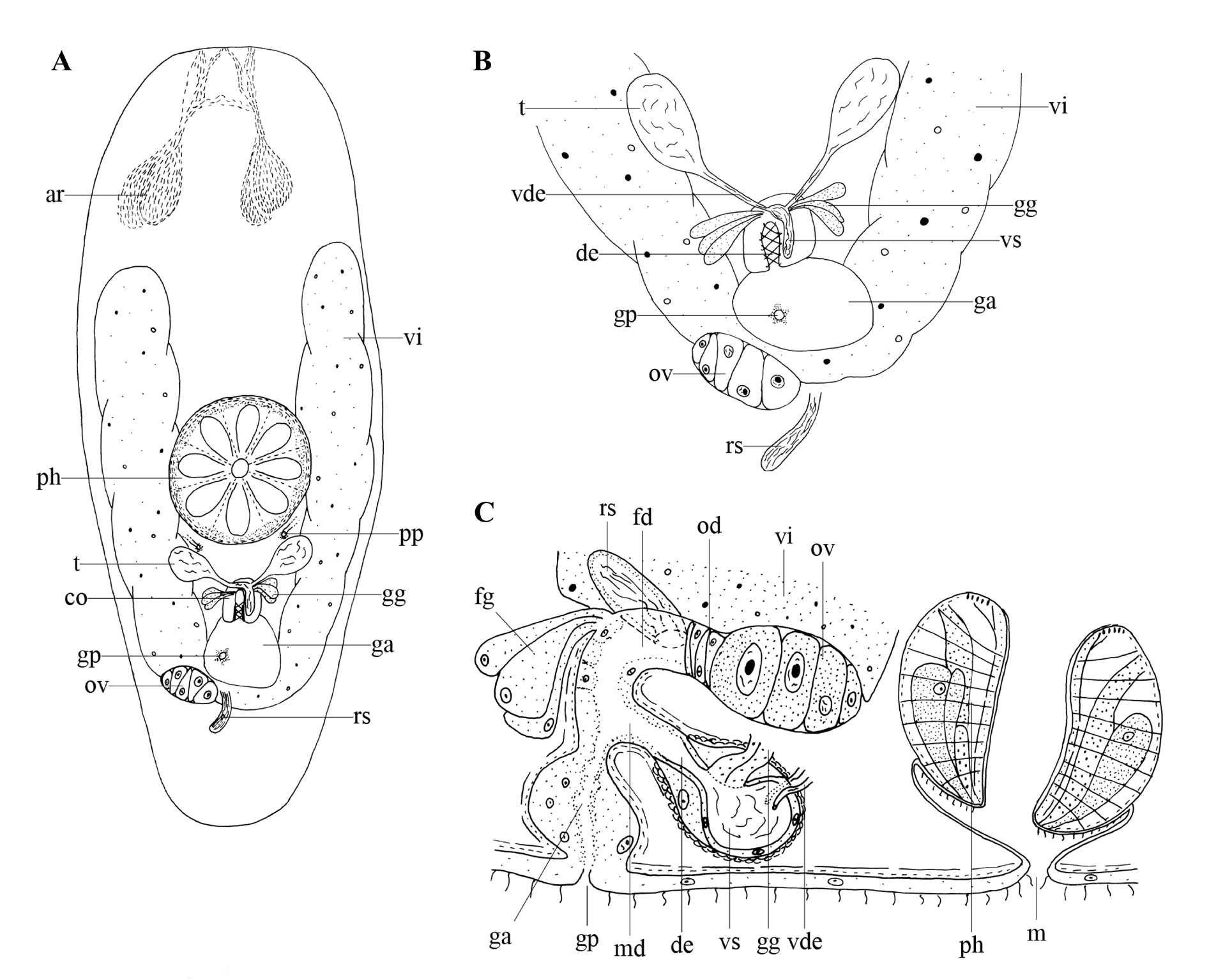
Fig. 11 View Fig
Material examined
Neotype GERMANY • 1 spec., live observations and sagittal sections; Kordel ; 49°49′24″ N, 06°38′06″ E; 24 Jul. 2011; A.M. Houben and W. Proesmans leg.; mosses growing on a wall; neotype no. 825 ; HU. GoogleMaps
Other material
GERMANY • 1 spec., serially sectioned; same collection data as for neotype; XIV.3.11; HU GoogleMaps • 2 specs, live observations, one of which serially sectioned; Bavaria, Oberau ; 07°33′33″ N, 11°06′57″ E; 13 Jul. 2011; A.M. Houben and W. Proesmans leg.; forest litter; XIV.3.12; HU GoogleMaps • 5 specs, live observations, three of which serially sectioned; Lanaken ; 50°56′03″ N; 05°39′36″ E; 27 Jul. 2011; A.M. Houben and W. Proesmans leg.; mosses at the forest edge; XIV.3.13–XIV.3.15; HU GoogleMaps • 4 specs, live observations, two of which serially sectioned; Lanaken, National Park ‘ Hoge Kempen’ ; 50°56′02″ N; 05°39′38″ E; 27 Jul. 2011; A.M. Houben and W. Proesmans leg.; moist forest soil; XIV.3.16–XIV.3.17; HU GoogleMaps .
Description
The studied specimens are about 0.8 mm long. The anterior and posterior body ends are rounded ( Fig. 11A View Fig ). Rhabdite glands are present and arranged into two groups behind the brain ( Fig. 11A View Fig : ar); the rhabdites themselves extend forward in two anastomosing tracts. Dermal rhabdites were not observed. The paired protonephridiopores ( Fig. 11A View Fig : pp) open laterally and somewhat caudal to the rosulate pharynx ( Fig. 11A, C View Fig : ph), which is situated just behind the middle of the body.
The gonopore ( Fig. 11A–C View Fig : gp) is located at ±80% of the body and connected to a genital atrium ( Fig. 11A–C View Fig : ga). The genital atrium is surrounded by an inner circular and outer longitudinal muscle layer.
The small, round testes ( Fig. 11A–B View Fig : t) lie posterior to the pharynx and ventral to the vitellaria ( Fig. 11A–C View Fig : vi). Both vasa deferentia ( Fig. 11B–C View Fig : vde) enter the copulatory organ ( Fig. 11A View Fig : co) separately from the lateral side. This 22 µm long, oval-shaped copulatory organ is surrounded by two layers of spiral muscles and contains an intracapsular seminal vesicle ( Fig. 11B–C View Fig : vs) with a low, nucleated epithelium and an ejaculatory duct ( Fig. 11B–C View Fig : de), which is surrounded by two layers of spiral muscles. Extracapsular, eosinophilic prostate glands ( Fig. 11A–C View Fig : gg) open laterally into the copulatory organ. A male duct ( Fig. 11C View Fig : md) with the same musculature as the genital atrium connects the copulatory organ to the latter.
The paired vitellaria extend from 25% of the body to the posterior end where they fuse via a broad anastomosis. A single ovary ( Fig. 11A–C View Fig : ov) is closely associated with one of the vitellaria forming an ovovitellarium. The female duct ( Fig. 11C View Fig : fd) is surrounded by the same musculature as the genital atrium and receives a club-shaped seminal receptacle ( Fig. 11A–C View Fig : rs) and ovovitelloduct at its proximal end. Fine-grained, eosinophilic glands ( Fig. 11C View Fig : fg) surround and enter the female duct.
Discussion
See the general discussion on the genus Protoplanella .
Remarks
Intensive surveys at the type locality yielded no specimens. The specimens from Oberau, Bavaria, Germany, which is relatively close to the original type locality in Graz, Austria, are of poor quality. Therefore, a specimen from Kordel, Germany was designated neotype, since all diagnostic features are exactly as described by Reisinger (1924).
Previously known distribution
In the vicinity of Graz, Austria in forest humus ( Reisinger 1924, 1954; An der Lan & Franz 1954; An der Lan 1963), on the Faroe Islands ( Steinböck 1931); near Poznań, Poland, in moss and litter ( Kolasa 1974).
General discussion on Protoplanella
As mentioned by Van Steenkiste et al. (2011), the identification of Protoplanella simplex is challenging. New material found during several sampling trips showed a need for type material to unravel the morphological differences between all known descriptions of P. simplex . Reisinger (1924) originally described animals with round testes; vasa deferentia that open separately from the lateral side of the copulatory organ; a vitellarium and ovovitellarium connected to each other over a broad anastomosis; and a seminal receptacle that opens into the proximal part of the female duct. Luther (1963) described a specimen with elongated testes; a bursa that could be a seminal receptacle near the male copulatory organ; and, judging from his illustration, most probably paired ovovitellaria. Also, Van Steenkiste et al. (2011) describe a sack-like protrusion at the genital atrium (which was not mentioned by Reisinger 1924); a female bursa (which was called a seminal receptacle by Reisinger 1924) containing remnants of sperm; and their illustration shows the vasa deferentia uniting while entering the copulatory organ at the proximal side.
The descriptions of Luther (1963) and Van Steenkiste et al. (2011) differ substantially from that of Reisinger (1924) and, therefore, we consider them not to refer to representatives of P. simplex . Here, we consider these descriptions as referring to a new species: Protoplanella leiae Houben, Proesmans & Artois sp. nov. Moreover, the description of Luther (1963) indicates some, albeit smaller, differences with that of Van Steenkiste et al. (2011). The illustration made by Luther (1963) is not detailed and only made from live specimens. Since Luther’s drawings are in general more detailed, we assume this illustration was based on sub-ideal circumstances, which yielded a quickly made sketch. For now, we consider the specimens described by Luther (1963) and Van Steenkiste et al. (2011) to be members of the same species since differences are small and, in the case of Luther, not based on thoroughly studied material.
No known copyright restrictions apply. See Agosti, D., Egloff, W., 2009. Taxonomic information exchange and copyright: the Plazi approach. BMC Research Notes 2009, 2:53 for further explanation.
|
Kingdom |
|
|
Phylum |
|
|
Order |
|
|
Family |
|
|
Genus |