
Trichomycterus alternatus ( Eigenmann, 1917 )
|
publication ID |
https://doi.org/ 10.11646/zootaxa.4585.1.6 |
|
publication LSID |
lsid:zoobank.org:pub:EA9C736C-95F0-4596-8912-BB715CAF9E72 |
|
persistent identifier |
https://treatment.plazi.org/id/038AA02B-026C-8C22-37A0-51A5FCDBFC15 |
|
treatment provided by |
Plazi |
|
scientific name |
Trichomycterus alternatus ( Eigenmann, 1917 ) |
| status |
|
Trichomycterus alternatus ( Eigenmann, 1917) View in CoL
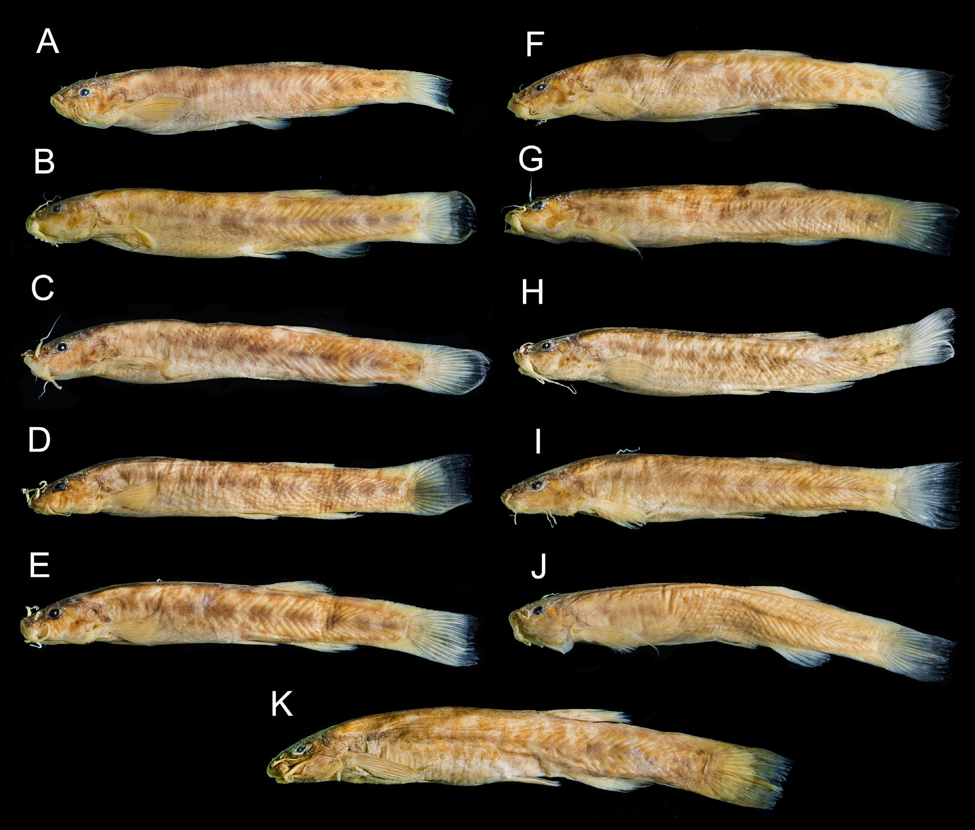
( Fig. 2 View FIGURE 2 , 3 View FIGURE 3 )
Pygidium alternatum Eigenmann, 1917: 700 View in CoL ; Rio Doce, Brazil [holotype: FMNH 58082 (ex CM 7079), paratypes: CAS 64575 (4), FMNH 58083 (62)]
Description of type specimens. Morphometric data for holotype and paratypes in Table 1 View TABLE 1 . See figures 2 and 3 for general external aspect. Body cross-section oval near head, gradually compressed posteriorly towards caudal fin. Body elongate with dorsal and ventral profiles slightly convex on trunk and nearly straight along caudal peduncle. Body depth approximately even along entire body, only slightly decreasing posterior to dorsal-fin origin.
* Including full length of first pectoral-fin ray
Head depressed, approximately as long as wide. Head width increasing slightly from snout to end of opercle, trapezoid in dorsal view. Mouth subterminal. Upper lip convex, covered with numerous small, short papillae on internal and external surfaces. Premaxillary teeth arranged in three or four well-defined and roughly regular rows. Dentary teeth arranged in one regular outer row, with two additional inner irregular rows near symphysis. Lower lip well developed, convex, with lateral fleshy folds adjacent to base of rictal barbel. Fleshy fold slender than, and half overall size of, lower lip, its insertion anterior to origin of rictal barbel. Anterior nostril closer to upper lip than to anterior margin of eye, surrounded by membrane forming short and slender tube continuous posterolaterally with base of nasal barbel. Posterior nostril anteriorly surrounded by thin and short flap of integument. Maxillary barbel reaching to base of pectoral fin or slightly beyond. Rictal barbel reaching middle of interopercular patch of odontodes or posterior margin of gill opening. Nasal barbel extending to anterior margin of opercular patch of odontodes.
Eye large, without free orbital rim, located at anterior half of head length and on dorsolateral region of head. Elliptical ocular capsule formed by thin and translucent skin not adhered to surface of eyeball. Interorbital space slightly convex and equivalent to twice orbital diameter.
Opercular patch of odontodes dorsolaterally located on head, surrounded by fold of integument, with posterior margin almost reaching vertical through anterior margin of base of first pectoral-fin ray, with 15 [1], 16* [2], 17 [4], 20 [2], 21 [1], 23 [1] claw-like odontodes. Interopercular patch of odontodes large, ventrolaterally located on head, about two thirds as deep as opercular patch, with 27 [1], 30 [2], 31 [2], 33 [1], 34 [2], 38* [2], 40 [1] clawlike odontodes. Branchiostegal membranes narrowly joined to isthmus medially and forming large free fold. Branchiostegal rays 6 [1], 7* [9], with bigger ones partly visible in outline on surface of skin.
Pectoral-fin rays I + 7 [11*]. Distal margin of pectoral fin straight or slightly convex; first ray prolonged as a long filament. Anterior portion of pectoral-fin base not covered by branchial membrane. Axillary-gland pore dorsal to base of first pectoral-fin ray, anteroventral to first lateral-line pore, bigger and adjacent to lateral-line pores. Pelvic-fin rays I+4 [11], plus pelvic splint. Distal margin of pelvic fin entirely covering anus. Base of pelvic fin separated by one eye diameter. Dorsal-fin rays iii+II+7* [6], ii+II+7 [3], II+7 [2]. Dorsal fin convex distally, on posterior half of SL, its origin slightly anterior to vertical through origin of anal fin. Dorsal-fin pterygiophores 8[11*]. First dorsal pterygiophore anterior to neural spine of 13 th [1], 15 th * [15], 16 th [35], 17 th [2] post-Weberian vertebrae. Anal-fin rays iii+II+5* [4], ii+II+5 [4], II+5 [3]. Distal margin of anal fin convex. Anal-fin pterygiophore 6[11*]. First anal pterygiophore anterior to hemal spine of 17 th [1], 18 th [5], 19 th *[32], 20 th [13] post- Weberian vertebrae. Caudal-fin rays 6+7 [11*]. Caudal fin shape variable, with margin ranging from truncate to slightly convex, in all cases with round corners. Post-Weberian vertebrae 32 [1], 33 [1], 34 [11], 35 [35], 36* [15], First post-Weberian vertebrae nearly half the length of subsequent ones. Second post-Weberian vertebrae, short parapophyses, with a wide tip. Ribs 8 [1], 9 [2], 10* [29], 11 [21], 12[8].
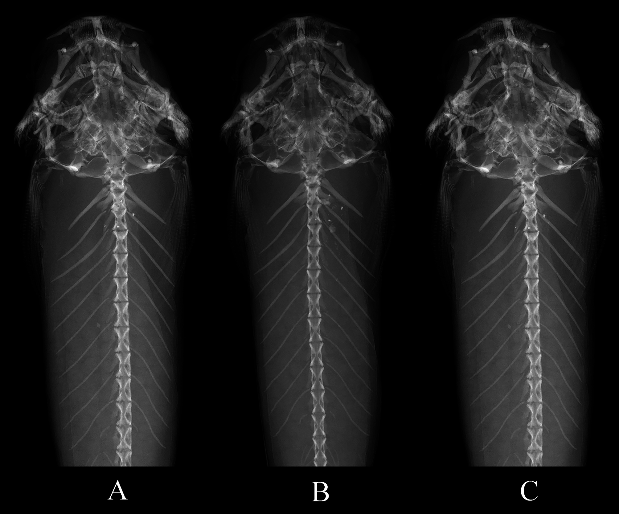
Cephalic lateral-line canals with simple, non-dendritic tubes ending in single pores. Supraorbital canal mostly within frontal bone. Supraorbital pores: s1, mesial to nasal-barbel base; s3 mesial to posterior nostril; and s6 median, positioned at interorbital area slightly posterior to transverse line through middle of eyes and at midlength of frontal. Holotype with abnormal asymmetrical supranumerary pores additional to single median s6 pore ( Fig. 8A View FIGURE 8 ), one of which positioned immediately anterior to s6 and another laterally to its right). Such additional pores are considered an abnormality, not seen in other specimens of the type series ( Fig. 3 View FIGURE 3 ). Infraorbital canal incomplete, with four pores, i1 (ventrolateral to nasal-barbel base) and i3 (ventrolateral to posterior nostril), i10 (posteroventral to eye) and i11 (posterior to eye). Otic canal without pores. Postotic pores po1 (anteromedial to opercular patch of odontodes), and po2 (medial to opercular patch of odontodes). Lateral line of trunk anteriorly continuous with postotic canal and reduced to short tube in all specimens, apparently with tubular ossification in holotype ( Fig. 4 View FIGURE 4 ). Lateral line pores ll1 and ll2 present dorsally to pectoral-fin base.
Coloration in ethanol ( Figs. 2 View FIGURE 2 and 3 View FIGURE 3 ). Pigmentation severely faded in all specimens due to long preservation history, but with general features still visible. Dark chromatophores distributed in inner and outer skin layers. Those on inner skin layer forming large roundish maculae responsible for the main color features of the body. Basic arrangement of maculae in four variably arranged rows. One row median, extending mid-dorsally along entire body, from occiput to base of caudal fin. Second row ventrolateral to that, extending from base of head through upper part of flanks, dorsal portion of caudal peduncle, to base of caudal fin. Third row most conspicuous of series, running along mid-lateral line, from immediately posterior to opercle to base of caudal fin. Fourth and ventralmost row shorter, extending from mid-length of abdomen through ventral margin of caudal peduncle to base of caudal fin. Basic four-row pattern disrupted by fusions (mostly along anterior part of body) and unaligned maculae, resulting in varied configurations. Mid-lateral row mostly independent but occasionally fused on dorsum. Head darkest on region corresponding to neurocranium, outlined by brain pigment seen by transparency. Dark spot normally at base of opercular patch of odontodes, with additional dark markings on cheeks. Light teardrop-shaded area extending from posterior margin of eye to base of opercular patch of odontodes, corresponding to levator operculi muscle. Base of nasal barbels surrounded with concentration of dark pigment, extending posteriorly as elongate dark field to anterior margin of eyes. Distal margin of integument fold of opercular patch of odontodes darkly-pigmented. Interopercular patch of odontodes white. Ventral side of the body lacking dark pigment. Fins with small brownish spots randomly distributed on fin-rays.


No known copyright restrictions apply. See Agosti, D., Egloff, W., 2009. Taxonomic information exchange and copyright: the Plazi approach. BMC Research Notes 2009, 2:53 for further explanation.
|
Kingdom |
|
|
Phylum |
|
|
Class |
|
|
Order |
|
|
Family |
|
|
Genus |
Trichomycterus alternatus ( Eigenmann, 1917 )
| Reis, Vinícius J. C. & Pinna, Mário De 2019 |
Pygidium alternatum
| Eigenmann, C. H. 1917: 700 |