
Trogolaphysa Mills, 1938
|
publication ID |
https://doi.org/10.11646/zootaxa.4032.1.1 |
|
publication LSID |
lsid:zoobank.org:pub:CB9720EB-7BB7-4199-A835-A3266B0DDA6B |
|
DOI |
https://doi.org/10.5281/zenodo.6096434 |
|
persistent identifier |
https://treatment.plazi.org/id/038AC52E-FFC6-A47C-FF3D-F99FFE267A5A |
|
treatment provided by |
Plazi |
|
scientific name |
Trogolaphysa Mills, 1938 |
| status |
|
Genus Trogolaphysa Mills, 1938
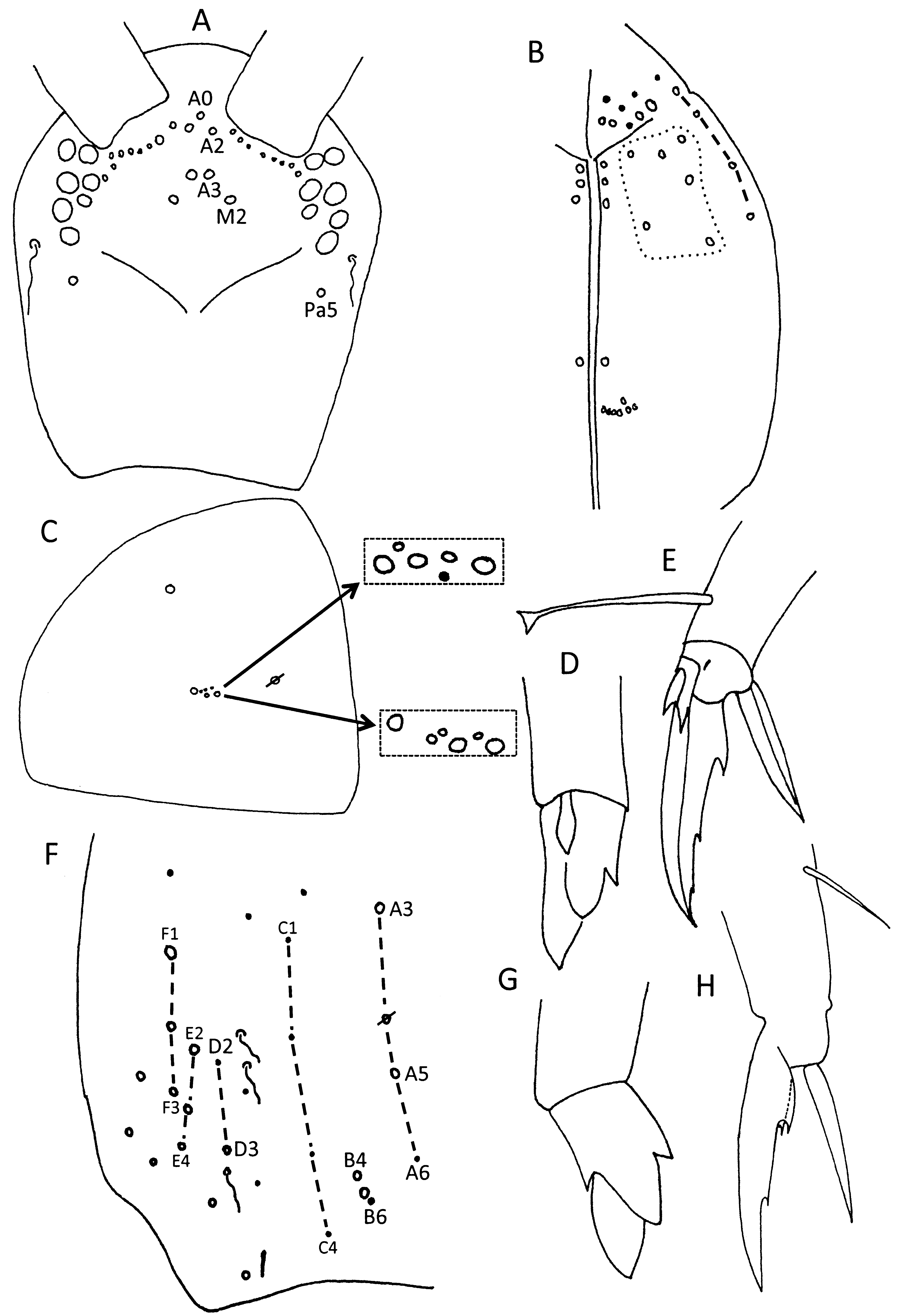
Diagnosis. Hyaline, finely denticulate scales covering Ant. 1–2, head, body, and ventral face of furcula; Ant. 4 not subdivided; eyes 0–8; labial chaetae L1 and L2 subequal; inner dorsal chaetotaxy of Th. 2 and Abd. 1 reduced to 4 and 3 chaetae, respectively; Abd. 2–4 with 2, 3, 3 bothriotricha; dens with 1–2 rows of spines; mucro square or rectangular but relatively short, with 3–5 teeth.
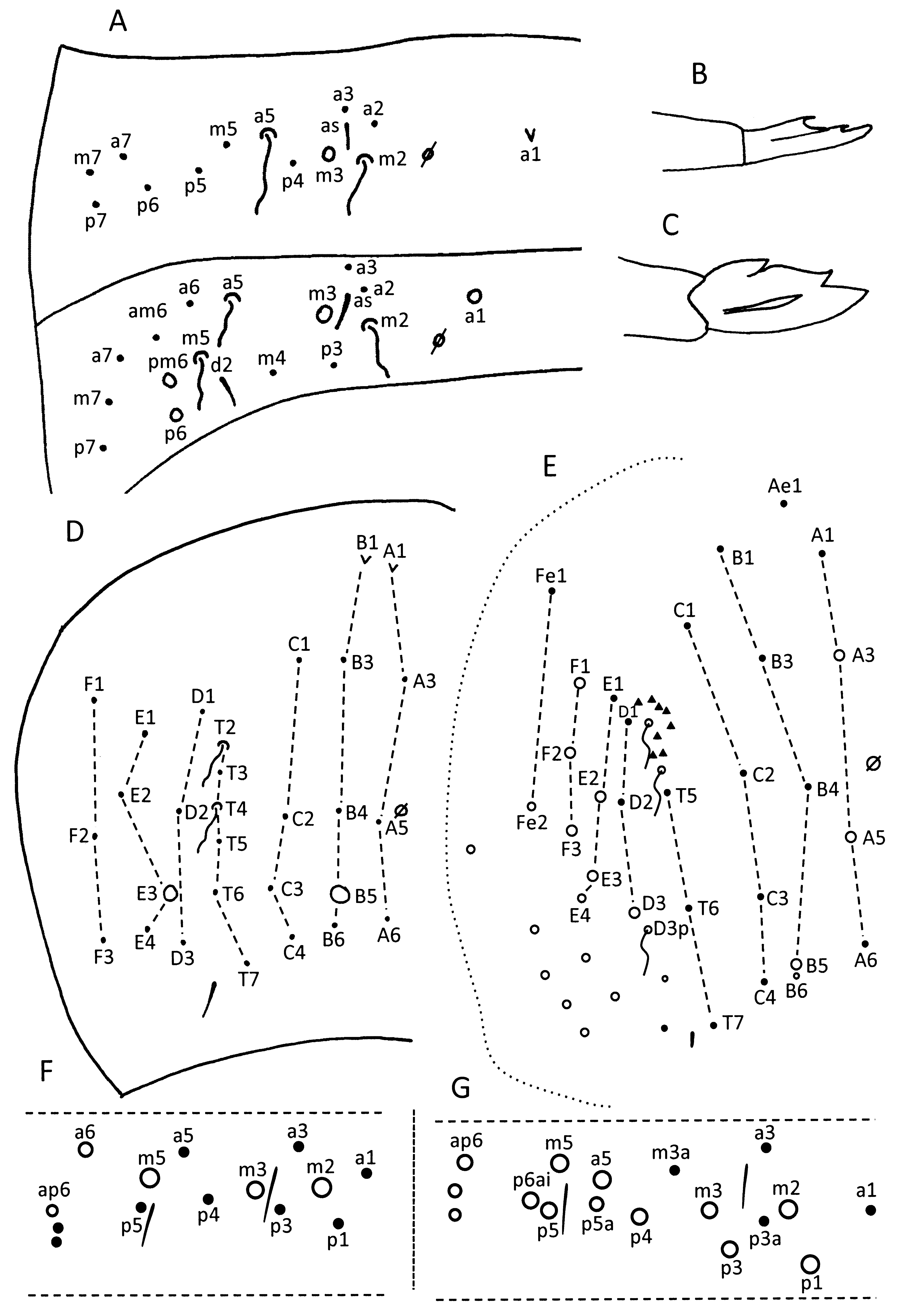
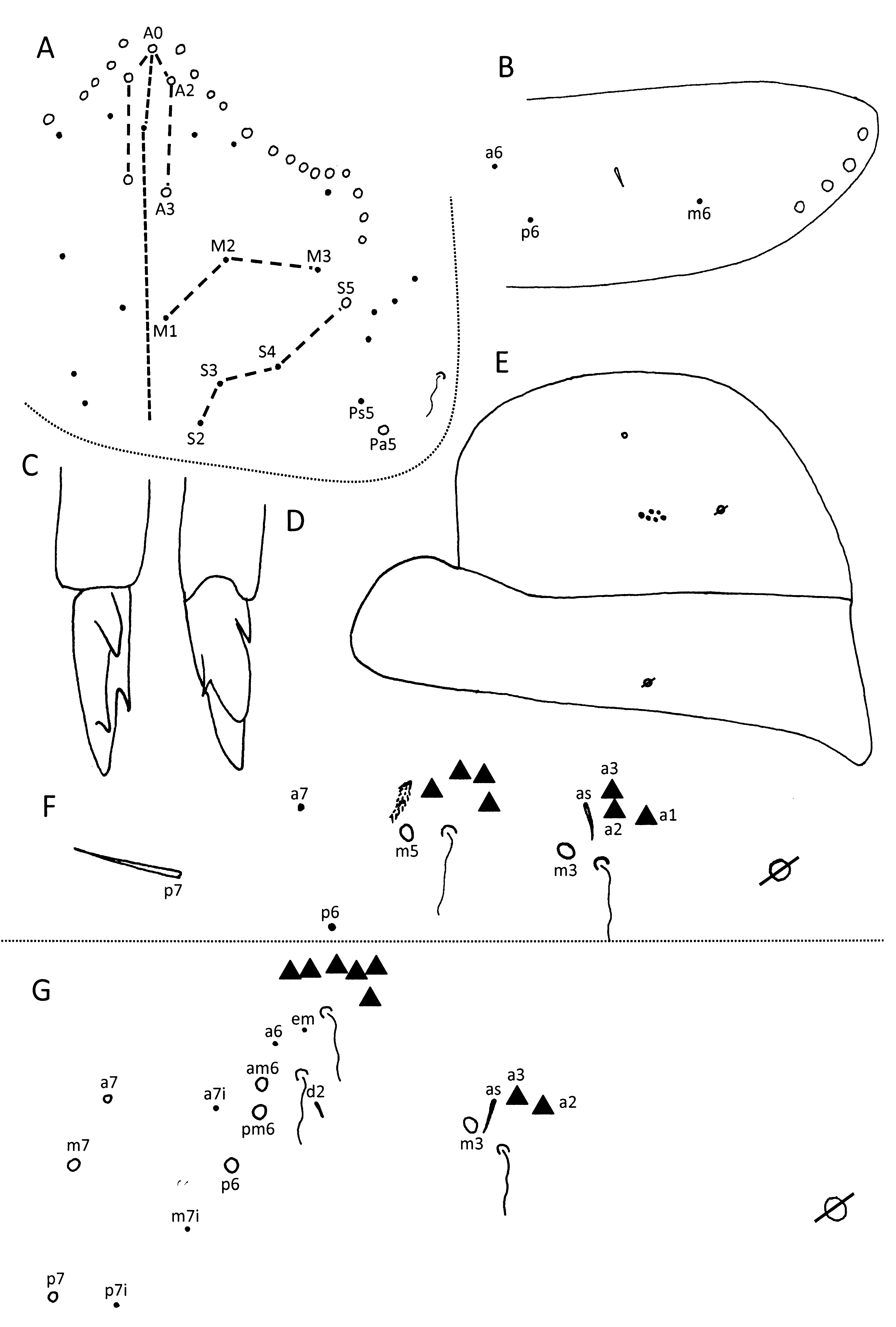
Remarks. The species described below share the following characters: scales present on Ant. 1–2 and basal fourth of Ant. 3; Ant. 4 with well organized whorls of chaetae on dorsal face, but indistinct on ventral face; labral chaetae smooth; sublobal plate of outer maxillary palp with two chaeta-like appendages ( Fig. 17 View FIGURE 17 C), one larger than the other; basal pleural chaeta coarsely ciliate, distal pleural chaeta short and smooth, spine-like (arrow on Fig. 17 View FIGURE 17 C); labial chaeta L2 as long as, or just slightly shorter than L1; all post-labial chaetae ciliate; post-labial column I with 3 proximal and 1 distal chaetae ( Fig. 14 View FIGURE 14 B); 6–8 ventral cervical chaetae; Th. 2–Abd. 5 with 11/011?2 Schaetae and 10/10100 S-microchaetae; Abd. 4 with mesochaeta B6; multiple (up to 16) lenticular organs present on membrane between fourth and fifth abdominal segments ( Fig. 20 View FIGURE 20 F); dens with two rows of spines; and mucro with four teeth.
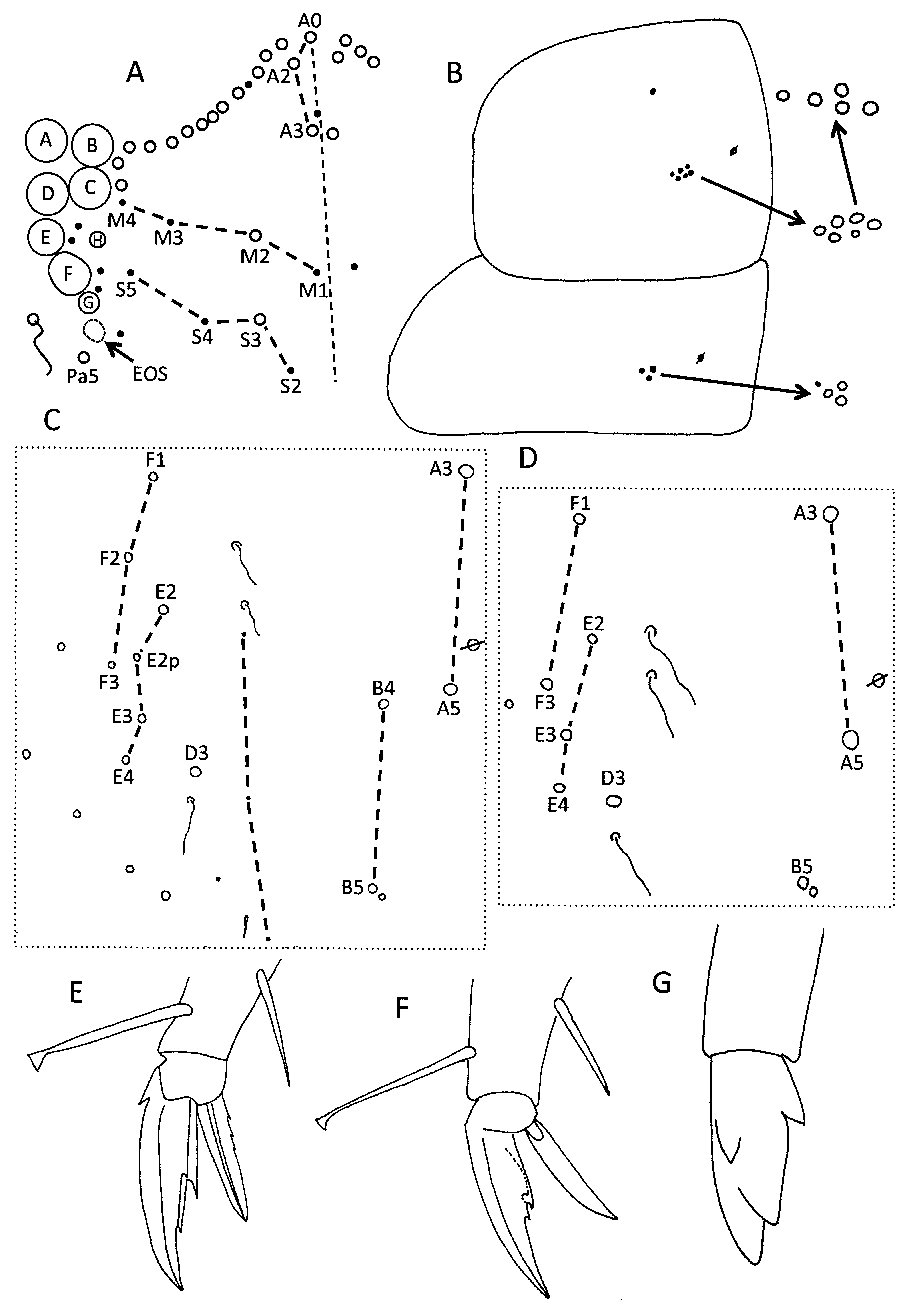
The organization of the dorsal chaetotaxy of head and body is typical for tribe Paronellini as described above and, as far as it can be observed in the 50+ years old specimens, most microchaetae are constant. All species carry four posterior microchaetae on the first abdominal segment and only the presence or absence of chaeta a6 is diagnostic. The general chaetotaxy of abdominal segments 2–3 is constant across all species. The second abdominal segment ( Fig. 12 View FIGURE 12 F) includes two bothriotricha, two macrochaetae (m3 and m5), S-chaeta as and three microchaetae external to bothriotrix a5. The third abdominal segment ( Fig. 12 View FIGURE 12 G) carries three bothriotricha, four macrochaetae (m3, am6, pm6 and p6), S-chaeta as, S-microchaeta d2 and six chaetae external to the lateral bothriotrichal complex. All bothriotricha are surrounded by supplementary chaetae. Most supplementary chaetae are fan-shaped or coarsely ciliate, but their diagnostic value was not explored because most specimens studied are opaque and it is not possible to score the characters unambiguously. There are usually six large lateral macrochaetae on Abd. 4 identified as D3, E2, E3, F1, F2 and F3. In the descriptions below these macrochaetae are individually listed when some are absent.
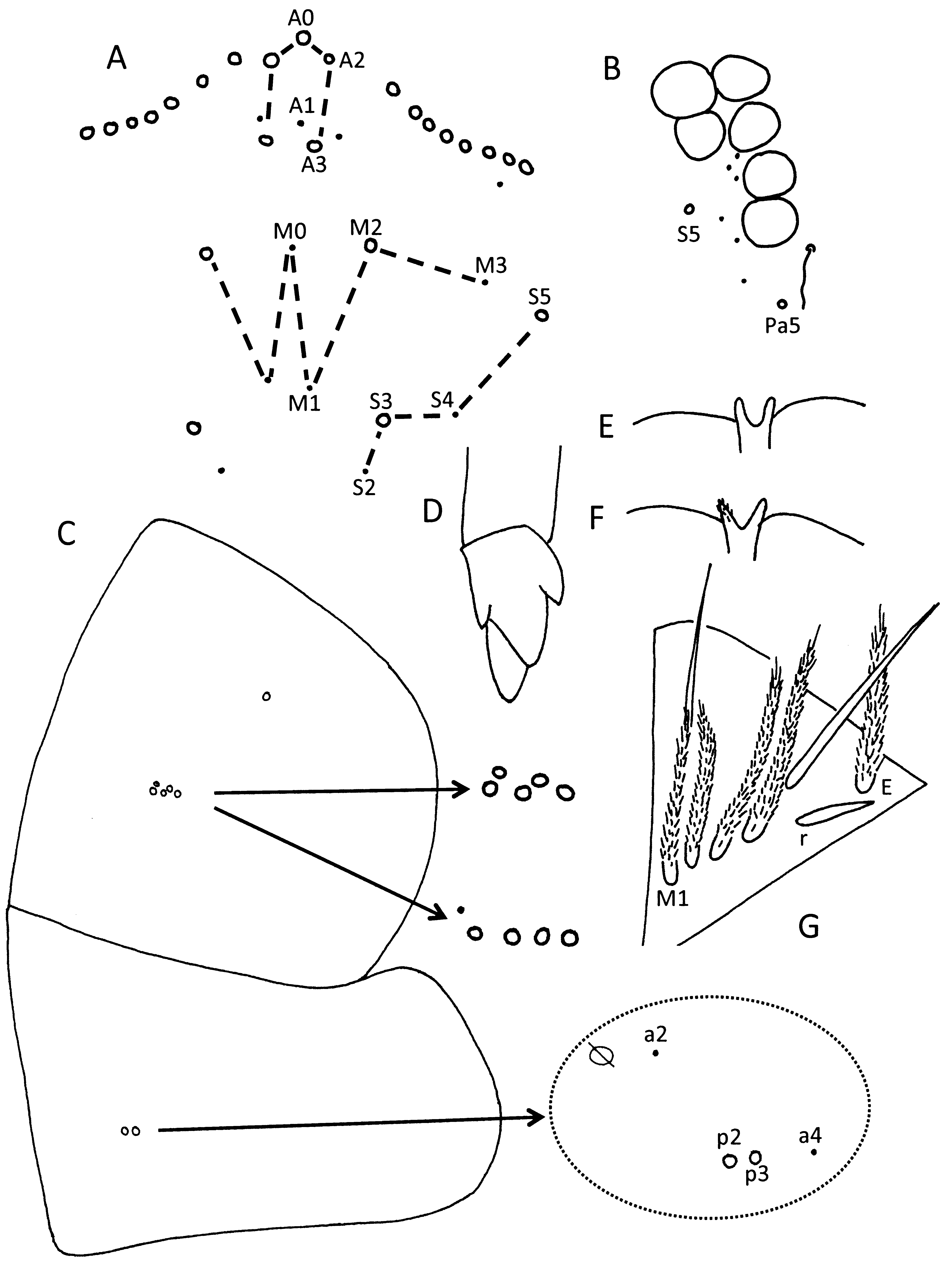
The number of chaetae in the trochanteral organ reported for most species is an approximation, as view of the organ is often obstructed; little diagnostic value should be attributed to this character. The chaetotaxy of the ventral tube was not considered because the opacity of most specimens obstructed observation of the chaetae. The number of spine-like chaetae on the dens is reported for the sake of completeness but most have fallen off in the material examined and only sockets were counted. Mucronal shape and relative position of basal teeth seem to be somewhat informative in species identification. Some species have a relatively square mucro (ratio mucro length to width of distal end of dens ≈2.2x or less, Figs. 2 View FIGURE 2 C, 5D) whereas in others the mucro appears rectangular or elongate (ratio 2.3 or more, Fig. 12 View FIGURE 12 C–D). In some species the basal mucronal teeth are sequential and do not overlap ( Figs. 7 View FIGURE 7 G, 8H), while in others the basal teeth are paired or almost paired ( Figs. 5 View FIGURE 5 D, 14G). These characters have to be evaluated carefully for each species because perception of shape and teeth placement is influenced by observation perspective and instar of the individual studied (cf. Figs. 8 View FIGURE 8 H–I). Illustrations of the mucro are provided for all species.
Variable characters, informative for species diagnosis in the taxa described below are: color pattern; eye number; number and relative insertion of macrochaetae on head, thorax and fourth abdominal segments; ornamentation of pre-labral chaetae; presence or absence and shape of spines on distal margin of labrum; number and ornamentation of posterior row of labial triangle chaetae; presence or absence of Abd. 1 chaeta a6; number of posterior chaetae on fourth abdominal segment; shape of tenent hair; number and shape of inner teeth of unguis, and shape of unguiculus.


No known copyright restrictions apply. See Agosti, D., Egloff, W., 2009. Taxonomic information exchange and copyright: the Plazi approach. BMC Research Notes 2009, 2:53 for further explanation.
|
Kingdom |
|
|
Phylum |
|
|
Class |
|
|
Order |
|
|
Family |