
Diura washingtoniana ( Hanson, 1940 )
|
publication ID |
https://doi.org/10.5281/zenodo.4761152 |
|
publication LSID |
lsid:zoobank.org:pub:6432D29B-B107-4809-8772-E4A65A12B5BD |
|
DOI |
https://doi.org/10.5281/zenodo.4763742 |
|
persistent identifier |
https://treatment.plazi.org/id/038B87F9-7570-FFA0-9925-ACA01CD5FC78 |
|
treatment provided by |
Felipe |
|
scientific name |
Diura washingtoniana ( Hanson, 1940 ) |
| status |
|
Diura washingtoniana ( Hanson, 1940) View in CoL , species propria
Presidential Springfly http://lsid.speciesfile.org/urn:lsid: Plecoptera .speciesfile.org: TaxonName:502549
Figs. 1 - 30 View Fig View Figs View Figs View Figs View Figs View Figs
Dictyopterygella washingtoniana Hanson 1940:147 View in CoL .
Holotype ♂, ( USNM), Lakes of the Clouds , Mt. Washington, New Hampshire, USA
Diura nanseni Brinck 1949:65 View in CoL , in part
Diura nanseni View in CoL “ subsp. washingtoniana View in CoL ” Brinck 1954:199
Diura nanseni Illies 1966:383 View in CoL , in part
Diura nanseni Zwick 1973:228 View in CoL , in part
Diura nanseni washingtoniana Hitchcock 1974:230 View in CoL
Diura nanseni Stark et al. 1998: 56 View in CoL
Diura nanseni Nelson 2001:616 View in CoL , in part
Diura nanseni Kondratieff 2004:152 View in CoL , in part
Diura nanseni DeWalt et al. 2017 2018 View in CoL : in part
Material examined. Specimens of D. washingtoniana examined for this study are consistent with previous descriptions and illustrations ( Hanson 1940, Brinck 1954, Hitchcock 1974, and Kondratieff 2004) of this species: USA; New Hampshire; Coos County, Lakes of the Clouds between Mt. Washington and Mt. Monroe, collector unknown, 4 July 1907, 1♂, 1♀ (paratopotypes) (CHNC). Same site, D. Arenburg,
17 June 1939, 5♂, 4♀ (paratopotypes) ( CHNC). Same site, C.H. Nelson and E.S. Nelson , 27 June 1996, 4 ♂, 11♀, 1 mature larva, 16 exuviae ( CHNC), 1♂, 1♀ ( BYU), [ CRN laboratory photo voucher 4] .
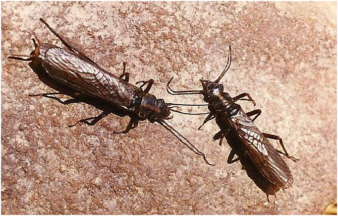
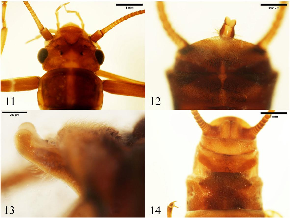
Adult habitus General color dark chestnut brown in life and in alcohol ( Fig. 1 View Fig , 11-14 View Figs ). The head dorsum exhibits an almost black X-band with an anterior margin that forms a distinct M-line ( Figs. 2 View Figs , 11 View Figs ). Anterior to the M-line is a lighter area that extends onto the clypeus; in some individuals it is shaped as a vague light M-band. Frontoclypeus with small orange-yellow oval-shaped spot near the base of each antenna and mandible. The interocellular area exhibits an orange-yellow subtriangular spot. The vertex and occiput is dark brown with a broad, longitudinal, orange-yellow, median band adjacent to, but clearly separated from, the interocellular area. Pronotum ( Figs. 2 View Figs , 11 View Figs ) dark brown and exhibiting a uniformly wide longitudinal, orangeyellow, median band. Meso- and metathoracic eusterna ( Fig. 3 View Figs ) dark brown with lateral areas slightly more darkly pigmented. The mesothoracic sternum ( Fig. 3 View Figs ) with a complete sternacostal suture (ss) separating the basisternum (bs) from the furcasternum (fs). Only the first two abdominal segments divided on each side by a pleural membrane into a separate tergum and sternum.
Male. Body Length 10.1 – 15.7 (𝑥 = 11.6) mm (n = 10). Macropterous, forewing length 7.0-8.3 (𝑥 = 7.7) mm (n=9). Tergum 8 ( Fig. 4 View Figs ) partially divided by a narrow, longitudinal, pale median strip that extends from near the posterior margin to near the anterior margin; setose and bearing a few blunt, stout sensilla basiconica near the hind margin on each side of the median strip. Tergum 9 ( Fig. 4 View Figs )
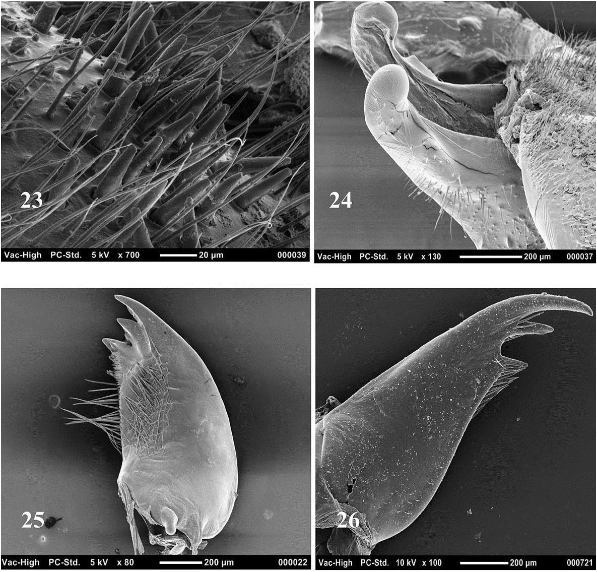
nearly divided by a pale, narrow, longitudinal, lightly sclerotized area that is a U- or V- shaped at the hind margin and extends anteriorly as a narrow strip to near the front margin; setose with a posterior mesal patch of blunt, stout sensilla basiconica on each side of the median strip ( Fig. 4 View Figs ). Tergum 10 in dorsal view ( Fig. 4 View Figs , 12 View Figs ) produced posteriorly; anterior half darker; divided by a paler, narrow, longitudinal, lightly sclerotized median strip that from the central broad triangular-shaped area at the posterior margin extends to the anterior margin; setose with a mesal patch of blunt, stout sensilla basiconica on each side of the median strip ( Figs. 4 View Figs , 23 View Figs ). Tergum 10 in lateral view posteromesally somewhat convex ( Fig. 5 View Figs ). Epiproct absent. Paraprocts extend from cerci on each side to meet at the median where they abruptly project caudally and are appressed or lie close to each other ( Figs. 4, 7 View Figs ). Inner surfaces of paraproct caudal projections membranous with outer surfaces largely sclerotized ( Figs. 6, 7 View Figs ) and setose bearing both short as well as long, slender setae. Caudal projections in dorsal and ventral views ( Figs. 4, 7 View Figs , 12 View Figs ) digitate with outer surfaces broadly convex, apices somewhat bulbous; in lateral view ( Figs. 5, 6 View Figs , 13 View Figs , 24 View Figs ) digitate, appearing boot-like as a result of upturned apices with broadly rounded margins.
Female. Body length 11.1 – 16.6 (𝑥 = 14.7) mm (n = 16). Macropterous, forewing length 8.9 – 11.7 (𝑥 =10.6) mm (n=16). Sternum 8 with mesal area darker than immediate lateral areas. Subgenital plate extends for a short distance over sternum 9 ( Figs. 8 View Figs , 14 View Figs ); shape variable ( Fig. 9 View Figs ), lateral margins either broadly rounded or become abruptly narrowed posteriorly, posterior margin either evenly rounded or slightly excavated mesally or truncate; ca 3.1 – 4.4 times as wide as long. Sternum 9 bearing two oblique brown bands ( Fig. 8 View Figs ). Spermatheca and vagina ( Fig. 10 View Figs ) membranous; elongate spermatheca (s) elongate, somewhat sausage-link-shaped, approximately 2.3X longer than wide; spermathecal duct (sd) approximately 0.8X length of vagina (v); spermatheca and duct bearing accessory glands (ag). Larva. Body length estimated from 1 mature larvae and 16 exuviae 14.5 - 16.5 (𝑥 = 15.5) mm. General body color dark brown patterned with light markings; antennae, legs and cerci light brown. Frontoclypeal region ( Fig. 15 View Figs ) dark brown, exhibiting a light anterior margin and a light mesolongitudinal spot just anterior to a transverse, light M-band connecting to the median ocellus. Interocellular region bears a mesal light ovalshaped spot and is bordered on each side by a light longitudinal band that extends toward the median ocellus from the lateral ocellus. Between the light longitudinal band and the antennal base of each side there is a small light round spot just anterior to the frontal suture. Vertex and occiput ( Fig. 15 View Figs ) dark brown with two oval light bands adjacent to the eyes and between them a transverse triangularshaped light median band; the three light bands are all patterned with dark brown lines. Labrum ( Fig. 15 View Figs ) dark with a mid-anterior light spot bordered on each side by a dark brown band. Left mandible ( Fig. 25 View Figs ) bearing three major and three minor teeth arranged as follows from dorsal aspect, a group consisting of one major tooth flanked on each side by a minor tooth, followed by a linear arrangement of a minor tooth and two major teeth; the ventralmost major tooth is the largest; teeth lack serrations; a row of acanthae arises from the bases of the three dorsal teeth. Maxillary lacinia triangular; bidentate ( Fig. 26 View Figs ), subapical tooth shorter, approximately one-half length of apical tooth; 2 axillary setae at base of subapical tooth; single marginal seta found just below the subapical tooth; inner margin below teeth forms a pronounced shoulder; shoulder and apical third of inner margin below the shoulder bearing setae; scattered sub-marginal setae present. Gills absent. Pronotum ( Fig. 15 View Figs ) with margins light; disc dark brown with light reticulate markings on each side of a light median band; anterior and posterior margins bearing row of short setae interspersed with a few long setae; lateral margins with few or no setae ( Fig. 27 View Figs ). Mesothoracic and metathoracic nota ( Fig. 16 View Figs ) dark brown and from anterior to posterior bearing four rows of light spots; first row bears two somewhat indistinct spots, the second and third each exhibit four spots and the fourth a single longitudinal band. Wingpads light with elongate dark brown markings. Legs ( Fig. 17 View Figs ) with dorsal margins of the femur, tibia, and tarsi fringed with long, silky setae. Femur ( Fig. 17 View Figs ) outer surface, except for narrow medial band, bearing numerous short thick setae. Tibia ( Fig. 17 View Figs ) outer surface with setae on dorsal and ventral areas. Only abdominal segments 1 & 2 divided by a pleural membrane of each side into separate terga and sterna. Abdominal tergal areas ( Fig 18 View Figs ) dark brown, each with anterior transverse row of light spots and a lighter posteromesal region. In lateral view paraprocts of mature male larva ( Fig. 22 View Figs ) with apical sections narrow, elongate and turned dorsally; those of mature female larva somewhat pyramidshaped with apices directed posteriorly, not narrowed or elongate. Stout setae lining eighth sternal margin of mature male larva interrupted mesally for a very short distance ( Fig. 20 View Figs ); interrupted in the mature female larva for longer distance, approximately 0.3 width of segment ( Fig. 21 View Figs ). Cercal segments ( Fig. 19 View Figs ) with dorsal margins bearing a fringe of long silky setae; posterior margins bearing a whorl of short setae, beginning with segment 16 posterior margin bearing one or two long setae.
Egg. Length ca. 450 µm; width ca. 298 µm (n=1). General shape oval ( Fig. 28 View Figs ); cross-section triangular ( Fig. 29 View Figs ). Collar ring-shaped and stalked ( Fig. 30 View Figs ). Anchor plate is mushroom-type of Isobe (1997), in Fig. 30 View Figs somewhat dorsally-ventrally compressed covering the collar margins and bearing globular processes. Chorionic surface dotted with numerous, slightly raised, similar-sized rounded protuberances ( Figs. 28-30 View Figs ).
Diagnosis. D. washingtoniana is distinguished by the following combination of features: (1) male tergum 8 partially divided by a narrow, membranous, pale median strip extending from near the posterior margin to near the anterior margin; (2) male tergum 9 nearly divided by a pale, narrow, longitudinal, lightly sclerotized area that is a U- or V- shaped at the hind margin and extends anteriorly as a narrow strip to near the front margin; (3) male tergum 10 divided by a narrow, membranous, pale median strip that from the central, broad membranous triangular-shaped area at the hind margin extends uninterrupted to the front margin; (4) male terga 8 – 10 bearing patches of blunt, stout sensilla basiconica; (5) male paraproct caudal projections in dorsal view with convex outer surfaces; (6) apices of male paraproct caudal projections broadly rounded and upturned to appear boot-like in lateral view.


| USNM |
Smithsonian Institution, National Museum of Natural History |
| BYU |
Monte L. Bean Life Science Museum |
No known copyright restrictions apply. See Agosti, D., Egloff, W., 2009. Taxonomic information exchange and copyright: the Plazi approach. BMC Research Notes 2009, 2:53 for further explanation.
|
Kingdom |
|
|
Phylum |
|
|
Class |
|
|
Order |
|
|
Family |
|
|
Genus |
Diura washingtoniana ( Hanson, 1940 )
| Nelson, Charles H. & Nelson, C. Riley 2018 |
Diura nanseni
| Kondratieff, B. C. 2004: 152 |
Diura nanseni
| Nelson, C. H. 2001: 616 |
Diura nanseni
| Stark, B. P. & S. W. Szczytko & C. R. Nelson 1998: 56 |
Diura nanseni washingtoniana
| Hitchcock, S. W. 1974: 230 |
Diura nanseni
| Zwick, P. 1973: 228 |
Diura nanseni
| Illies, J. 1966: 383 |
Diura nanseni
| Brinck, P. 1954: 199 |
Diura nanseni
| Brinck, P. 1949: 65 |
Dictyopterygella washingtoniana
| Hanson, J. F. 1940: 147 |
Diura nanseni
| Diura nanseni DeWalt et al. 2017 2018 : in part |