
Agyneta sandia, Dupérré, Nadine, 2013
|
publication ID |
https://doi.org/10.11646/zootaxa.3674.1.1 |
|
publication LSID |
lsid:zoobank.org:pub:981F80ED-96D7-40C7-8A3C-677954416A2E |
|
DOI |
https://doi.org/10.5281/zenodo.6162378 |
|
persistent identifier |
https://treatment.plazi.org/id/038D6700-FFE7-5648-118C-0064AB8CB3FA |
|
treatment provided by |
Plazi |
|
scientific name |
Agyneta sandia |
| status |
sp. nov. |
Agyneta sandia View in CoL new species
Figs 283–290 View FIGURES 283 – 289 View FIGURES 290 – 299 , map 19
Type material: Male holotype from New Mexico, Bernalillo County, Sandia Mountains, C.C. Hoff ( AMNH). EXAMINED.
Etymology: The specific name is a noun in apposition taken from the type locality Sandia Mountains, New Mexico, USA.
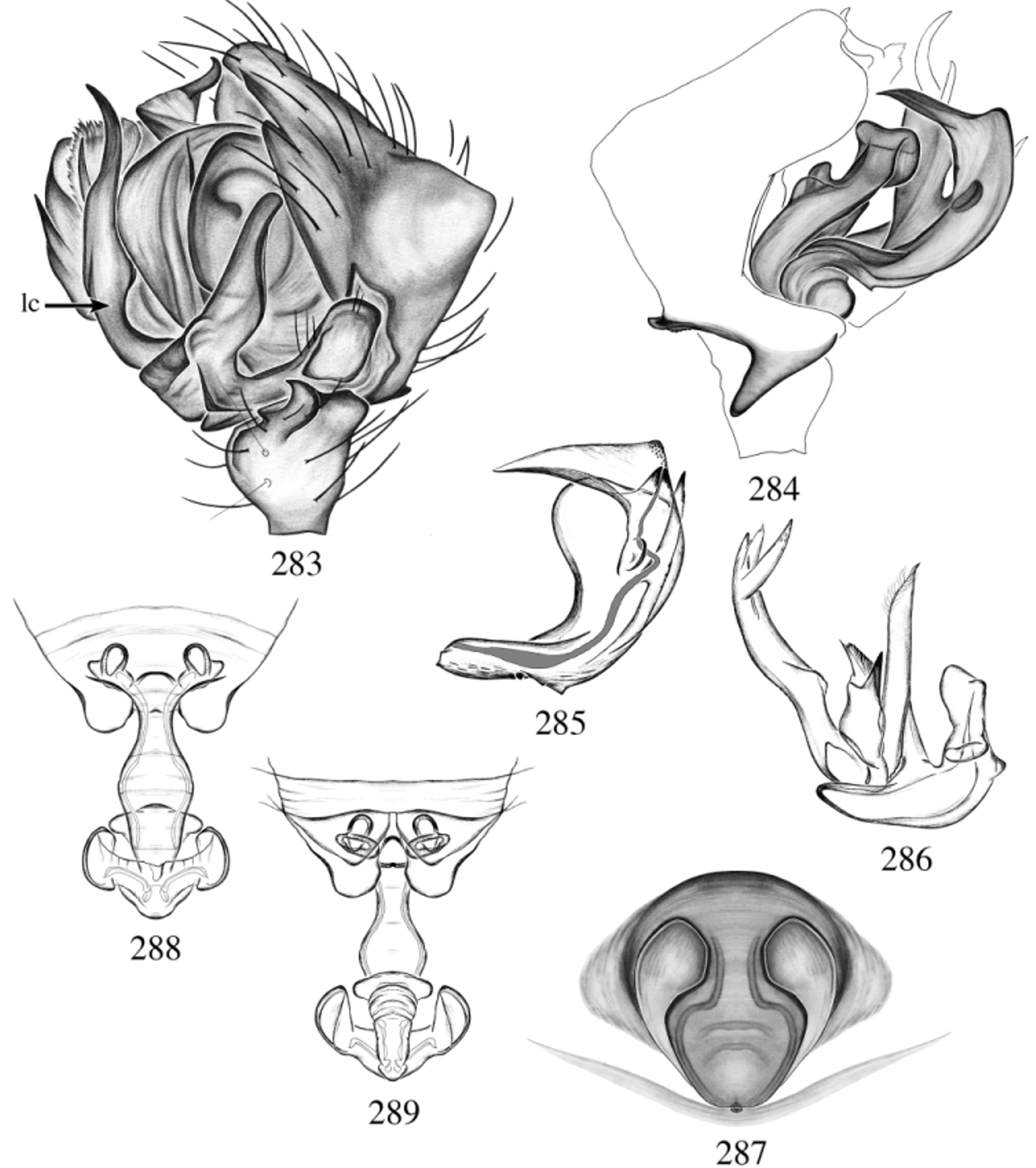
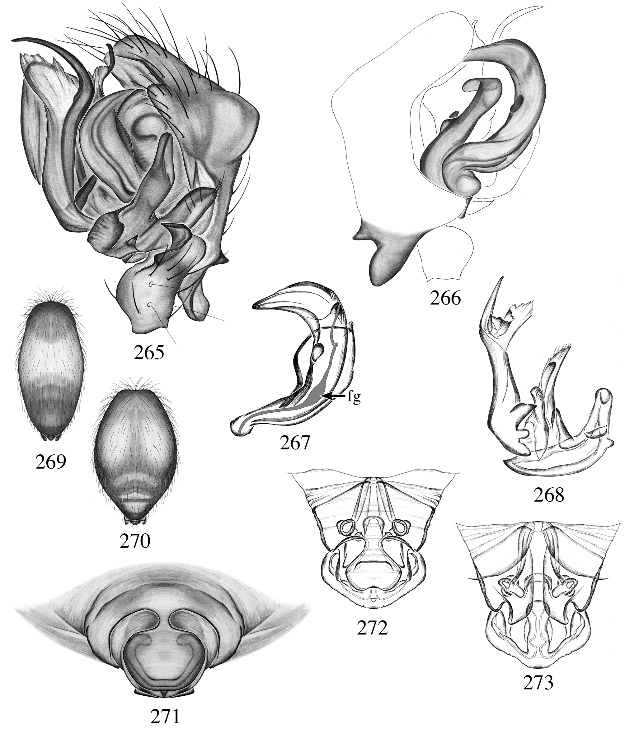
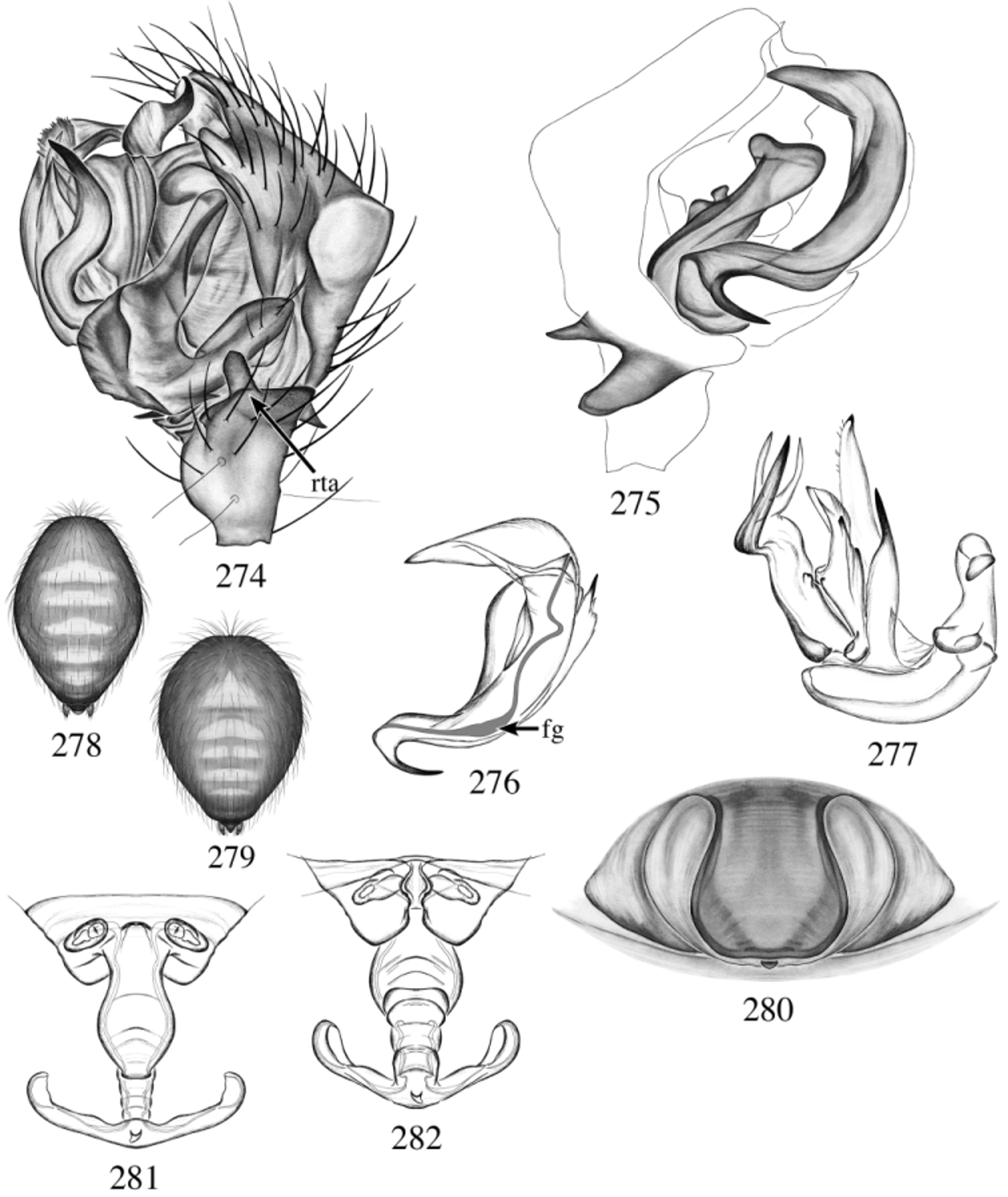
Diagnosis: Males are diagnosed from all Agyneta species by their straight, Y-shaped lamella characteristica ( Fig. 283 View FIGURES 283 – 289 ). From other species of the group by the large gap between the dorsal and ventral cymbial tubercles ( Fig. 284 View FIGURES 283 – 289 ), narrower in all other species ( Figs 255 View FIGURES 254 – 264 , 266 View FIGURES 265 – 273 , 275 View FIGURES 274 – 282 ). Females are diagnosed from all Agyneta by their basally narrow proximal part of scape enlarging in a spatula-shaped plate ( Fig. 287 View FIGURES 283 – 289 ). To distinguish it from A. barfoot see diagnosis of the latter.
Description: Male: Total length 1.90; carapace length 0.84, width 0.72.
MAP. 19. Localities of Agyneta sandia n. sp.
CEPHALOTHORAX: Carapace yellow, shiny, finely reticulate; margin, radiating lines suffused with dark gray. Sternum suffused with dark gray. Clypeus height 2. Chelicerae yellow, excavated; ~ 11 seta-tipped tubercles; promargin two teeth, retromargin three tiny denticles; both margins with rounded projection at base of fang, smaller on promargin. Cheliceral stridulatory organ ~ 58 striae, well spaced slowly getting close together basally. ABDOMEN: Oval, uniformly light to dark gray. LEGS: Yellow; leg I total length: 3.63; leg III total length: 2.47; Tm I: 0.20, Tm IV absent. GENITALIA: Palpal retrolateral tibial apophysis curved, smooth; dorsal tibial apophysis absent; two retrolateral, one dorsal trichobothria ( Fig. 283 View FIGURES 283 – 289 ). Cymbium triangular; glabrous depression present ( Fig. 283 View FIGURES 283 – 289 ); dorsal cymbial turbercle sharp and rugose; ventral cymbial tubercle elongated, pointed and smooth; prolateral notch shallow ( Fig. 284 View FIGURES 283 – 289 ). Paracymbium apical pocket medium, anterior pocket short and curved, posterior pocket angled ( Fig. 283 View FIGURES 283 – 289 ). Embolus tip pointed and thin; small spines apically; basally with two spikes; Fickert’s gland basal, slightly enlarged; ventral lamella slender and pointed; thumb short, reaching below the embolus proper ( Fig. 285 View FIGURES 283 – 289 ). Embolus proper set apically, of equal part ( Fig. 285 View FIGURES 283 – 289 ). Anterior terminal apophysis long, narrow with thin long protrusions, no extra process; posterior terminal apophysis with two sharp pointed tip; lamella characteristica long, ending in two large well sclerotized point, with a transparent lamella in between ( Fig. 286 View FIGURES 283 – 289 ).
Female: Total length 1.99; carapace length 0.75, width 0.59.
CEPHALOTHORAX: Coloration as in male. Chelicerae promargin five teeth, retromargin four denticles. Cheliceral stridulatory organ ~ 51 striae, narrowly spaced. ABDOMEN: Oval, uniformly light to dark gray. LEGS: Coloration as male; palpal tarsal claw absent; leg I total length: 3.00; leg III total length: 2.13; Tm I: 0.22, Tm IV: absent. GENITALIA: Epigynum with proximal part of scape narrow basally, enlarging (2.5x) and spatula-shaped; epigynal slits large, not reaching the anterior part of epigynum; pit hook depression absent ( Fig. 287 View FIGURES 283 – 289 ); lateral lobes long and folded; stretcher minuscule; pit deep ( Fig. 288 View FIGURES 283 – 289 ). Median part of scape narrow, short, wrinkled; genital pores situated at base of lateral lobes pockets ( Fig. 289 View FIGURES 283 – 289 ). Internal genitalia with an oval ventral receptacula and an elongated horizontally positioned dorsal one ( Figs 288, 289 View FIGURES 283 – 289 ).
Other material examined: USA: Arizona: 45km E Douglas, Guadalupe Canyon, 13.vii.1972, 4Ƥ ( CAS); Chiricahua Mountains, 11.vi. 1973, 2743m, 23, V. Roth ( CAS); Chiricahua Mountains, Barfoot Meadows, 05.vii.1975, 131Ƥ, D. Ubick ( DUC); Chiricahua Mountains, Cave Creek Canyon, 28.viii.1982, 1554– 1615m, pitfall, 23, V. Roth ( CAS); Chiricahua Mountains, Cave Creek Canyon, outside of Woodcutter’s Cave, 26.vii.1996, 5200ft, 2Ƥ, P. Craig, V. Roth, D. Ubick ( DUC); Chiricahua Mountains, E Turkey Creek, 10.viii.1991, under rocks, 237Ƥ, A. Jung ( DUC); Chiricahua Mountains, Upper Cave Creek, 1828-2286m, can traps, 27.vii.1984, 73, 22.viii.1984, 23, V. Roth ( CAS); Flagstaff, 16.vi.1934, 13, W. Ivie, H.A.R ( AMNH); Hualpal Mountains, 6500ft, 04.viii.1956, 1Ƥ, R. Schick ( AMNH); Santa Catalina Mountains, Nugget Cave, 21.vii.1993, 13, R. Pape ( DUC); Santa Catalina Mountains, Mount Bigelow, 24.vii.1965, 1Ƥ, W. Gertsch, R. Hastings ( AMNH). New Mexico: Emory Pass Summit, Mimbres Mountains, 06.ix.1941, 13, W. Ivie ( AMNH); Sandia Mountains, 13, C. Hoff ( AMNH). Texas: 6.4km N Lockhart, 13.iv.1963, 6Ƥ, W. Gertsch, W. Ivie ( AMNH); Bastrop State Park, 24– 27.v.1983, oak woods, 1Ƥ, S., J. Peck ( AMNH); Bill Haney Pecan Orchard, FM 1476, 3.2km E US 377/67, 02 - 09.v.2001, pitfall in pecan orchard, 232Ƥ, A. Calixto ( TAMU); Constant Sorrow Cave, Camp Bullis, 17.iv.2001, 1Ƥ, J. Reddell, J. Reyes ( TMM); Get a Rope Cave, Camp Bullis, 23.viii.2000, 1Ƥ, G. Veni (JCC); Mastodon Pit, 17.vi.1993, 1Ƥ, S. Harden, G. Veni (JCC). Utah: Verdure, 12.v.1933, 13, W. Ivie ( AMNH).
Distribution: Southwestern USA (Arizona to Texas).
The parva View in CoL group is the largest and includes eleven species, the group contains some Eastern species, A. parva ( Banks 1896) View in CoL , A. evadens ( Chamberlin 1925) View in CoL , A. unimaculata ( Banks 1892) View in CoL , A. barrowsi ( Chamberlin & Ivie 1944) View in CoL , and A. regina ( Chamberlin & Ivie 1944) View in CoL , and some Southwestern species, A. spicula View in CoL n. sp., A. grandcanyon View in CoL n. sp., A. chiricahua View in CoL n. sp., A. crista View in CoL n. sp., A. tuberculata View in CoL n. sp. and A. catalina View in CoL n. sp.
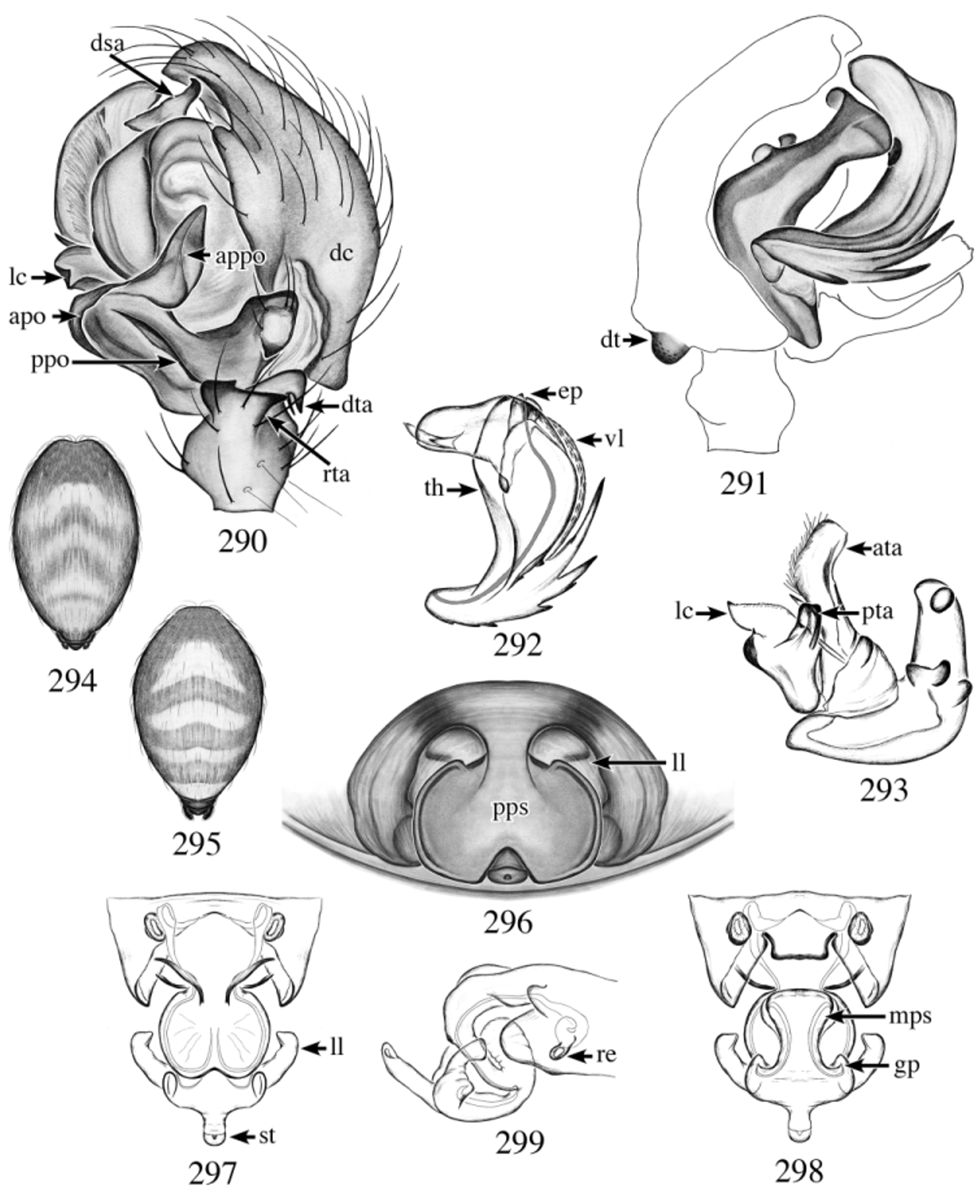
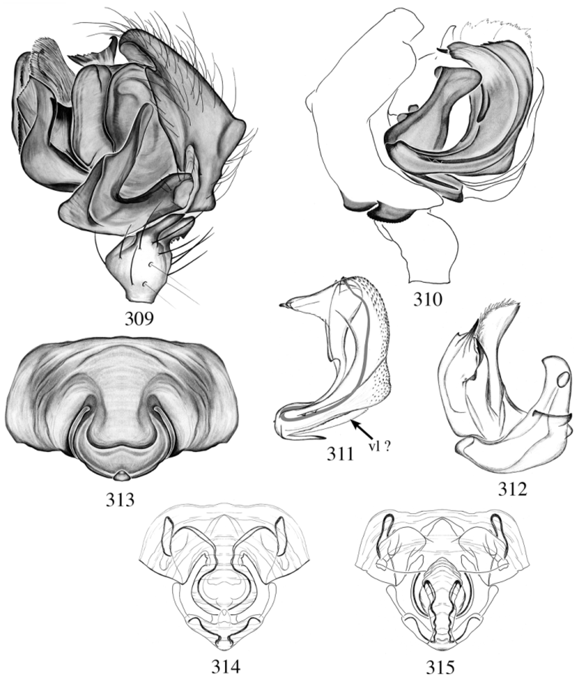
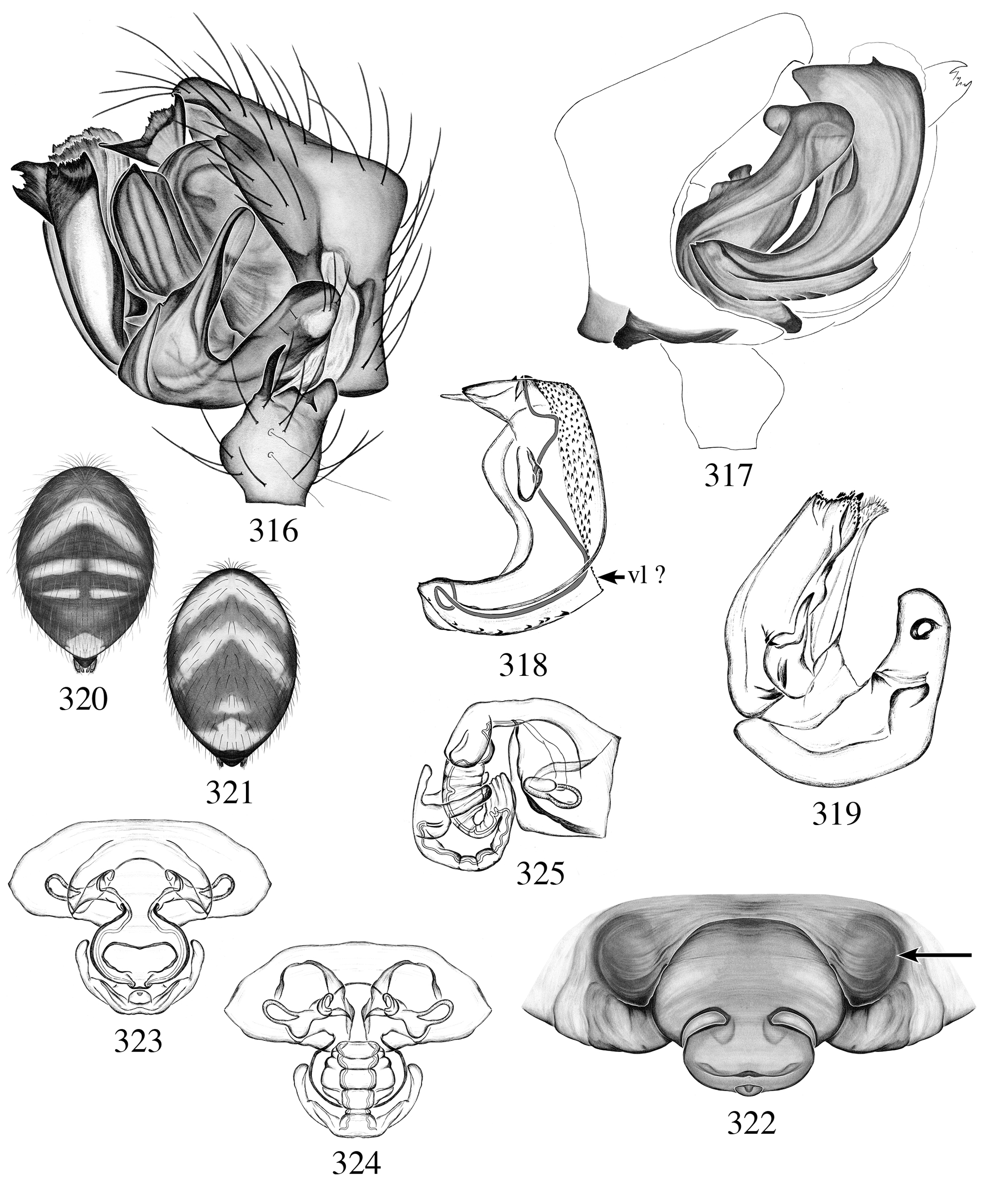
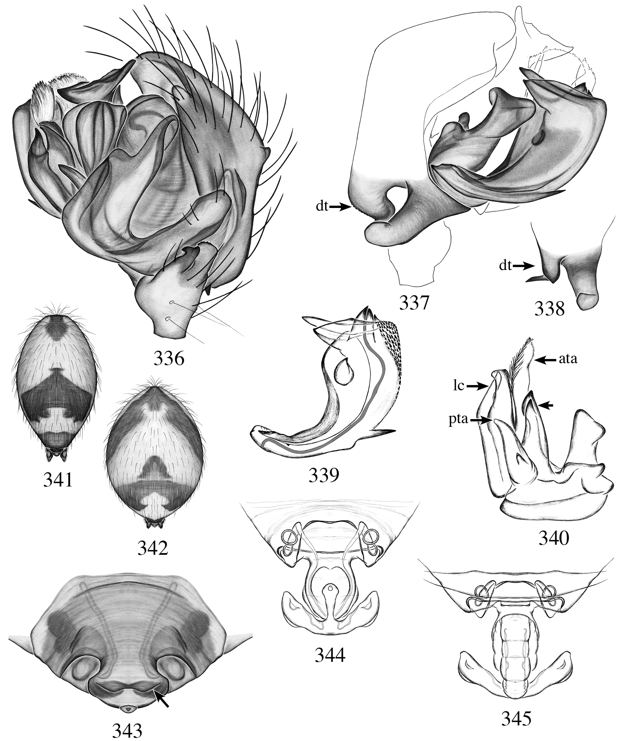
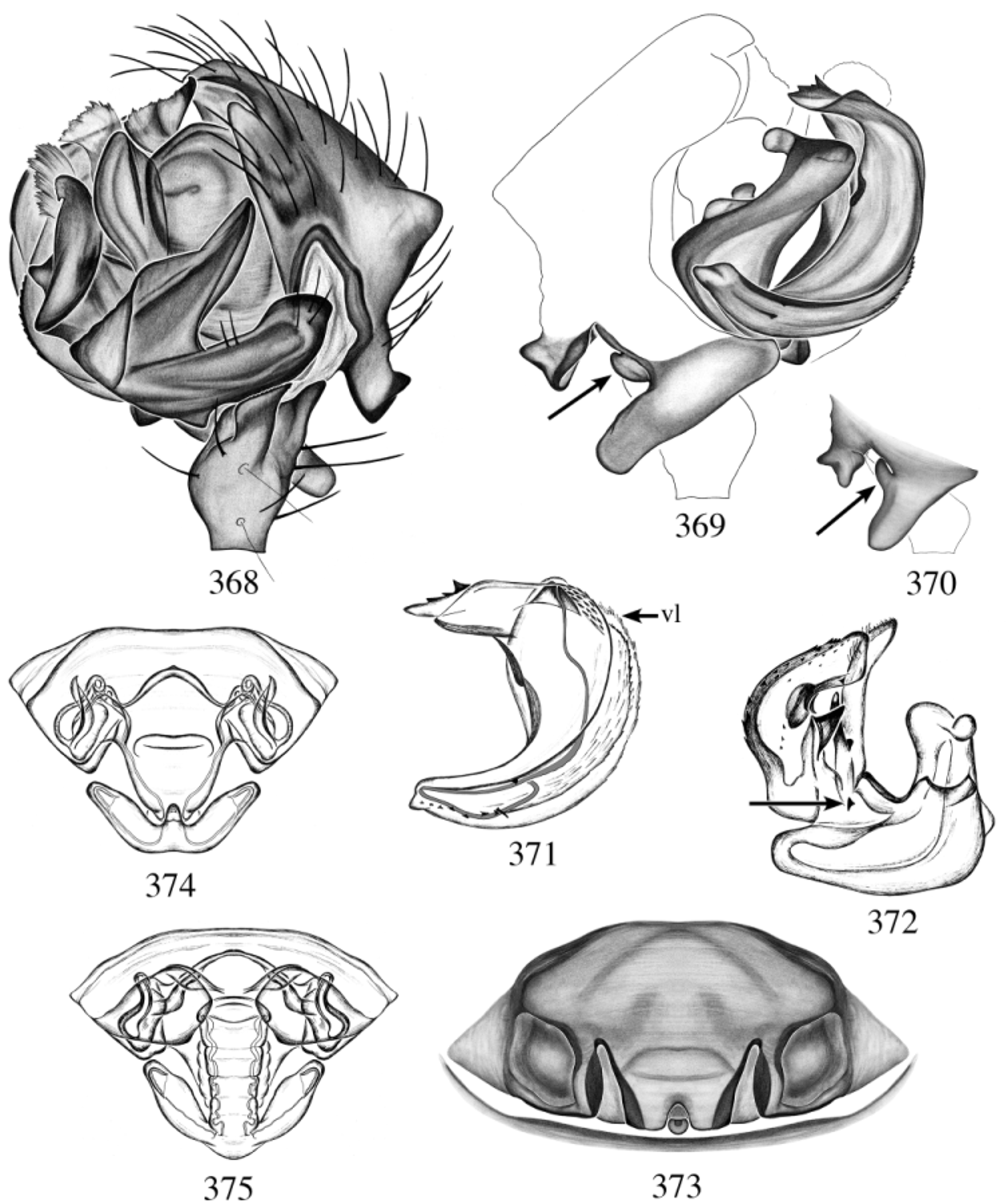
The group is defined by two unique characters. The first is the presence of a prong (serrated or not) at the tip of embolus ( Figs 292 View FIGURES 290 – 299 , 311 View FIGURES 309 – 315 , 318 View FIGURES 316 – 325 , 339 View FIGURES 336 – 345 , 371 View FIGURES 368 – 375 ; note that this prong is not an extension of the embolus proper like in the A. fillmorana View in CoL group). Second, the embolus proper set vertically and not related to the tip of the embolus ( Fig. 292 View FIGURES 290 – 299 ).
Within the group we can easily cluster some similar species, A. parva ( Banks 1896) View in CoL , A. evadens ( Chamberlin 1925) View in CoL , A. unimaculata ( Banks 1892) View in CoL , A. spicula View in CoL n. sp., A. grandcanyon View in CoL n. sp., A. chiricahua View in CoL n. sp. and A. regina ( Chamberlin & Ivie 1944) View in CoL . They all share non-excavated chelicerae without seta-tipped tubercles in males and the none serrated prong at the tip of the embolus.
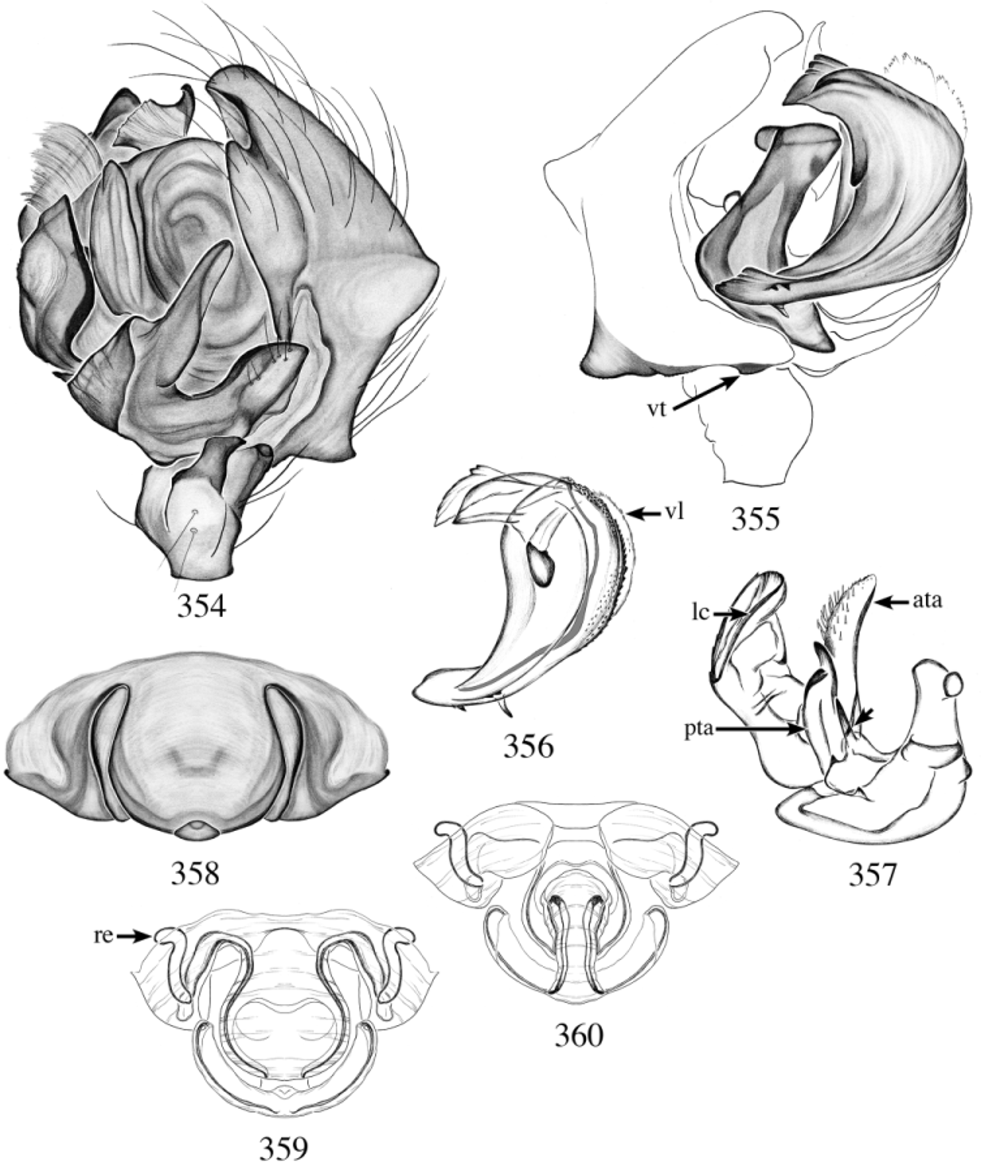
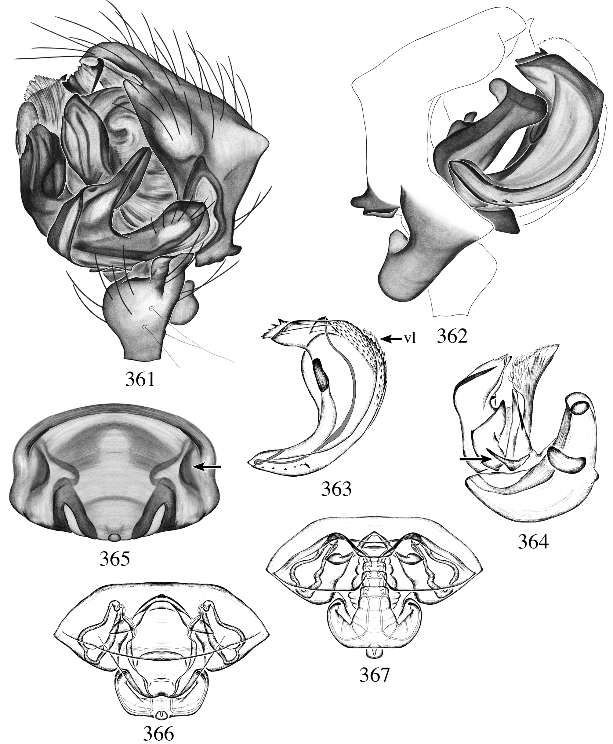
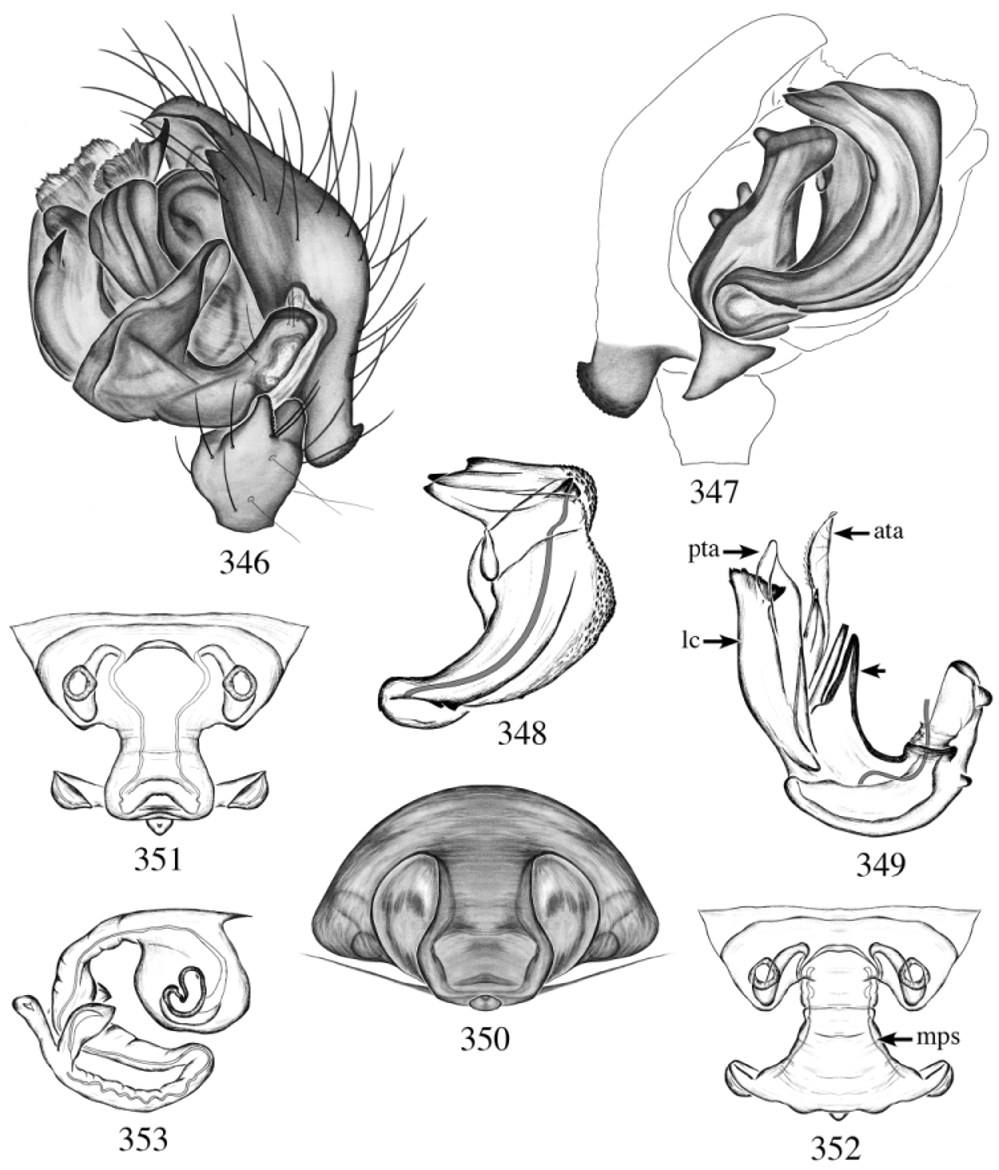
In A. barrowsi ( Chamberlin & Ivie 1944) View in CoL , A. crista View in CoL n. sp., A. tuberculata View in CoL n. sp. and A. catalina View in CoL n. sp., males have excavated chelicerae with seta-tipped tubercles, a serrated prong at the tip of the embolus ( Figs 356 View FIGURES 354 – 360 , 363 View FIGURES 361 – 367 , 371 View FIGURES 368 – 375 , 378 View FIGURES 376 – 379 ) and an associated prong at the base of the posterior terminal apophysis ( Figs 357 View FIGURES 354 – 360 , 364 View FIGURES 361 – 367 , 372 View FIGURES 368 – 375 , 379 View FIGURES 376 – 379 ).
Members of the parva View in CoL group can be characterized as such, palpal tibia with two retrolateral trichobothria and a dorsal one; very well developed retrolateral and dorsal tibial apophyses ( Fig. 290 View FIGURES 290 – 299 ); embolus with apical prong; Fickert’s gland absent; ventral lamella reduced to absent. Note that the ventral lamella in this group is sometimes difficult to observe. Females have long lateral lobes (sometimes folded) and long stretchers ( Figs 297 View FIGURES 290 – 299 , 314 View FIGURES 309 – 315 , 353 View FIGURES 346 – 353 ).


No known copyright restrictions apply. See Agosti, D., Egloff, W., 2009. Taxonomic information exchange and copyright: the Plazi approach. BMC Research Notes 2009, 2:53 for further explanation.
|
Kingdom |
|
|
Phylum |
|
|
Class |
|
|
Order |
|
|
Family |
|
|
SubFamily |
Micronetinae |
|
Genus |
Agyneta sandia
| Dupérré, Nadine 2013 |
A. barrowsi (
| Chamberlin & Ivie 1944 |
A. regina (
| Chamberlin & Ivie 1944 |
A. regina (
| Chamberlin & Ivie 1944 |
A. barrowsi (
| Chamberlin & Ivie 1944 |
A. evadens (
| Chamberlin 1925 |
A. evadens (
| Chamberlin 1925 |
A. parva (
| Banks 1896 |
A. parva (
| Banks 1896 |
A. unimaculata (
| Banks 1892 |
A. unimaculata (
| Banks 1892 |