
Asteroporpa ( Astrohelix ) bellator ( Koehler, 1904 )
|
publication ID |
https://doi.org/10.11646/zootaxa.4227.4.4 |
|
publication LSID |
lsid:zoobank.org:pub:D71F8124-BD24-4FDC-A659-9ECC496150BD |
|
DOI |
https://doi.org/10.5281/zenodo.5688478 |
|
persistent identifier |
https://treatment.plazi.org/id/038E87A0-4E7B-D36D-FF7F-FF68E5FAFDB1 |
|
treatment provided by |
Plazi |
|
scientific name |
Asteroporpa ( Astrohelix ) bellator ( Koehler, 1904 ) |
| status |
comb. nov. |
Asteroporpa ( Astrohelix) bellator ( Koehler, 1904) View in CoL comb. nov.
( Figs 1–2 View FIGURE 1 View FIGURE 2 )
Astrotoma bellator Koehler, 1904 View in CoL , 154–144, pl. 19, fig. 8, pl. 23, fig. 1, pl. 28, fig. 8–9.
Astrohelix bellator Döderlein, 1930 View in CoL , 376–377, pl. 1, fig. 9–9b (redescription of holotype of Astrotoma bellator View in CoL ).
Type material. ZMA E2044, holotype of Astrotoma bellator , collected by the Siboga Expedition , station 105, off northern Sulu Island, Philippine, 6˚8’N, 121˚19’E, 275 m deep, 4 July 1899.
Diagnosis. Epidermal ossicles are cone-shaped on aboral disc, cone and plate-shaped on the oral plates, and cone-shaped with terminal projections at the edges of the disc in the interradii; cone-shaped with relatively long terminal projections, at the lateral interradial disc; hooklet-bearing plates on basal portion of arms are completely in contact.
Redescription of holotype. Size is 15.9 mm in disc diameter, at least 70 mm in arm length.
Disc. Disc is five-lobed in shape with notched interradial edges ( Fig. 1 View FIGURE 1 A–B). On the aboral side, radial shields and their surrounds are tumid ( Fig. 1 View FIGURE 1 A). There are no conspicuous raised rows of hooklet-bearing plates and the hooklet-bearing plates are scattered on the periphery of each radial shield ( Fig. 1 View FIGURE 1 E). Each hooklet-bearing plate possesses ten to twelve tubercle-shaped articulations for hooklets. Except for the hooklet-bearing plates, the aboral disc is covered by cone-shaped epidermal ossicles with one or two terminal projections which are approximately the same length of the height of the cone-shaped epidermal ossicles ( Fig. 1 View FIGURE 1 C–D). The cone-shaped epidermal ossicles are approximately 200–300 µm in length at the periphery of the disc ( Fig. 1 View FIGURE 1 E), and approximately 180–280 µm in length at the center of the disc ( Fig. 1 View FIGURE 1 D). Radial shields are completely concealed by epidermal ossicles, oval shape, approximately 7.5 mm in length, 2.5 mm in width distally and 1.0 mm in width proximally, and almost reach the disc center ( Fig. 1 View FIGURE 1 A).
The oral surface of the disc is covered both by flat, polygonal plate-shaped epidermal ossicles and by coneshaped epidermal ossicles with mostly one, or rarely two terminal projections similar to those on aboral disc ( Fig. 1 View FIGURE 1 G). The cone-shaped epidermal ossicles are scattered on the periphery of the disc and on the oral plates ( Fig. 1 View FIGURE 1 G– H). The plate-shaped epidermal ossicles are approximately 300–330 µm in length on the periphery of the disc, and approximately 180–270 µm in length on the oral plates ( Fig. 1 View FIGURE 1 G–H). The cone-shaped epidermal ossicles are approximately 300–330 µm in length ( Fig. 1 View FIGURE 1 H). Oral shields, adoral shields, oral plates and ventral arm plates are completely concealed by epidermal ossicles ( Fig. 1 View FIGURE 1 B, G). Uniformly acute and spiniform teeth are situated on the jaws ( Fig. 1 View FIGURE 1 G). The length of the teeth are different dependent on their location on each jaw. At the top of jaws, they are approximately 1 mm in length, and toward the basal part of the jaws, the teeth gradually decrease in length to approximately 350 µm ( Fig. 1 View FIGURE 1 G).
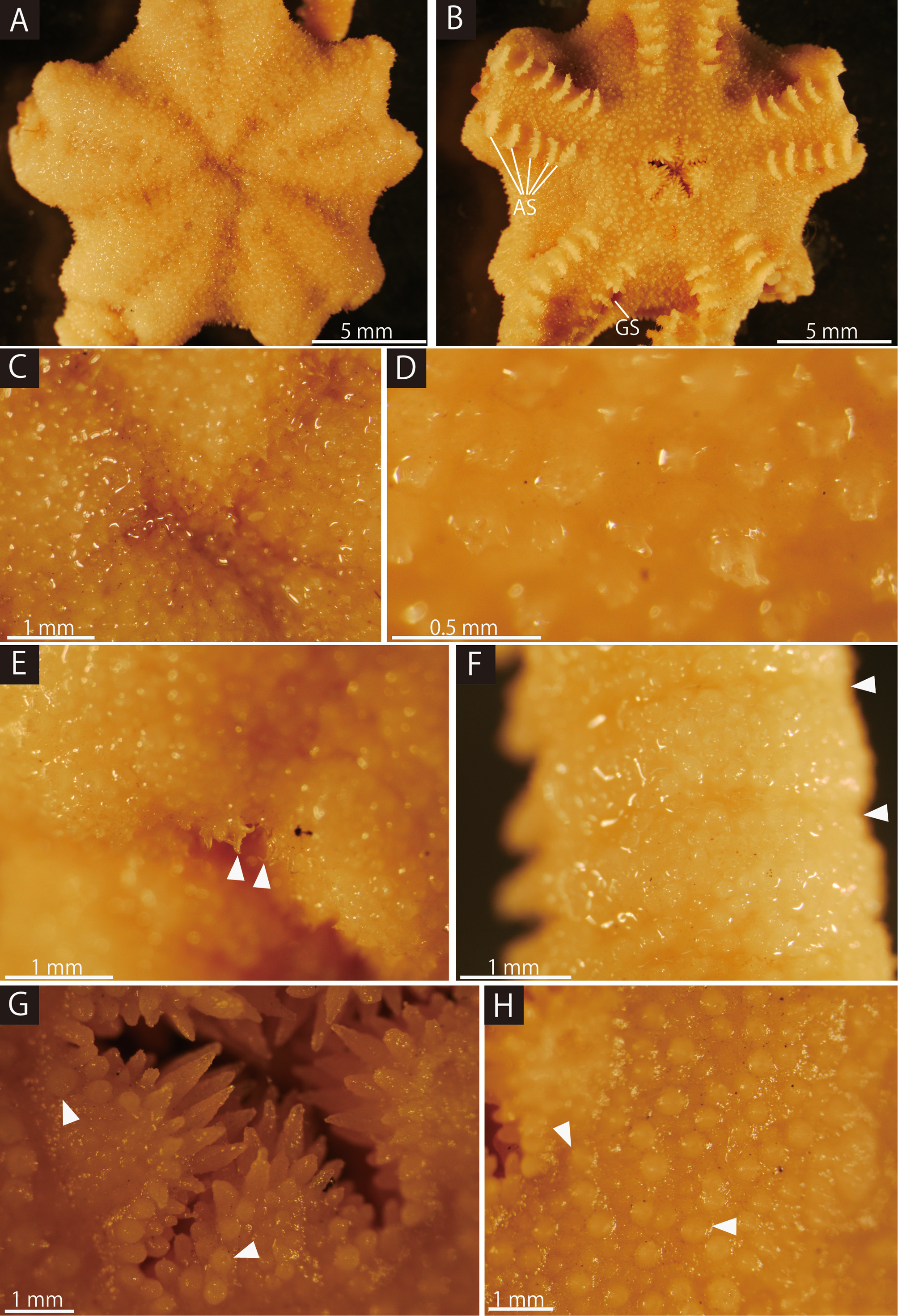
The lateral interradial surface of the disc is slightly inclined towards the oral side and covered only by coneshaped epidermal ossicles with mostly one or rarely two terminal projections which are approximately twice the length of the height of the cone-shaped epidermal ossicles ( Fig. 2 View FIGURE 2 B). The cone-shaped epidermal ossicles are approximately 130–200 µm in length ( Fig. 2 View FIGURE 2 B). Two genital slits ( 1.8 mm long and 0.05 mm wide) are present in each interradius ( Fig. 1 View FIGURE 1 B). One circular madreporite is situated on the oral interradius, approximately 0.7 mm in diameter ( Fig. 2 View FIGURE 2 A).
Arms. Arms are simple, five in number, have no abrupt change in thickness. The basal portion of the arm is 3.2 mm wide and 2.8 mm high, with an arched aboral surface and flattened oral surface. Arms taper gradually toward the arm tip ( Fig. 2 View FIGURE 2 D–H).
On the aboral and lateral surface, each arm segment is surrounded by single annual transverse row of hookletbearing plates ( Fig. 2 View FIGURE 2 D). Those hooklet-bearing plates are completely in contact with each other along the entire arm length ( Fig. 2 View FIGURE 2 D, E, G). Except for the hooklet-bearing plates, the aboral and lateral surface of the basal arm is completely covered by cone-shaped and polygonal plate-shaped epidermal ossicles are approximately 210–280 µm in length ( Fig. 2 View FIGURE 2 D). The oral surface is covered by cone-shaped and polygonal plate-shaped epidermal ossicles, similar to those on the oral disc, and approximately 170–210 µm in length ( Fig. 2 View FIGURE 2 C). In the middle portion of the arm, the aboral and lateral surfaces are covered by cone-shaped epidermal ossicles, approximately 150–180 µm in length, and domed and round granule-shaped epidermal ossicles, approximately 130–150 µm in length ( Fig. 2 View FIGURE 2 E). The oral surface is also covered by relatively flat and round granule-shaped epidermal ossicles approximately 90– 110 µm in length. In the distal portion of the arm, the aboral and lateral surface is covered by flat and round granule-shaped epidermal ossicles approximately 60 µm in length ( Fig. 2 View FIGURE 2 G). The oral surface is covered by flat and Epidermal ossicles Hooklet-bearing Disc
Species plates at base of Locality References
diameter On aboral disc On oral plates At lateral interradial disc arms
round plate-shaped epidermal ossicles approximately 80 µm in length. Over the entire arm, lateral arm plates and ventral arm plates are completely concealed by epidermal ossicles ( Fig. 2 View FIGURE 2 E, H).
First tentacle pores have no arm spine; the second ones have five, the third ones have five or six, and from the fifth to the middle arm segments there are six arm spines ( Figs 1 View FIGURE 1 B; 2C). The number of arm spines decrease gradually to a single spine toward the arm tip. Throughout the arm, all the arm spines are approximately half the length of the corresponding arm segment, and are covered by a thin integument ( Fig. 2 View FIGURE 2 C, F, H). Arm spines in the basal one-fourth of the arm are ovoid, carrying two or three terminal projections. In the middle portion, one-fourth to three-fourths of the arm, arm spines have three terminal projections. In the distal quarter of the arm, arm spines become hook-shaped. These hook-shaped spines have one inner tooth.
Color. In ethanol specimen, body color is uniformly creamy brown. No living color information for original description ( Koehler, 1904).
Distribution. Philippines: north off Sulu Island, 275 m ( Koehler, 1904), southern Philippines.

| ZMA |
Universiteit van Amsterdam, Zoologisch Museum |
No known copyright restrictions apply. See Agosti, D., Egloff, W., 2009. Taxonomic information exchange and copyright: the Plazi approach. BMC Research Notes 2009, 2:53 for further explanation.
|
Kingdom |
|
|
Phylum |
|
|
Class |
|
|
Order |
|
|
Family |
|
|
Genus |
|
|
SubGenus |
Asteroporpa |
Asteroporpa ( Astrohelix ) bellator ( Koehler, 1904 )
| Okanishi, Masanori 2017 |
Astrohelix bellator Döderlein, 1930
| Doderlein 1930 |
Astrotoma bellator
| Koehler 1904 |