
Suwallia talalajensis Zhiltzova, 1976
|
publication ID |
https://doi.org/ 10.11646/zootaxa.3994.4.4 |
|
publication LSID |
lsid:zoobank.org:pub:FFDE53F2-3AC0-4667-B262-384509133310 |
|
DOI |
https://doi.org/10.5281/zenodo.6103083 |
|
persistent identifier |
https://treatment.plazi.org/id/038E87D1-1327-FF80-FF09-FBDFDB5EFDE9 |
|
treatment provided by |
Plazi |
|
scientific name |
Suwallia talalajensis Zhiltzova, 1976 |
| status |
|
Suwallia talalajensis Zhiltzova, 1976 View in CoL
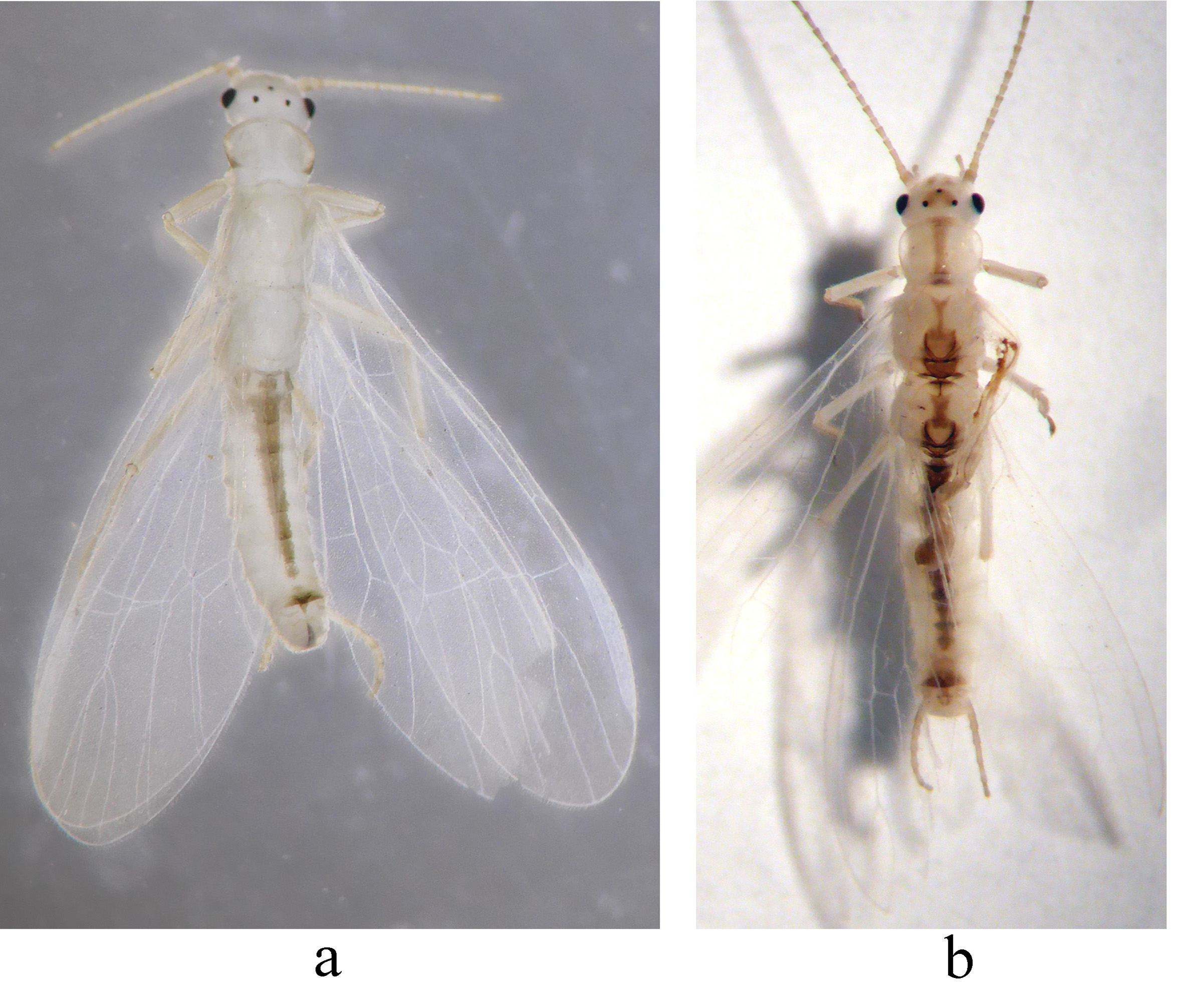
( Figs. 1 View FIGURE 1 b, 5)
Suwallia talalajensis Zhiltzova, 1976 View in CoL : Levanidova & Zhiltzova 1976. Presnovodnaia fauna Chukotkogo poluostrova, Akademia Nauk SSSR Trudy Biologo-pocvenogo Instituta, Vladivostok, 36(139): 25; Alexander & Stewart 1999. Transactions of the American Entomological Society, 125: 220; Teslenko & Zhiltzova 2009. Key to the Stoneflies (Insecta, Plecoptera View in CoL ) of Russia and Adjacent Countries. Imagines and Nymphs: 86.
Material examined. 1♂ ( HNHM): CHINA: Inner Mongolia Autonomous Region, Genhe City, Hanma National Nature Reserve, Central Protection Station at bridge of Bonuo River, 1.viii.2014, leg. Li Shi, Mingrun Tian, Yuxuan Zhu, Xuefeng Gao and Chao Chen.
Description of the Chinese specimens. Male ( Figs. 1 View FIGURE 1 b, 5). Head pale with subquadrate pigmentation in ocellar triangle and two brown spots on frons. Pronotum with thin dark lateral margins and darker longitudinal median stripe ( Fig. 5 View FIGURE 5 a); the median abdominal stripe ends at the posterior margin of tergum 8 ( Fig. 1 View FIGURE 1 b).
Terminalia ( Fig. 5 View FIGURE 5 ). Tergum 9 with wide brown to dark brown medial pigment ( Fig. 5 View FIGURE 5 b), posterior margin produced into a triangular protrusion in lateral view ( Fig. 5 View FIGURE 5 d). Tergum 10 with a sclerotized “turtle-like” medial area before base of epiproct; large, paired longitudinal lateral sclerites nearly touching epiproct, lightly sclerotized. Hemitergal processes nearly straight and directed posteriorly ( Figs. 5 View FIGURE 5 c–d). Epiproct covered with dense fine hairs ( Fig. 5 View FIGURE 5 c). Aedeagus is membranous and lacking distinct armature, basal portion robust and nearly parallel-sided, apical part upcurved and triangular in outline, the apex beak-like in lateral view ( Fig. 5 View FIGURE 5 e).
Diagnosis and remarks. In addition to characteristic head pattern and epiproct, the aedeagus of this species lacks armature. Terga 9 and 10 also has distinct pattern not mentioned by Alexander & Stewart (1999). This character is helpful in providing identification. Our male specimen still shows slight variation as compared to Russian specimens in some details of pigmentation of tergum 9 and median sclerite of tergum 10 (compared to figs. 524–527 in Teslenko & Zhiltzova 2009). No larvae were associated with the adult male and are still unknown.

| HNHM |
Hungarian Natural History Museum (Termeszettudomanyi Muzeum) |
No known copyright restrictions apply. See Agosti, D., Egloff, W., 2009. Taxonomic information exchange and copyright: the Plazi approach. BMC Research Notes 2009, 2:53 for further explanation.
|
Kingdom |
|
|
Phylum |
|
|
Class |
|
|
Order |
|
|
Family |
|
|
Genus |
Suwallia talalajensis Zhiltzova, 1976
| Li, Weihai, Murányi, Dávid & Shi, Li 2015 |
Suwallia talalajensis
| Zhiltzova 1976 |