
Ascidia collini, Bonnet, Nadia Y. K. & Rocha, Rosana M., 2011
|
publication ID |
https://doi.org/10.5281/zenodo.277398 |
|
DOI |
https://doi.org/10.5281/zenodo.6186565 |
|
persistent identifier |
https://treatment.plazi.org/id/038F878E-FFA4-FFFE-0BB5-FC0BFBFD4BB7 |
|
treatment provided by |
Plazi |
|
scientific name |
Ascidia collini |
| status |
sp. nov. |
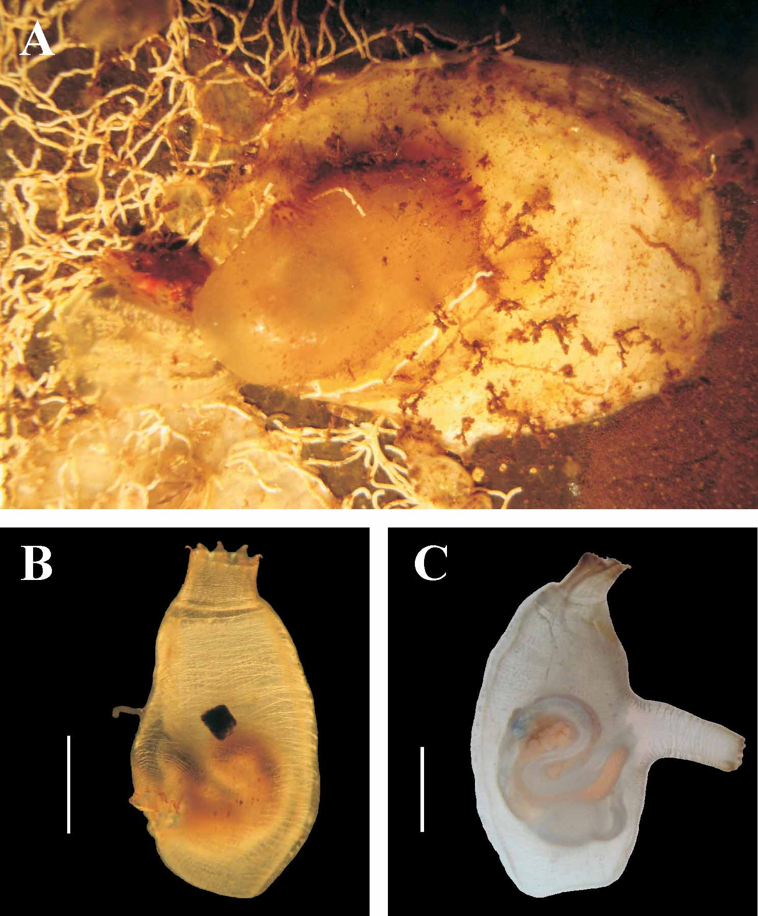
Ascidia collini sp. nov.
( Figs. 4–5 View FIGURE 4 View FIGURE 5 )
Material examined. Holotype: MZUSP 00014—1 ind.; Casa Blanca; on polyethylene recruitment plate; 05.vii.2009.
Paratypes: DZUP ASC 162—1 ind.; STRI Bay; on recruitment plate; 18.x.2008; DZUP ASC 139—2 ind.; STRI Bay; 0.5 m, on recruitment plate; 09.iii.2009; DZUP ASC 140—1 ind.; STRI Bay; 0.5 m, on recruitment plate; 25.iii.2009; DZUP ASC 141—2 ind.; STRI Bay; 0.5 m, on recruitment plate; 09.iv.2009.
Etymology. The name is in homage to Dr. Rachel Collin, director of Bocas del Toro Research Station of the Smithsonian Tropical Research Institute, who enthusiastically supported this study at the research station.
Diagnosis. Small individuals; brown lobes in the siphons; body musculature comprising complete transverse fibers; reduced number of longitudinal vessels in the pharynx; small alimentary canal, with isodiametric intestine; ovary ramified.
Individuals attach to the substrate on the left side of the body, with fine sediment and sometimes with epibionts (algae and hydrozoans) covering the free surface. Tunic uncolored, translucent, smooth and thin ( 0.1–0.16 mm) of cartilaginous consistency with transparent vessels.
The largest specimen is 2.1 cm and oblong. Without the test is 0.9–2.0 cm (including oral siphon) by 0.3–0.7 cm. Siphons are 0.2–0.4 cm long; the oral siphon has eight smooth lobes, the atrial has 7–10 smooth lobes and is 0.4–0.7 cm posterior from the ring of tentacles. Lobes in both siphons are brown with a yellow spot between each.
The body wall is colorless; on the right side the musculature comprises mostly transverse muscle fibers, which completely cross the body from the ventral to the dorsal margin and a few oblique fibers. On the left side, the musculature comprises short transverse fibers in the dorsal margin. The few longitudinal muscle fibers of the siphons are somewhat concentrated towards each lobe and extend for only a short distance posterior to the base of the siphons.
The 62–82 oral tentacles are of four lengths with the largest of 0.7–2.0 mm. The prepharyngeal groove is double, with smooth margin, 0.3–0.8 mm from the ring of tentacles; the area between the prepharyngeal groove and the tentacles has a few large papillae. The peritubercular area is rounded; the dorsal tubercle aperture U-shaped, without enrolled ends. The neural ganglion is very close to the dorsal tubercle. The dorsal lamina is double anteriorly, with small finger-like projections formed by the ends of the left transverse vessels, between which are additional small projections. The dorsal lamina continues past the esophageal aperture on the left to the end of the pharynx; usually papillae are absent on the right side. A toothed lamina is on the right side of the esophageal aperture. The pharynx has 27–31 longitudinal vessels on the right, 24–28 on the left and 37–67 transverse vessels; it is slightly pleated and the meshes have 4–6 stigmata; the primary papillae are bilobed. Parastigmatic vessels and secondary papillae are absent.
The alimentary canal occupies less than half of the body wall in the middle of the body. The stomach is rounded, with 5–7 internal longitudinal folds; the intestine is isodiametric, with accentuated primary and secondary loops; anus with bilobed margin, about 3.0– 7.1 mm from the oral tentacles. Renal vesicles are not conspicuous, 0.04–0.09 mm diameter, covering the stomach and primary intestinal loop. There are small and irregular papillae on the digestive tube.
The ramified ovary is within the primary intestinal loop and extends over the intestine; it is completely visible from the side of the atrial cavity; from the outside it is visible only in and around the primary intestinal loop; oocytes are 0.08–0.10 mm in diameter. Testis follicles are elongated and spread out over the intestine, but not on the stomach and rectum. Gonoducts open posteriorly to the anus.
Remarks. In the Atlantic Ocean, only A. achimotae Millar, 1953 (from the west coast of Africa) is similar to A. collini sp. nov.: it has undeveloped body musculature, a dorsal tubercle that is U-shaped close to the nervous ganglion, isodiametric intestine and few internal folds in the stomach ( Millar 1953). However, A. achimotae differs from A. collini sp. nov. because it has well developed longitudinal muscles in the siphons, 2–3 stigmata per mesh in the pharynx, and smooth dorsal lamina (individuals described were young and so they had no gonads) ( Millar 1953).
In the Pacific Ocean, A. citrina Nishikawa & Tokioka, 1975 is described as having eight or more lobes in the short siphons, toothed dorsal lamina, 5–6 stigmata per mesh, a small digestive tract and isodiametric intestine ( Nishikawa & Tokioka 1975), and our samples also fit that description. But A. citrina has yellow coloration when alive, body musculature formed by a net of fibers (mainly transverse), around 30 oral tentacles, no papillae between the ring of tentacles and the prepharyngeal groove, and 48–54 longitudinal vessels in each side ( Nishikawa & Tokioka 1975), differing from A. collini sp. nov. Ascidia melanostoma Sluiter, 1885 , is another Pacific species that is similar to ours, with a brown tunic, numerous lobes and longitudinal muscles forming bands in the siphons, small digestive tract, few folds in the stomach, isodiametric intestine and open second intestinal loop ( Monniot 1987; Nishikawa 1986). However, A. melanostoma and A. collini sp. nov. may be separated by color, body musculature formed by a net of fibers, the long longitudinal muscles fibers in the left side of the body and the lobed ovary.
Ascidia lambertae Monniot, 2007 was recently described and is a new species from the Pacific Ocean of Mexico. This species is small (the largest is 3.0 cm long), with light brown tunic (darkest at the siphons), with about 60 oral tentacles, papillae between the ring of tentacles and the prepharyngeal band, 38 longitudinal vessels on the right side of the pharynx and 29 on the left side, isodiametric intestine and ramified ovary ( Monniot 2007). Thus, it is similar to A. collini sp. nov. However, the black spots between the lobes in the siphons, body musculature formed by an irregular network of fibers on the right side, dorsal lamina with long papillae on the right side and a larger digestive tube ( Monniot 2007) all separate A. lambertae from A. collini sp. nov.

No known copyright restrictions apply. See Agosti, D., Egloff, W., 2009. Taxonomic information exchange and copyright: the Plazi approach. BMC Research Notes 2009, 2:53 for further explanation.
|
Kingdom |
|
|
Phylum |
|
|
SubPhylum |
Tunicata |
|
Class |
|
|
Order |
|
|
Family |
|
|
Genus |
Ascidia collini
| Bonnet, Nadia Y. K. & Rocha, Rosana M. 2011 |
Ascidia lambertae
| Monniot 2007 |