
Ascidia monnioti, Bonnet, Nadia Y. K. & Rocha, Rosana M., 2011
|
publication ID |
https://doi.org/10.5281/zenodo.277398 |
|
DOI |
https://doi.org/10.5281/zenodo.6186573 |
|
persistent identifier |
https://treatment.plazi.org/id/038F878E-FFAD-FFE5-0BB5-FAC8FE484D2B |
|
treatment provided by |
Plazi |
|
scientific name |
Ascidia monnioti |
| status |
sp. nov. |
Ascidia monnioti sp. nov.
( Figs. 10–11 View FIGURE 10 View FIGURE 11 )
Material examined. Holotype: MZUSP 00019—1 ind.; Isla Pastores; 0.5 m, coral reef; 13/vi/2009.
Paratypes: DZUP ASC 138—Casa Blanca; coral reef; 05/vii/2009. DZUP ASC 134—1 ind.; Hospital Point; 8.0 m, coral reef; 23/xii/2008. DZUP ASC 137—2 ind.; Crawl Cay; 0.5 m, coral reef; 14/vi/2009. DZUP ASC 132—2 ind.; Pastores island; coral reef; 17/viii/2006; DZUP ASC 133—1 ind.; Pastores island; coral reef; 20/viii/2006; DZUP ASC 135—2 ind.; Pastores island; 1.5 m, coral reef; 25/ii/2009; MZUSP 00020—3 ind.; Pastores island; 0.5 m, coral reef; 13/vi/2009. DZUP ASC 136—1 ind.; Big Bight; 1.0 m, coral reef; 05/vi/2009.
Etymology. The name is in homage to Claude and Françoise Monniot for their great contributions to the study of ascidian diversity in the world.
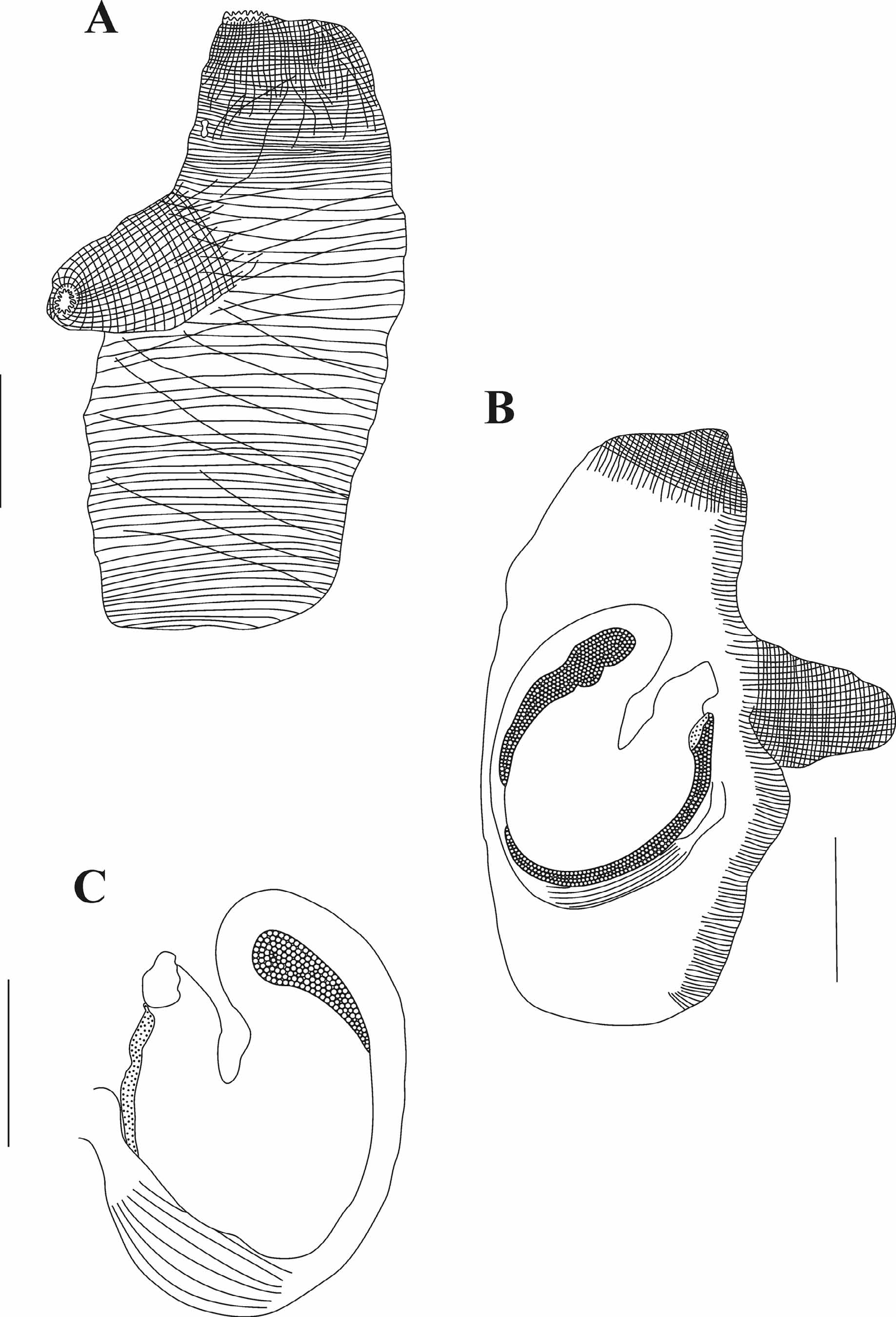
Diagnosis. Live animals yellow with both siphons close together; tunic 1.0–2.0 mm, smooth without projections or incrustations, gelatinous and completely transparent; musculature on the right side comprising thick transverse fibers that may cross each other, sometimes as long as the dorsal-ventral width, sometimes divided forming two or more longitudinal patches; 10–16 round lobes in each siphon; 50–90 oral tentacles, slender and side by side; pharynx with 40–70 longitudinal vessels on each side; 4–8 stigmata per mesh; stomach with many internal folds; large alimentary canal, with sac-like dilation; smooth anus rim; elongated ovary inside the primary intestinal loop.
Animals attach on the left side to living or dead coral in a vertical position, siphons pointing up. The tunic is very delicate and usually it tears when taking the animal from the substrate. It can be wrinkled in contracted animals.
The body is elongate, cylindrical, slightly flat on both sides, 3.0– 7.5 cm (including the oral siphon) by 1.0– 2.1 cm, without tunic. The oral siphon is 0.5–1.0 cm long and the atrial is 1.0– 1.3 cm and displaced towards the oral siphon. Thus, in living animals, the apertures are close together. Both have conspicuous circular and longitudinal musculature, not forming bands. There are 10–16 triangular lobes on both siphons, with smooth or slightly indented margins, and a yellow or orange spot between the lobes.
The body wall is colorless. On the right side, the body wall musculature comprises short and thick transverse fibers, parallel or even crossing each other and thin longitudinal fibers; fibers sometimes are not complete forming two or more longitudinal patches. On the left side, there are short transverse fibers along the dorsal margin.
The 50–90 oral tentacles are of four sizes, the largest 2.0–6.0 mm long, wide at the base, slender and close to one another. Tentacles project from a conspicuous but thin muscular ring. The double prepharyngeal groove has an anterior lamina with long filiform projections. The area between tentacles and prepharyngeal groove is 0.3–1.5 mm wide and has small papillae (in variable density). The peritubercular area is U-shaped; the aperture of the dorsal tubercle is heart or U-shaped with or without enrolled ends. The neural ganglion varies in position but is usually closer to the atrial siphon and away from the dorsal tubercle. The dorsal lamina is double anteriorly (5.0 mm) and with small finger-like projections formed by the ends of the left transverse vessels, between which are additional small projections; the dorsal lamina wall close to the esophageal aperture lacks projections. The dorsal lamina is uniformly wide throughout and passes on the left of the esophageal aperture to the end of the pharynx. The ends of the right transverse vessels form a narrow membrane on the right side of the esophageal aperture. The pharynx has 40–60 longitudinal vessels on each side and as many as 232 transverse vessels; it is not pleated and meshes have 4– 8 stigmata. The primary papillae comprise one small lobe (some papillae have another lobe at the base), may have a lateral expansion on each side; pharynx lacks intermediate papillae, but there are few (if any) parastigmatic vessels.
The alimentary canal is large, occupying half of the left side of the body. It has a thin, transparent wall through which internal contents may be seen. The stomach is oval and wide with 10–18 internal folds. The intestine forms two loops and the descending portion is dilated as a sac-like pouch. The large, plain-rimmed anus is 1.0– 2.7 cm from the oral tentacles at the level of the anterior extremity of the primary loop of the intestine. Irregularly-shaped papillae cover the stomach wall and the ascending portion of the intestine on the side of the atrial cavity.
The lobed ovary occupies the space inside the primary intestinal loop and may be seen from both, outside and the atrial cavity, although it is usually more external. The oviduct opens by a large slit close and posterior to the anal aperture. The testis is extensively branched and comprises small pyriform follicles, covering the alimentary canal wall (including the rectum) on the atrial side; difficult to see externally. When well developed it also covers the primary intestinal loop making it impossible to see the ovary from inside. The sperm duct aperture is between the oviduct aperture and the anus, but smaller.
All samples had many copepods and some amphipods inside the pharynx and one had a large isopod between the body wall and the tunic.
Remarks. The yellow color in life is very conspicuous in the field, though only the siphons are exposed. In the Caribbean region, two known Ascidia species have many lobes in the siphons: A. xamaycana Millar & Goodbody, 1974 and A. interrupta Heller, 1878 , in addition to Ascidia panamensis sp. nov. All also have a dilated rectum and ovary within the primary intestinal loop. However, A. xamaycana is green in life, body musculature on the right side as a net, few oral tentacles (12–30), 2–3 stigmata per mesh, small alimentary canal without the secondary intestinal loop ( Millar & Goodbody 1974); A. interrupta and Ascidia panamensis sp. nov. have dark vessels in the tunic, a net of muscles fibers on the right side of the body and a big saculiform dilation in the rectum. Ascidia gemmata Sluiter, 1895 is also similar to Ascidia monnioti sp. nov.: it has a yellow tunic, numerous lobes in the oral siphon, area between prepharyngeal groove and tentacles with many papillae, toothed dorsal lamina and dilated rectum ( Kott 1985). However, in the former the body musculature forms a net, the prepharyngeal groove is smooth, there are few internal folds in the stomach and the anus is bilobed.
Within the Ascidiidae , Phallusia julinea Sluiter, 1915 also has many siphon lobes, a toothed lamina on the right side of the esophageal aperture, 6–8 stigmata per mesh and dilated rectum that forms a sac-like pouch. Additionally, P. julinea may not have accessory openings ( Monniot 1987), as do the Ascidia . In contrast to Ascidia monnioti sp. nov., P. julinea has indented lobes in the siphons, 70–90 longitudinal vessels on each side of the pharynx, and anus with multilobed rim.

No known copyright restrictions apply. See Agosti, D., Egloff, W., 2009. Taxonomic information exchange and copyright: the Plazi approach. BMC Research Notes 2009, 2:53 for further explanation.
|
Kingdom |
|
|
Phylum |
|
|
SubPhylum |
Tunicata |
|
Class |
|
|
Order |
|
|
Family |
|
|
Genus |