
Paragourretia sandrae, Felder & Dworschak, 2023
|
publication ID |
https://doi.org/ 10.11646/zootaxa.5284.3.6 |
|
publication LSID |
lsid:zoobank.org:pub:E742B6DC-A5CB-4CB9-8DA2-D464FE3EC249 |
|
DOI |
https://doi.org/10.5281/zenodo.7929445 |
|
persistent identifier |
https://treatment.plazi.org/id/03916A48-A244-FFA7-FF3C-F94BFBF83F97 |
|
treatment provided by |
Plazi |
|
scientific name |
Paragourretia sandrae |
| status |
sp. nov. |
Paragourretia sandrae View in CoL n. sp.
https://zoobank.org/ urn:lsid:zoobank.org:act:4E6884D8-4B64-4A1F-B00E-529BE672BD98
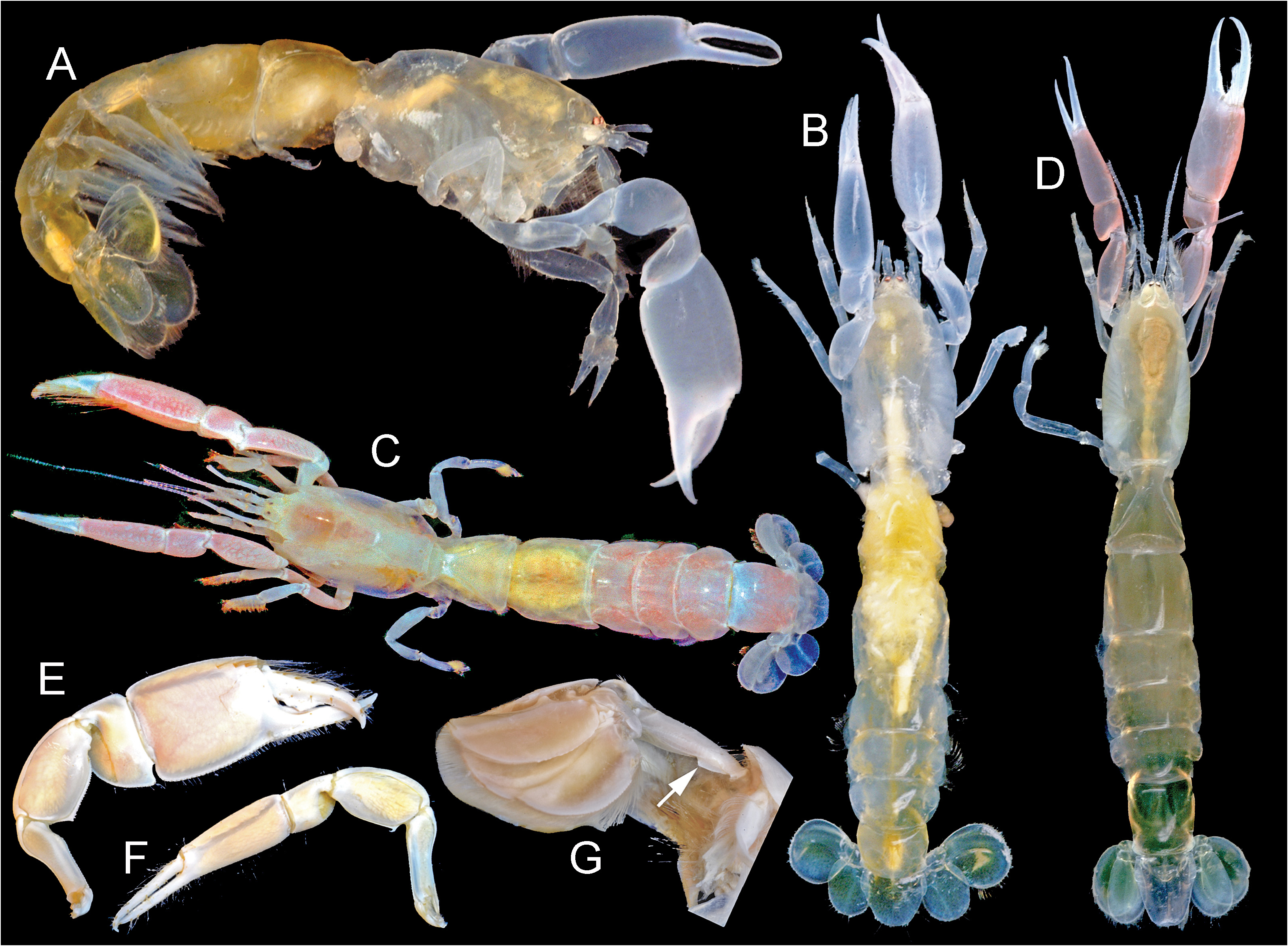
( Figs 1 View FIGURE 1 , 2 View FIGURE 2 , 3A–C View FIGURE 3 )
Type material. Holotype (southwestern Gulf of Mexico): male, cl 4.4 mm; dredge station NSF-II-096, muddy eroded calcareous rubble, Campeche Banks , 22° 08.04´N, 91° 23.67´W, 53 m, 17 June 2005; coll D.L. Felder, R. Robles, H. Bracken-Grissom, S. Fredericq, E. Garcia and colleagues aboard R/V Pelican; USNM 1541885 About USNM (= ULLZ 7304 View Materials , photograph voucher). GoogleMaps
Diagnosis. Carapace with narrow triangular spiniform rostrum reaching beyond midlength of eyestalks. Eyestalk with subterminal cornea in distal third, diameter spanning over half of eyestalk width. Major chela with merus inferior keel bearing proximal hooked spine and adjacent ancillary spine, otherwise microdenticulate. First pleonal tergite crossed dorsally by smooth transverse furrow in anterior half, posterior sclerite narrowing anteriorly to acute middorsal terminus, sixth with neither angular nor arched projection in anterior third of lateral margin. Telson length slightly exceeding width, lateral margins converging posterior to weak lateral lobes, posterior margin truncate, lacking median spine. Uropodal endopod elongate ovoid with low longitudinal ridge; exopod anterodorsal plate obscure to obsolescent, lateral margin lacking incision. Diagnostic sequence data for the COI mitochondrial gene is provided under GenBank www.ncbi.nlm.nih.gov/genbank) accession number OQ413315.
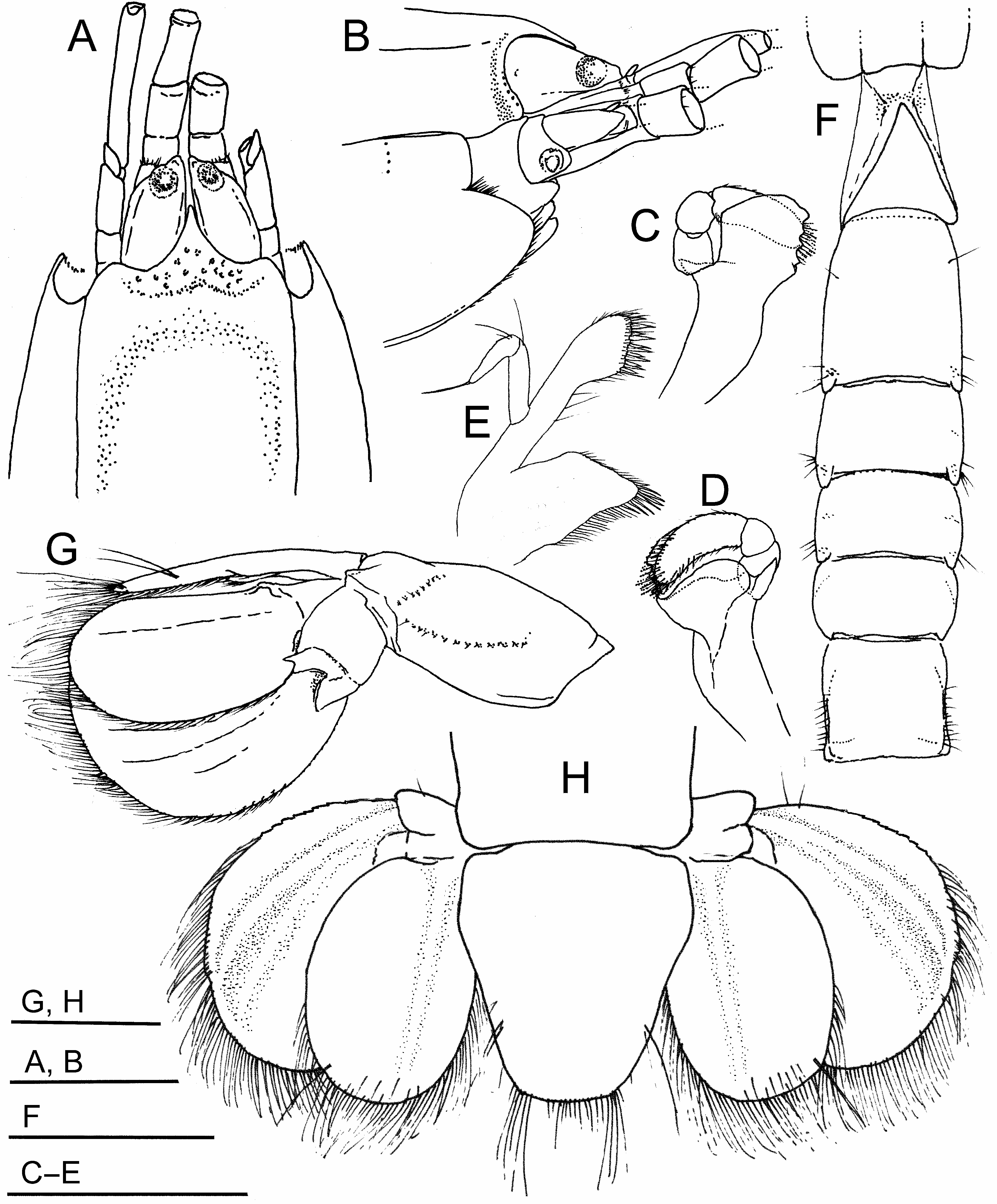
Description. Carapace front ( Figs 1A, B View FIGURE 1 ; 3A, B View FIGURE 3 ) with acute, narrowly triangular rostrum, deflected between eyestalks in lateral view, terminally spiniform, reaching beyond mid-length of eyestalks, rostral base flanked laterally by low, obtuse shoulders forming outer angles of orbits, orbital margin laterally lined by several small setal pits; postantennal carapace margin transverse, near vertical, strongly inset from front, slightly arched transverse row of four small setal pits posterior to margin; dorsal oval not defined; linea thalassinica extending full length of carapace; hepatic boss weakly developed; cardiac prominence poorly defined.
Eyestalks ( Fig. 1A, B View FIGURE 1 ) elongate, narrowing distally to subtriangular tips not reaching to second (penultimate) article of antennular peduncle, carried slightly deflected, swollen proximally in lateral view, distal part dorsoventrally flattened, lateral margin developed into narrow crest laterally; subterminal cornea defined as rounded dorsal swelling in distal third of eyestalk, diameter spanning over half of eyestalk width, poorly faceted, pigmentation ( Fig. 3A, B View FIGURE 3 ) filling cornea in life, dispersed into multiple coalesced dark spots with preservation.
Antennular peduncle ( Fig. 1A, B View FIGURE 1 ) much shorter and distinctly heavier than antennal peduncle, reaching approximately to distal end of fourth article of antennal peduncle; first (basal) article swollen, more than twice length of second, third article approximately 1 and 1.5 times length of second; second and third articles bearing few minute marginal setae. Antennular flagella missing. Antennal peduncle (fifth articles and flagella missing) distinctly overreaching antennular peduncle; first article with short produced ventrolateral process bearing excretory pore; length of second article about twice width, distal articulation to third article overreached dorsally by short bladelike scaphocerite; fourth article slightly exceeding combined lengths of first two, all sparsely setose.
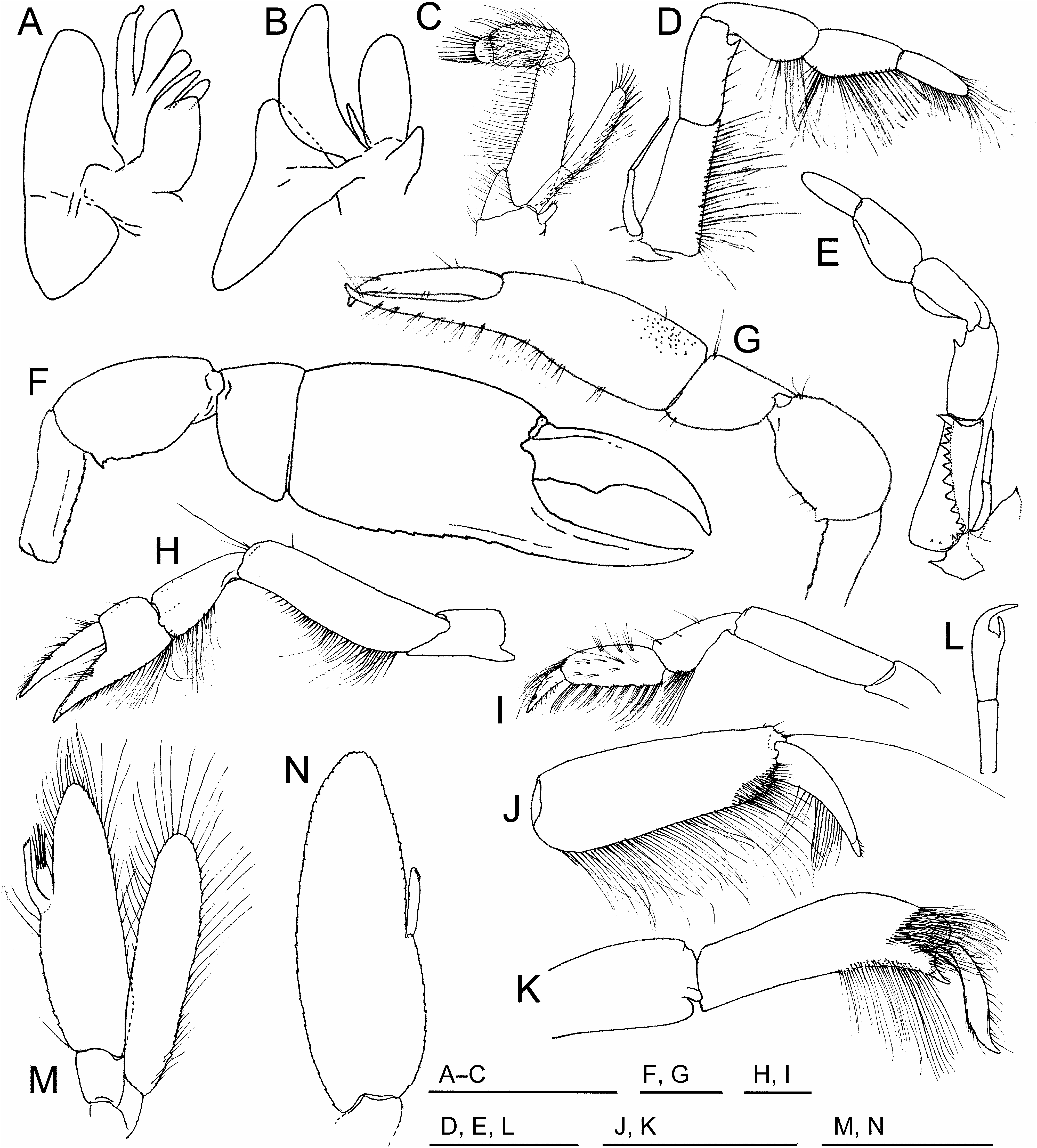
Mandible ( Fig. 1C, D View FIGURE 1 ) with palp of three articles, proximal articles short, little if any longer than broad, third article elongate, heavy, arched, distal setae elongate, densely plumose; gnathal lobe subquadrate, distolateral shoulder angular, incisor process with 4 well-defined subacute triangular teeth on cutting margin, tips corneous, molar process on internal surface forming internal lip of concavity accommodating flexed palp, lip originating proximal and internal to incisor teeth. Maxillule ( Fig. 1E View FIGURE 1 ) endopodal palp narrow, biarticulate, distal article deflected, over half length of proximal, bearing long terminal seta; proximal endite forming broad subangular lobe, uniformly fringed by dense row of setae along most of mesial margin, setae heavier, some spiniform, at distal prominence; distal endite elongate, terminally broadened, setation of mesial margin including closely set overlain rows of strongly spiniform setae. Maxilla ( Fig. 2A View FIGURE 2 ) margins setose, endopod constricted distally to form narrow terminus; first and second endites each longitudinally subdivided, mesially directed distal margins fringed by long stiff setae; exopod forming broad scaphognathite.
First maxilliped ( Fig. 2B View FIGURE 2 ) margins setose, endopod small, narrow, less than half length of exopod, partially concealed between base of distal endite and exopod; proximal endite a subangular lobe, field of dense stiff setae terminally; distal endite subrectangular, margins and most of external surface densely setose, strongest setae mesially directed, originating distal and along mesial margin; exopod elongate, arcuate, margins setose, strongest setae terminal and along convex lateral margin; epipod broad, not reaching beyond exopod, anterior and posterior lobes subtriangular.
Second maxilliped ( Fig. 2C View FIGURE 2 ) much smaller than third, both rami setose; endopod merus subrectangular, slightly broadened proximally, weakly arched, length about 3 times width, length about twice combined length of propodus and dactylus; propodus robustly subcylindrical, most of surface setose, length about 1 ½ times breadth; dactylus short, broader than long, rounded terminus bearing long stiff setae; exopod narrow, straplike, carried closely against internal surface of endopod, not reaching end of endopod merus, terminally rounded with stiff marginal setae; short digitiform epipod evident near base of exopod.
Third maxilliped ( Fig. 2D, E View FIGURE 2 ) with narrow biarticulate exopod, length approximately equal to ischium, bearing long stiff terminal seta; basis bearing spine on inner surface just proximal to articulation with ischium; endopod fringed by long setae, densest on mesial margins of ischium, propodus, and dactylus, ischium subrectangular, length more than twice breadth, internal surface with slightly arched longitudinal row of 13 spiniform teeth forming strong crista dentata, distalmost tooth forming spine, several additional small ancillary teeth trailing row proximally; merus subquadrate, length approximately twice width, length over three-fourths that of ischium, flexor margin with few long setae and strong distal spine; carpus almost as broad as propodus, both much longer than broad, flexor margins of both distinctly convex; dactylus subcylindrical, digitiform, length exceeding two times breadth, with dense subterminal to terminal field of long setae originating primarily from internal surface.
First pereopods ( Figs 2F, G View FIGURE 2 ; 3A, B View FIGURE 3 ) strongly heterochelous; major (right) cheliped ( Fig. 2F View FIGURE 2 ) ischium slender, superior margin sinuous, inferior marginal carina armed by row of small denticles, merus superior margin smoothly convex, inferior (flexor) margin forming keel with triangular proximal spine, smaller more distal sharp spine, denticulate margin followed by smooth margin over distal half; carpus short, subtriangular, much broader than long, superior and inferior margins smoothly keeled, superior terminating distally at low rounded corner, inferior terminating distally in obtusely angular rounded corner; propodus heavy, more inflated than carpus, upper margin of palm 3 times length of carpus, length of fixed finger about 2/3 to 3/4 length of palm, palm superior margin keeled proximally, propodus inferior marginal keel weakly serrate, extending onto fixed finger, fixed finger weakly bowed ventrally, prehensile edge with few proximal microdenticles, otherwise unarmed, weak submarginal longitudinal furrow to internal side of tip not notably upturned; dactyl superior margin smooth arched over most of length, prehensile edge with minute notch in proximal 1/4, broadly triangular tooth centered proximal to mid-length, remainder mostly smooth, tip weakly deflected.
Minor cheliped ( Fig. 2G View FIGURE 2 ) ischium narrowly elongate, superior margin sinuous, inferior margin weakly serrate; merus subovoid, superior margin strongly convex, inferior margin with smooth keel with prominent proximal spine (possibly doublet, as damaged); carpus superior margin nearly straight, inferior margin arcuate, article length subequal to breadth; propodus palm longer than fixed finger, palm length over twice breadth, 2.5 times as long as carpus; fixed finger and dactylus with unarmed prehensile edges, gape minimal, weakly hooked tips acute, crossed distally when closely opposed.
Second pereopod ( Fig. 2H View FIGURE 2 ) chelate, superior margin of merus terminating distally in setal tuft including long stiff seta, flexor margin of merus, distal flexor margin of carpus, inferior margin of propodus lined by long regularly spaced setae, those of fixed finger becoming distally short, stiff, more hooked; outer surface of dactylus and fixed finger bearing tufts of stiff setae.
Third pereopod ( Fig. 2I View FIGURE 2 ) merus length near four times width; carpus elongate, length over twice breadth, inferior margin distally with dense field of long stiff setae; propodus with superior margin ending distally in dense tuft of long setae, inferior margin weakly subdivided into eight low lobes, each surmounted by tuft of long stiff setae, proximal lobe slightly produced to form rounded heel, most of outer surface covered by tufts of setae; dactylus lanciform, slightly hooked, tip slightly twisted from arc of flexure, outer and inner surface with dense distal tuft of short stiff setae.
Fourth pereopod not subchelate, coxa mobile, ( Fig. 2J View FIGURE 2 ), propodus length approximately three times width, inferior (flexor) margin lined by closely spaced long setae, inferodistal corner with dense field of stiff setae, superodistal corner including long stiff seta greatly exceeding full length of dactylus; dactylus approximately half length of propodus, weakly hooked, tapering distally to corneous tip, flexor margin will dense row of stiff setae.
Fifth pereopod ( Fig. 2K View FIGURE 2 ) obscurely subchelate terminally amid dense setation, propodus length slightly exceeding three times breadth, with dense inner and outer fields of long setae proximal to and overlying articulation of dactylus, another along distal half of inferior margin proximal to fixed finger, fixed finger slightly hooked, narrowly acute, terminating in corneous spine; dactylus elongate, approximately half length of propodus, distally twisted from arc of flexure, ending distally in upturned tip, appearing sinuous in lateral outline.
Gills limited to paired arthrobranchs on third maxilliped and each of first through fourth pereopods.
Pleonal tergites glossy smooth, enamel-like dorsally ( Fig. 1F View FIGURE 1 ; 3A, B View FIGURE 3 ), few setae and obscure tracks of setal pits laterally. First pleonal tergite crossed by smoothly depressed transverse furrow in anterior half, posterior 2/3 covered by triangular dorsal sclerite with acute anterior, middorsal terminus. Second tergite slightly longer than first, almost twice length of third. Third to fifth tergites slightly decreasing in length to posterior. Sixth tergite approximately 2/3 length of second, dorsally with posterolateral track of fine short setae extending anteriorly as row of setal pits converging anteriorly, ventrolaterally with neither angular nor arched projection or keel in anterior half of marginal to submarginal region.
First pleopod uniramous ( Fig. 2L View FIGURE 2 ), less than half length of third pleopod, biarticulate, distal article slightly longer than proximal, broadened distal end of article terminating in strong hooked spine over-reaching shorter straight distally directed spine. Second pleopod ( Fig. 2M View FIGURE 2 ) biramous, approximately twice length of first; exopod narrowly ovoid, not reaching end of endopod when flexed against it, margins bearing long setae; endopod broader than exopod, lateral and distal margins with long setae, mesial margin with long narrow appendix interna bearing minute terminal hooks and shorter robust appendix masculina bearing long distal setae. Third ( Fig. 2N View FIGURE 2 ) to fifth pleopods larger than second, forming biramous, posteriorly cupped fans, endopod of each an elongate ovoid with elongate digitiform appendix interna projecting distinctly from mesial margin, opposed surfaces on appendix internae of two sides each with small field of microscopic hook setae.
Telson ( Figs 1G, H View FIGURE 1 ; 3A, B View FIGURE 3 ) elongate subhexagonal, narrowing posteriorly, length slightly exceeding width, broadest where lateral margins protrude as weak lobes in anterior 1/3, margins beyond converging posteriorly, dorsally with each posterolateral region bearing 2 submarginal tufts of long setae, posterior margin truncate, lacking median spine, posterolateral corners each with short track of long setae, shorter setae along margin between corners.
Uropods ( Figs 1G, H View FIGURE 1 ; 3A, B View FIGURE 3 ) with endopod broad, ovoid, about 1.5 times longer than broad, dorsal surface with weak longitudinal ridge, sparse submarginal setation distally, and single tuft of several stiffer longer setae posterolaterally, posterior margin with continuous fringe of long setae; exopod broadly ovoid, length approximately 1.3 times breadth, anterodorsal plate obscure to obsolescent, pair of weak, arcuate longitudinal ridges diverging distally from common basal origin, anterolateral of two ridges producing slight vertical offset where intersecting lateral margin (obscure in lateral view), margin lacking distinct notch or incision, anterolateral margin weakly serrate, posterior margin with continuous fringe of long setae.
Color. Primarily translucent pale yellowish integument ( Fig. 3A, B View FIGURE 3 ), more opaque portions of chelipeds and other pereopods whitish, especially at thickened articulations, traces of pinkish violet proximal to fingers of chelipeds.
Habitat and Distribution. Known from only 53 m depth on the Campeche Banks of the western Yucatan shelf, southwestern Gulf of Mexico, in muddy calcareous rubble comprised in part of dead coral and molluscan shells.
Etymology. The species name “sandrae” honors Sandra Cheryl Collier, for her generous contributions to Smithsonian Institution research efforts in Florida and Belize.
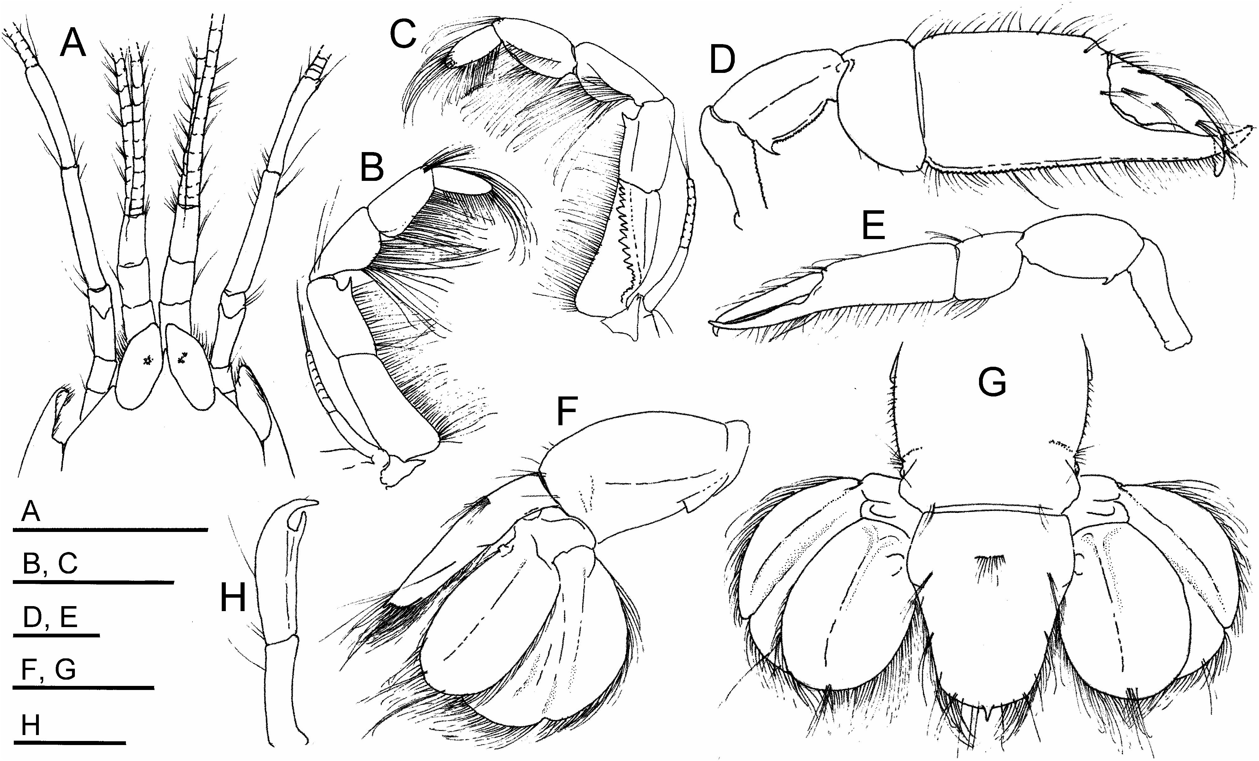
Remarks. In accord with recent revisions ( Poore et al. 2019), Paragourretia and Gourretia de Saint Laurent, 1973 are separated from Dawsonius , the only other ctenochelid genus known to occur in the western Atlantic, by the presence of an exopod on the third maxilliped. Species of these genera also differ from Dawsonius in lacking a lateral projection or keel in the anterior half of the sixth pleomere, except for Paragourretia biffari in which it occurs in a somewhat ventrolateral position ( Figs 3G View FIGURE 3 , 4F View FIGURE 4 ). All members of Paragourretia can in turn be separated from Gourretia by their lacking sharp proximally directed teeth along the cutting edges of the minor cheliped fingers characteristic of that genus ( Poore et al. 2019: fig. 17i). As in most other members of the genus, the carapace in P. sandrae n. sp. appears to have a dorsal cardiac prominence of its carapace, albeit weakly expressed. As in its congeners, the antennular peduncle is both heavier and shorter than the antennal peduncle, and the antenna bears a short bladelike dorsal scaphocerite where its second article joins to the third. Typical of the genus, its second maxilliped is comparatively small, with a straplike exopod carried against the internal surface of the slightly longer endopodal merus, and a small epipod originating at its base.
The description of P. sandrae n. sp. brings known world membership of the genus Paragourretia to ten, six of which are found only in the Indo-West Pacific. Among the known species, the majority differ from P. sandrae n. sp. and resemble P. biffari in having a distinct marginal notch or indentation in the margin of the uropodal exopod. From illustrations accompanying descriptions, this feature appears to be conspicuous in at least P. aungtonyae ( Sakai, 2002) , P. coolibah ( Poore & Griffin, 1979) , P. galathea ( Sakai, 2017) , and P. laevidactyla ( Liu & Liu, 2010) . However, it appears to be weakly evident in P. crosnieri ( Ngoc-Ho, 1991) and not evident in illustrations of P. phuketensis ( Sakai, 2002) and P. lahouensis ( Le Loeuff & Intès, 1974) . In P. sandrae n. sp., the anterolateral margin of the uropodal exopod is intersected by the anteriormost of two dorsal longitudinal ridges producing a very slight vertical offset in the margin, most evident when viewed obliquely under magnification. The ridge itself appears to demarcate the edge of a very slightly offset dorsal plate on the exopod that is found in most members of the genus ( Poore et al. 2019: 118, fig. 16e).
The absence of an obvious marginal notch in the uropodal exopod of P. sandrae n. sp, a feature shared with regional ctenochelids of the genera Dawsonius and Gourretia , readily separates the new species from P. biffari . Like Dawsonius latispina (see Rabalais et al. 1981: fig. 3I), P. sandrae n. sp. also lacks a median terminal spine on the telson ( Fig. 1H View FIGURE 1 ), a character now documented to occur in P. biffari following re-examination of the holotype and study of additional specimens. Specimens from the western Gulf of Mexico and the Caribbean coast of Panama (see Materials and methods, above) were assigned to Paragourretia biffari only after direct comparisons to the female holotype (USNM 259410). These add to the original report from Venezuela and Honduras, suggesting that the species is widely distributed in offshore tropical to subtropical waters of the western Atlantic. Illustrations accompanying the original description ( Blanco Rambla & Liñero Arana 1994) were of limited detail and did not show the now confirmed characteristic posterior median spine on the telson ( Fig. 4G View FIGURE 4 ), which was also not mentioned in the accompanying text. The authors did specifically mention the lack of a sharp lateral projection on the sixth pleomere, which, along with the presence of an exopod on the third maxilliped, was postulated to distinguish the species from Dawsonius latispina . However, on close study of the holotype this sharp ridge-like projection or keel is present, albeit in a somewhat ventrolateral position, as is also the case in some Gulf of Mexico specimens. Varying in the degree to which it projects laterally, perhaps due to effects of preservation, it is not always evident in dorsal view. Gulf of Mexico and Panamanian materials share this keel, the median telson spine, dentition of the chelae, and other morphology with the Venezuelan holotype of this species. However, the female holotype is much larger (cl 17.3 mm) than known Gulf of Mexico and Panamanian materials (cl 5.7–13 mm).
No known copyright restrictions apply. See Agosti, D., Egloff, W., 2009. Taxonomic information exchange and copyright: the Plazi approach. BMC Research Notes 2009, 2:53 for further explanation.
|
Kingdom |
|
|
Phylum |
|
|
Class |
|
|
Order |
|
|
InfraOrder |
Axiidea |
|
Family |
|
|
Genus |