
Scolopendra angulata Newport, 1844
|
publication ID |
https://doi.org/ 10.11646/zootaxa.3821.2.1 |
|
publication LSID |
lsid:zoobank.org:pub:372CEC90-946B-4352-8996-835F33BE05D7 |
|
DOI |
https://doi.org/10.5281/zenodo.6126276 |
|
persistent identifier |
https://treatment.plazi.org/id/0392244D-FF99-9361-FF6B-F940FF40FD1F |
|
treatment provided by |
Plazi |
|
scientific name |
Scolopendra angulata Newport, 1844 |
| status |
|
Scolopendra angulata Newport, 1844 View in CoL
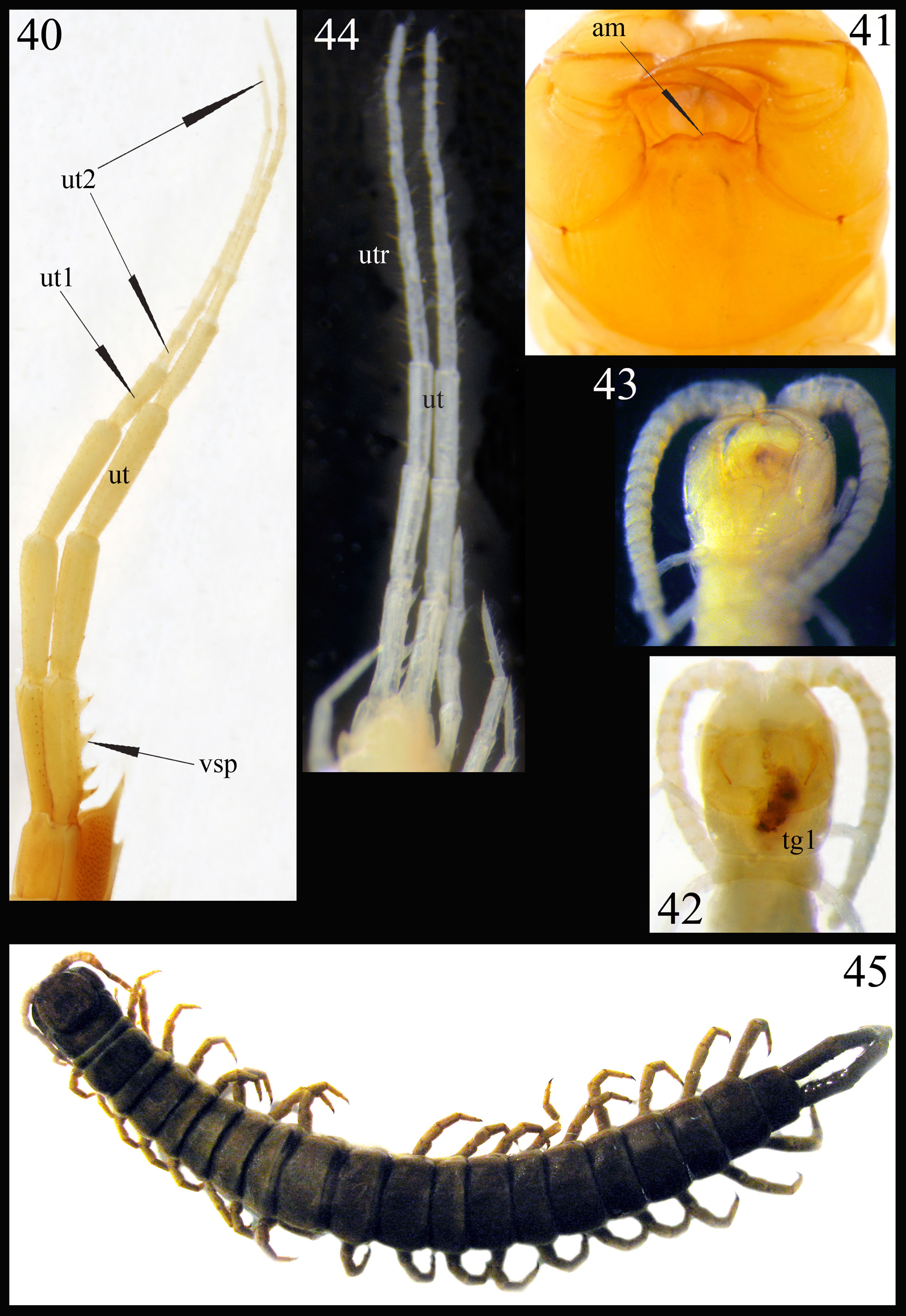
Figs 45–48 View FIGURES 40 – 45 View FIGURES 46 – 50
Scolopendra conjungens Muralewicz, 1913: 198 –200 nov. syn.; Scolopendra angulata: Attems, 1930: 40 View in CoL ;
Scolopendra angulata: Schileyko, 2002: 497 View in CoL .
Locus typicus: Trinidad.
Material. Venezuela, [Aragua State], [loc.7], m[ounain]. Victoria, 0 3.1891, 1 ♀, [col.] A.Teplow, N 6815. Additional material: Brazil, Para, Rio Tocantins, 3 spec, NN 6976, 6977, 7212.
Diagnosis. Antennae of 17 articles, 4 basal practically glabrous; subsequent articles covered by very short setae, arranged in longitudinal rows. Cephalic plate with complete paramedian sutures. Forcipular coxosternite punctate, with short median and incomplete transverse suture. Tergite 1 with anterior transverse suture crossed by posteriorly diverging paramedian sutures; tergites 2–20 with paramedian sutures, tergites 7–21 marginate. Sternites 3–20 with incomplete paramedian sutures. Legs 2–20 with tarsal spur; prefemora and femora of legs 18–20 with dorso-distal spines. Femur of ultimate legs with 2–3 dorso-medial and 1 medial spine plus 1 corner spine.
Re-description of holotype of Scolopendra conjungens Muralewicz, 1913 (adult N 6815).
Length of body ca 112 mm (105 mm according to the original description). Color in ethanol: entire animal uniformly greenish-brown ( Fig. 45 View FIGURES 40 – 45 ).
Antennae composed of 17 articles, nearly reaching the middle (or anterior margin) of tergite 5 when reflexed. 4 basal articles practically glabrous both dorsally and ventrally, subsequent articles densely covered by very short setae, arranged in longitudinal rows. All articles cylindrical, definitely longer than wide.
Cephalic plate relatively small ( Fig. 45 View FIGURES 40 – 45 ), narrowed and convex anteriorly; its posterior margin with rounded corners. Cephalic plate with complete paramedian sutures and transverse suture which crosses their posterior ends.
Second maxillae: article 2 of telopodite distally with a well-developed dorsal spur. Dorsal brush welldeveloped. Pretarsus with 2 long accessory spines.
Forcipular segment ( Fig. 46 View FIGURES 46 – 50 ) punctate, with a seta in each punctum. Coxosternite with short median suture and nearly complete transverse suture in its anterior half; chitin-lines absent. Tooth-plates somewhat longer than wide; each plate with 4 teeth, the lateral tooth is isolated and other 3 are fused to various degree. A single short strong seta in a small rounded depression approximately in the middle of tooth-plate. The basal sutures of the tooth-plates ( Fig. 46 View FIGURES 46 – 50 ) form an obtuse angle (ca 100°). Trochanteroprefemur with large process, which has two (apical and median) well-developed tubercles; the process is considerably longer than the tooth-plates. Tarsungula of normal length; their interior surface with a single sharp longitudinal ridge.
Tergites: anterior margin of tergite 1 covered by cephalic plate. Tergite 1 with curved anterior transverse suture and paramedian sutures which cross the transverse suture and diverge strongly posteriorly. The left paramedian suture is complete and the right one is absent in the posterior third of the tergite. Paired bifurcate sutures start from the ends of anterior transverse suture stretching to the middle of this tergite. Tergite 2 very short (its length less than 1/3 of tergite 1). Tergites 2–20 with well-developed paramedian sutures, tergites 4–17 with well-developed oblique sutures. Tergite 21 ( Fig. 47 View FIGURES 46 – 50 ) lacks sutures, definitely wider than long and not narrowed posteriorly; its sides curved and posterior margin obtusely angled. Tergites from 7 with incomplete and 18–21 with complete margination.
Sternites: 3–21 narrowed posteriorly ( Fig. 48 View FIGURES 46 – 50 ). Sternite 1 much wider than long, procoxae of legs clearly visible ( Fig. 46 View FIGURES 46 – 50 ). Sternites 3–20 with incomplete anterior paramedian sutures (no longer than ½ the length of the sternite). Sternites 2–20 with numerous fine anastomosing transverse sutures in anterior third, less numerous in the posterior body third. Ultimate sternite ( Fig. 48 View FIGURES 46 – 50 ) much narrower than penultimate, longer than wide and distinctly narrowed towards slightly convex posterior margin. Endosternites not recognizable.
Legs ( Figs 47, 48 View FIGURES 46 – 50 ): leg 1 with two tarsal spurs, 1 tibial, 1 femoral and 1 prefemoral spur; legs 2–20 with one tarsal spur. Tibial spurs absent; pretarsus of legs 1–20 with two accessory spines. Prefemora and femora of legs 18–19 each with 1 small (or rudimentary) dorso-distal spine (absent in femur of right leg 18). Prefemora and femora of legs 20 each with 2 dorso-distal spines (femur of left leg 20 with 1 such spine); also 1 small spine in the middle of dorsal surface of femur ( Fig. 47 View FIGURES 46 – 50 ).
Coxopleuron (excluding coxopleural process) approximately 1.5 times as long as sternite 21 ( Fig. 48 View FIGURES 46 – 50 ). Coxopleuron densely and almost completely covered with coxal pores of various sizes—only coxopleural process and a narrow area bordering posterior and inner margins of coxopleuron poreless. Coxolpeural process very short, conical with 3 apical spines. Posterior margin of coxopleuron straight with 2 (right side) or 1 (left side) spines. Coxopleural surface without setae, the posterior margin straight.
Ultimate legs ( Figs 47, 48 View FIGURES 46 – 50 ) ca 22–23 mm long (prefemur: 7, femur: 6, tibia: 4, tarsus 1: 3.5, tarsus 2: 1.7 mm) long, rather slender (width of prefemur ca 2 mm). Prefemur and femur somewhat flattened dorsally, other articles cylindrical. Left prefemur with 3+3 ventro-lateral, 2+1+2 ventro-medial, 2+3+3 dorso-medial spines plus 3 corner spines. Right prefemur with 2+4 ventro-lateral, 2+3 ventro-medial, 2+1+4 dorso-medial spines plus 3 corner spines; 1 rudimentary spine is disposed dorso-laterally. Left femur with 3, right femur with 2 dorso-medial and 1 medial (=inner) spine plus 1 corner spine ( Fig. 47 View FIGURES 46 – 50 ). Tibia and tarsus very sparsely covered by minute setae; pretarsus long (somewhat longer than ½ of tarsus 2) with two small accessory spines.
Range. Caribbean: Grenada; St.Vincent; St.Thomas; Trinidad-Tobago. US Virgin Islands (doubtful record). Bolivia. Ecuador. Brazil: State Para (Santarém; Rio Tocantins); State Amazônas (Rio Xingu); State Roraima (Rio Branco); State Mato Grosso.
In Venezuela. Aragua State, La Victoria, Municipio José Félix Ribas; “Orenoque, Rio Caroni”; Venezuela (without locality).
Remarks. 5–17 antennal articles are densely covered by short setae, arranged in longitudinal rows—this condition is not usual among Scolopendromorpha .
According to my experience the oblique sutures are present in Scolopendra on a few anterior tergites only but their distribution on tergites 4–17 (see above) and tergites 4(5)–16 and 3(4)– 17 in Brazilian specimens NN 6976 and 6977 respectively, is not usual for this genus or for Scolopendridae generally.
Discussion. In 1913 Muralewicz described Scolopendra conjungens for a single specimen from Venezuela, Victoria. We read at p. 198: “Nr. 20. 1 ♀. Venezuela, 1891, III.—[col.] A. Teplow)” and at p. 199: “Aus Venezuela (Victoria), von A. Teplow in Marz 1891 gefunden”. According to the original description (which is detailed enough but lacks illustrations), this species seems to be close to S. armata and S. alternans . So far as I know, nobody mentioned this species until now; it is also absent in Chilobase.
The ZMMU specimen described here (old inventory number 1258) was labeled “ Scolopendra conjugens ” by Muralewicz (not conjungens as in the original description). In the primary (field?) label, written in pencil and in Russian, reads—“г. Виктория”, i.e. “m.(ount) Victoria”. There is no indication that this specimen is a type, but according to the ICZN as S. conjungens was described from a single specimen it is the holotype. I re-identify this specimen as Scolopendra angulata Newport, 1844 because of the following key characters: presence of welldeveloped anterior transverse suture at tergite 1, presence of dorso-distal spines on prefemur and femur of legs 18–20, presence of spines on femur of ultimate legs ( Fig. 47 View FIGURES 46 – 50 ), absence of ventral spines at prefemur of leg 20 ( Fig. 48 View FIGURES 46 – 50 ), tergites from 7 marginate, 4 basal antennal articles practically glabrous.
Thus Scolopendra conjungens is regarded here as a junior synonym of Scolopendra angulata Newport, 1844 , n. syn.

No known copyright restrictions apply. See Agosti, D., Egloff, W., 2009. Taxonomic information exchange and copyright: the Plazi approach. BMC Research Notes 2009, 2:53 for further explanation.
|
Kingdom |
|
|
Phylum |
|
|
Class |
|
|
Order |
|
|
Family |
|
|
Genus |
Scolopendra angulata Newport, 1844
| Schileyko, Arkady A. 2014 |
Scolopendra angulata:
| Schileyko 2002: 497 |
Scolopendra conjungens
| Attems 1930: 40 |
| Muralewicz 1913: 198 |