
Mallinella bifida, Dankittipakul, Pakawin, Jocqué, Rudy & Singtripop, Tippawan, 2010
|
publication ID |
https://doi.org/10.5281/zenodo.276153 |
|
DOI |
https://doi.org/10.5281/zenodo.6204689 |
|
persistent identifier |
https://treatment.plazi.org/id/03938749-FF8E-FFD5-05BA-BCCCFC7FFD89 |
|
treatment provided by |
Plazi |
|
scientific name |
Mallinella bifida |
| status |
sp. nov. |
Mallinella bifida View in CoL sp. nov.
Figs 7–8 View FIGURES 5 – 8 , 32–36 View FIGURES 32 – 36
Types: Holotype: 3, INDONESIA, East Kalimantan Province, Berua District, near Kampung Suaran, ca. 40 km south of Tanjungredeb ( 1º59'42''S, 117º36'03''E), 50 m, primary forest on lime stone, 1.x.2008, leg. P.J. Schwendinger [MHNG, IND-08/11]. Paratypes: 13, 3♀, dataas holotype [MHNG, IND-08/11]; 23, 3♀, East Kalimantan Province, Sungai Wain protection Forest, ca. 15 km north of Balikpapan ( 1º08'36''S, 116º50'59''E), 80 m, secondary forest adjacent to primary forest, 5./ 7.x.2008, leg. P.J. Schwendinger [MHNG, IND-08/15]; 3♀, East Kalimantan Province, Berua District, Hutan Wisata Sei Tangap, ca. 8 km west of Tanjungredeb ( 2º08'04''N, 117º24'39''E), 30 m, primary forest, 2.x.2008, leg. P.J. Schwendinger [MHNG, IND-08/13]; 23, 4♀, South Kalimantan Province, ca. 23 km east of Banjarbaru ( 3º30'59''S, 115º01'00''E), 50 m, primary forest, 29.-30.ix.2008, leg. P.J. Schwendinger [MHNG, IND-08/26]; 23, 1♀, South Kalimantan Province, ca. 25 km east of Banjarbaru near Riamkanan Dam ( 3º30'59''S, 115º01'00''E), 50 m, secondary forest and primary forest, 16./ 17.x.2008, leg. P.J. Schwendinger [MHNG, IND-08/21]; 73, 1♀, South Kalimantan Province, Pagat, ca. 6 km east of Barabai, Gunung Batu Benewa ( 2º38'40''S, 115º24'46''E), 110 m, secondary forest on limestone, 11.-14.x.2008, leg. P.J. Schwendinger [MHNG, IND-08/18].
Etymology: The specific epithet is derived from the Latin bifidus (= divided or split into two parts) and refers to the embolus which is provided with two slender rami.
Diagnosis: Males of M. bifida sp. nov. can be easily recognized by the bifurcated embolus bearing two long rami and by the bifid TA on the male palp. Females can be identified by the ovoid median plate of the epigyne and by the spermathecae with a peculiar arrangement of the internal ducts.
Description: Male ( holotype). Total length 5.37; prosoma 2.75 long, 2.20 wide; opisthosoma 2.62 long, 1.80 wide. Eye sizes and interdistances: AME<PME>ALE=PLE; ratio: AME 1.0, ALE 0.90, PME 1.15, PLE 1.10, AME-AME 0.38, AME-ALE 0.98, PME-PME 0.90, PME-PLE 1.82; MOQ: 1.00 anterior width, 1.10 posterior width, 0.86 long. Leg formula: 4123. Leg measurements: I 11.5 (3.2, 3.1, 2.8, 2.4), II 11.0 (3.1, 3.0, 2.6, 2.3), III 10.5 (3.0, 3.0, 2.5, 2.0), IV 11.9 (3.3, 3.3, 2.8, 2.5).
Pattern and coloration ( Fig. 7 View FIGURES 5 – 8 ): Carapace pear-shaped, longer than wide, in profile highest half length between PME and longitudinal fovea; tegument smooth and shiny, dark reddish brown in color. Chelicerae dark brown. Labium triangular, yellowish brown, basal and lateral margins slightly darker. Endites brown, apices yellowish brown, with anteromesal brush of black hairs. Sternum brown, triangular, with bluntly pointed extensions fitting coxal and intercoxal concavities; anterior margin straight, posterior margin protruding; pairs of circular pits running along lateral margins. Legs coloration: coxa and trochanter orange brown; other leg segments brown, except for distal half of metatarsus and tarsus yellowish brown.
Opisthosoma elongated oval. Dorsal scutum lightly sclerotized, represented by thin, longitudinal band occupying cardiac region. Dorsum purplish, covered with fine pubescence. Dorsal pattern: anterior pairs greatly reduced, first pair absent; second pair irregular pale spots obliquely aligned; third and fourth pairs paired transverse oval bands; fifth and sixth pairs transverse chevrons. Venter purplish. Posterior ventral spines thin and elongate, apices sharply pointed, arranged in a single row.
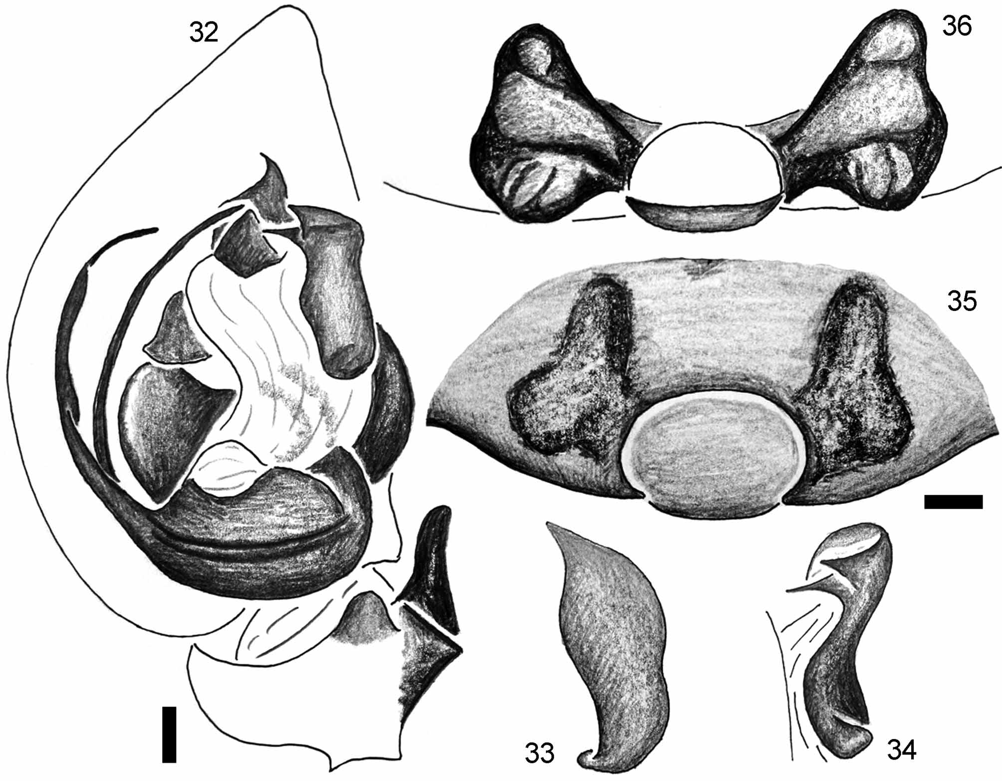
Palp ( Figs 32–34 View FIGURES 32 – 36 ): Palpal tibia with sharply pointed triangular mesolateral apophysis (MTA) directed retrolaterad. RTA digitiform, apex blunt. Cymbial fold broad, occupying approximately half the length of cymbium. TA more or less columnar, apico-prolaterally with triangular fold and triangular process directed mesad. Conductor beak-shaped, apex sharp, pointing laterad. Embolic base with anterio-median concavity accommodating anterior membrane. Embolus originating at 270°, bifurcated: lateral ramus thin, flange-like; mesal ramus filiform, without modification, its apex blunt.
Female ( paratype). Total length 6.17; prosoma 2.92 long, 2.08 wide; opisthosoma 3.25 long, 2.12 wide. Eye sizes and interdistances: AME=PME>ALE=PLE; ratio: AME 1.0, ALE 0.80, PME 1.10, PLE 0.86, AME-AME 0.40, AME-ALE 0.98, PME-PME 1.34, PME-PLE 1.88; MOQ: 1.00 anterior width, 1.10 posterior width, 1.20 long. Leg formula: 4123. Leg measurements: I 11.8 (3.3, 3.2, 2.8, 2.5), II 11.3 (3.2, 3.1, 2.5, 2.5), III 10.8 (3.1, 3.0, 2.4, 2.3), IV 13.5 (3.3, 3.3, 3.2, 2.7).
Pattern and coloration ( Fig. 8 View FIGURES 5 – 8 ): Carapace brown, tegument smooth. Chelicerae dark brown. Sternum yellowish. Legs bicoloured: coxa and trochanter yellowish white; other leg segments yellowish brown.
Opisthosoma ovoid, background purplish, dotted with numerous minute pale spots. Dorsal pattern similar to male. Dorsal scutum absent. Venter purplish. Posterior ventral spines cylindrical, apices bluntly pointed, arranged in a single row.
Genitalia ( Figs 35–36 View FIGURES 32 – 36 ): Median plate of epigyne ovoid. Lateral lobes as part of epigyne, sharply pointed, directed inwards. IDs short, originating anterior to spermathecae. Spermathecae more or less triangular, compacted; anteriorly with large simple coils, posteriorly more complex, strongly convoluted and heavily sclerotized.
Variation: In some specimens or juveniles, the first and second pairs of pale spots on the dorsum are fused and form a pair of longitudinal, oblique bands.
Natural history: The type specimens were collected by sifting decomposing leaves and organic litter in primary lowland forests along the costal lines.
Distribution: East and South Kalimantan, Indonesia ( Fig. 51 View FIGURE 51 ).


No known copyright restrictions apply. See Agosti, D., Egloff, W., 2009. Taxonomic information exchange and copyright: the Plazi approach. BMC Research Notes 2009, 2:53 for further explanation.
|
Kingdom |
|
|
Phylum |
|
|
Class |
|
|
Order |
|
|
Family |
|
|
Genus |