
Meri, Rheims & Jäger, 2022
|
publication ID |
https://doi.org/ 10.11646/zootaxa.5135.1.1 |
|
publication LSID |
lsid:zoobank.org:pub:0CC0D586-E099-4593-9032-EA1885F00F3B |
|
DOI |
https://doi.org/10.5281/zenodo.6820284 |
|
persistent identifier |
https://treatment.plazi.org/id/039787EF-FFBB-C92E-FF32-FAD5FE22FAE6 |
|
treatment provided by |
Plazi |
|
scientific name |
Meri |
| status |
gen. nov. |
Meri View in CoL gen. nov.
Type species. Sadala pictitarsis Simon, 1880 .
Etymology. The generic name refers to Meri , a folk hero and sun God of the Bororó people of Brazil; gender is masculine (last syllable is stressed).
Diagnosis. Species of the genus Meri gen. nov. resemble those of Nungara , Caayguara and Sadala in having the chelicerae with three promarginal teeth and intermarginal denticles ( Fig. 95 View FIGURES 95–98 ; Jäger et al. 2009: figs 10, 89, 109; Rheims 2010a: fig. 7, 2010b: fig. 2; Guala et al. 2012: fig. 22, Pinto & Rheims 2016: fig. 7) and a short-toothed female palpal claw ( Figs 97 View FIGURES 95–98 ; Rheims 2010a: fig. 7; Pinto & Rheims 2016: fig. 16). They are distinguished from Caayguara by the presence of 5–10 escort setae at base of fang ( Fig. 95 View FIGURES 95–98 ) (only one in Caayguara ), three pairs of spines on ventral tibiae I–II (only two in Caayguara ) and epigyne with MS bearing medial pocket (e.g., Figs 154 View FIGURES 150–156 , 173 View FIGURES 173–178 , 289 View FIGURES 285–291 ) (smooth in Caayguara ); from Sadala by the male palp with embolus with only 0.25 turn around tegulum (e.g., Figs 239 View FIGURES 238–244 , 266 View FIGURES 265–268 , 298 View FIGURES 297–303 ) (0.5 to 0.75 turn around tegulum in Sadala ); epigyne with MS bearing medial pocket (e.g., Figs 184 View FIGURES 184–190 , 208 View FIGURES 208–216 , 250 View FIGURES 250–252 ) (with triangular scape-like projection in Sadala ) and internal ducts with one turn (e.g., Figs 143–144 View FIGURES 138–144 , 177–178 View FIGURES 173–178 , 258–259 View FIGURES 253–259 ) (two in Sadala ); and from Nungara by the male palp with oval tegulum, with no grooves and embolus with 0.25 turn around tegulum ( Figs 104 View FIGURES 103–106 , 188 View FIGURES 184–190 , 266 View FIGURES 265–268 ) (tegulum circular with strong groove and embolus with 1 turn around tegulum in Nungara ) and by the epigyne with lateral lobes touching posteriorly (except in M. pictitarsis comb. nov., M. vanini spec. nov. and M. yaciba spec. nov. in which LL are posteriorly covered by MS) and vulva with GP arising from ducts at first turn and SP well-defined, not convoluted (LL not touching posteriorly, GP arising from ducts close to copulatory opening, not at first turn, and spermathecae convoluted, ill-defined in Nungara ).
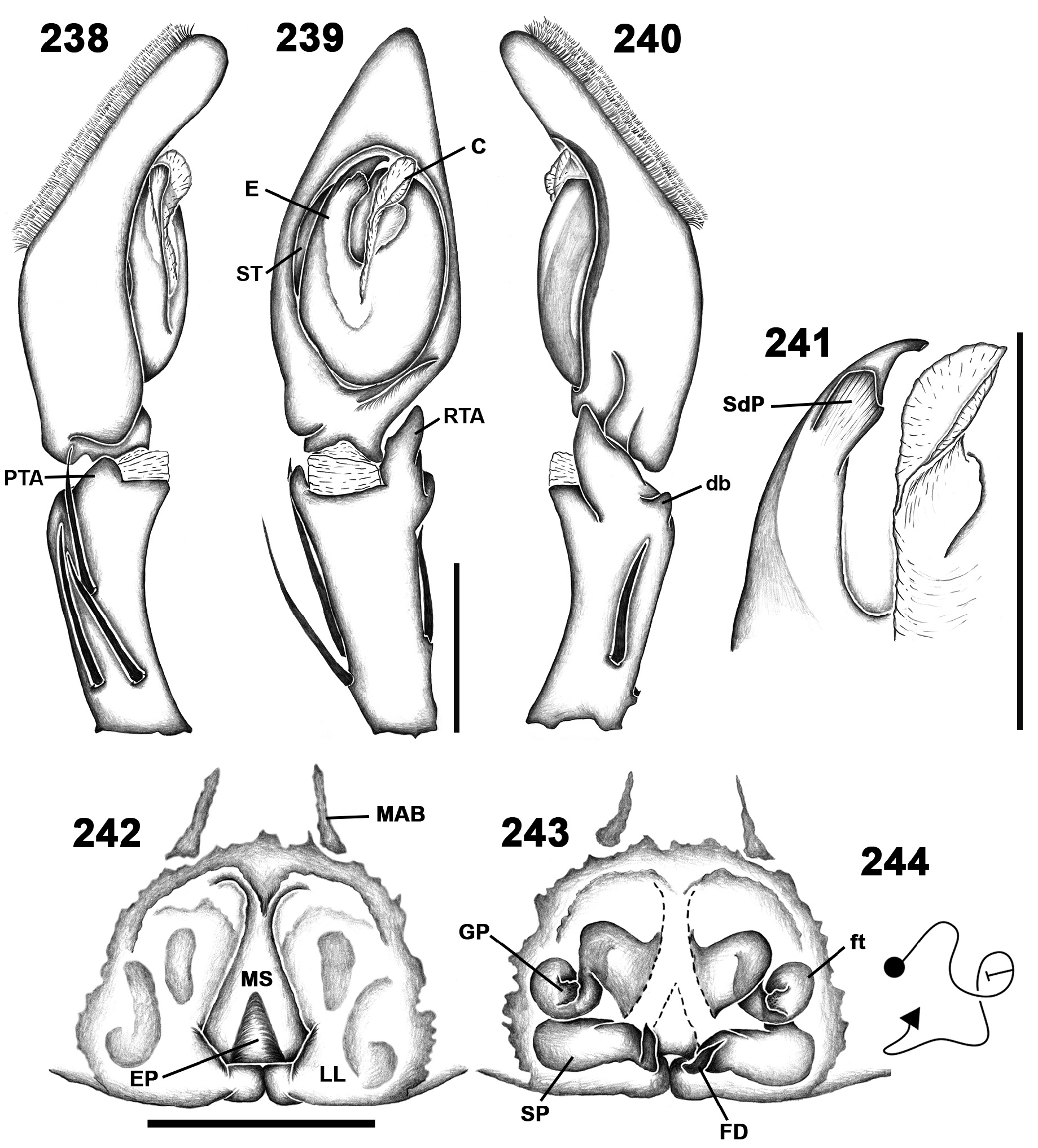
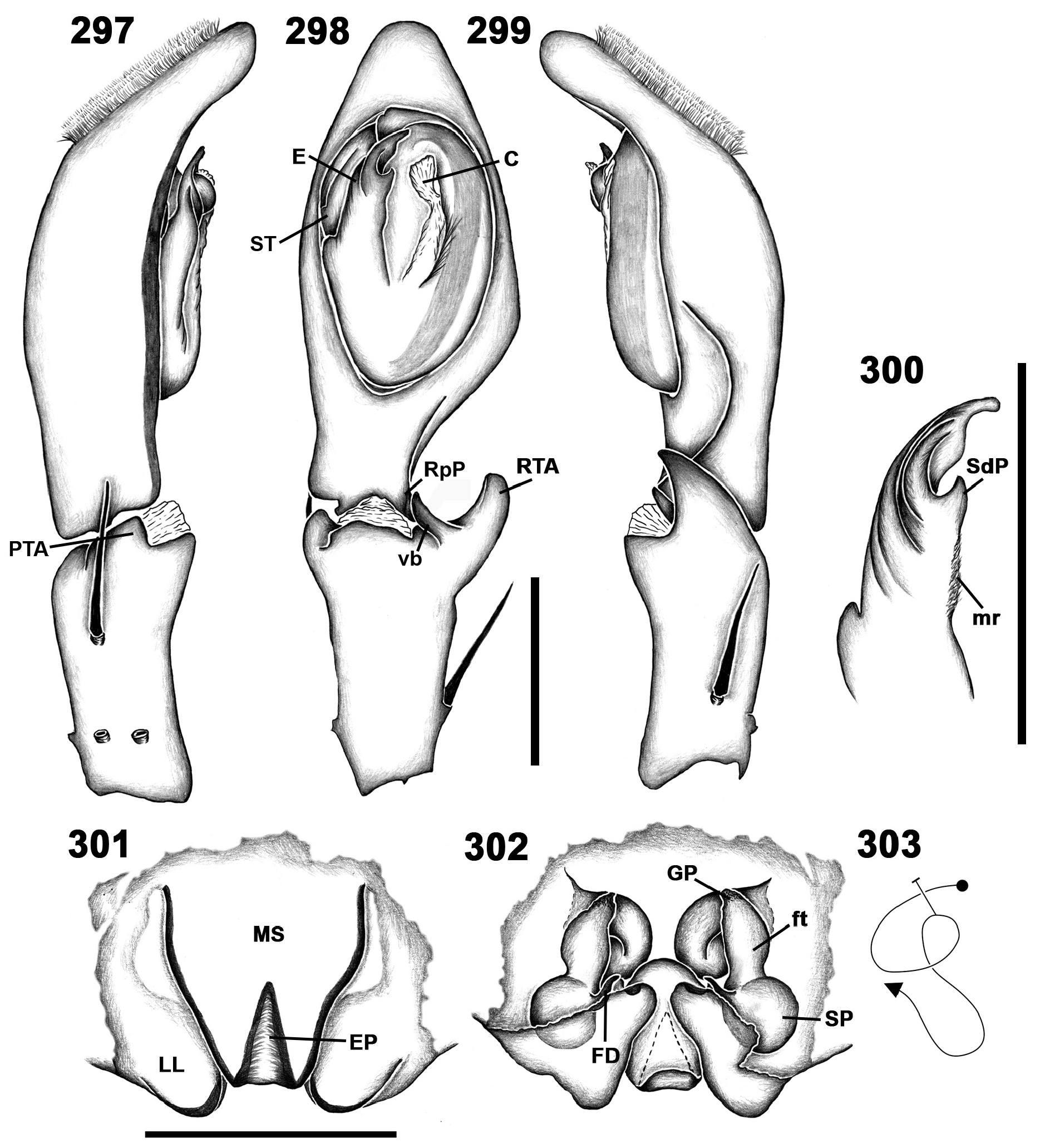
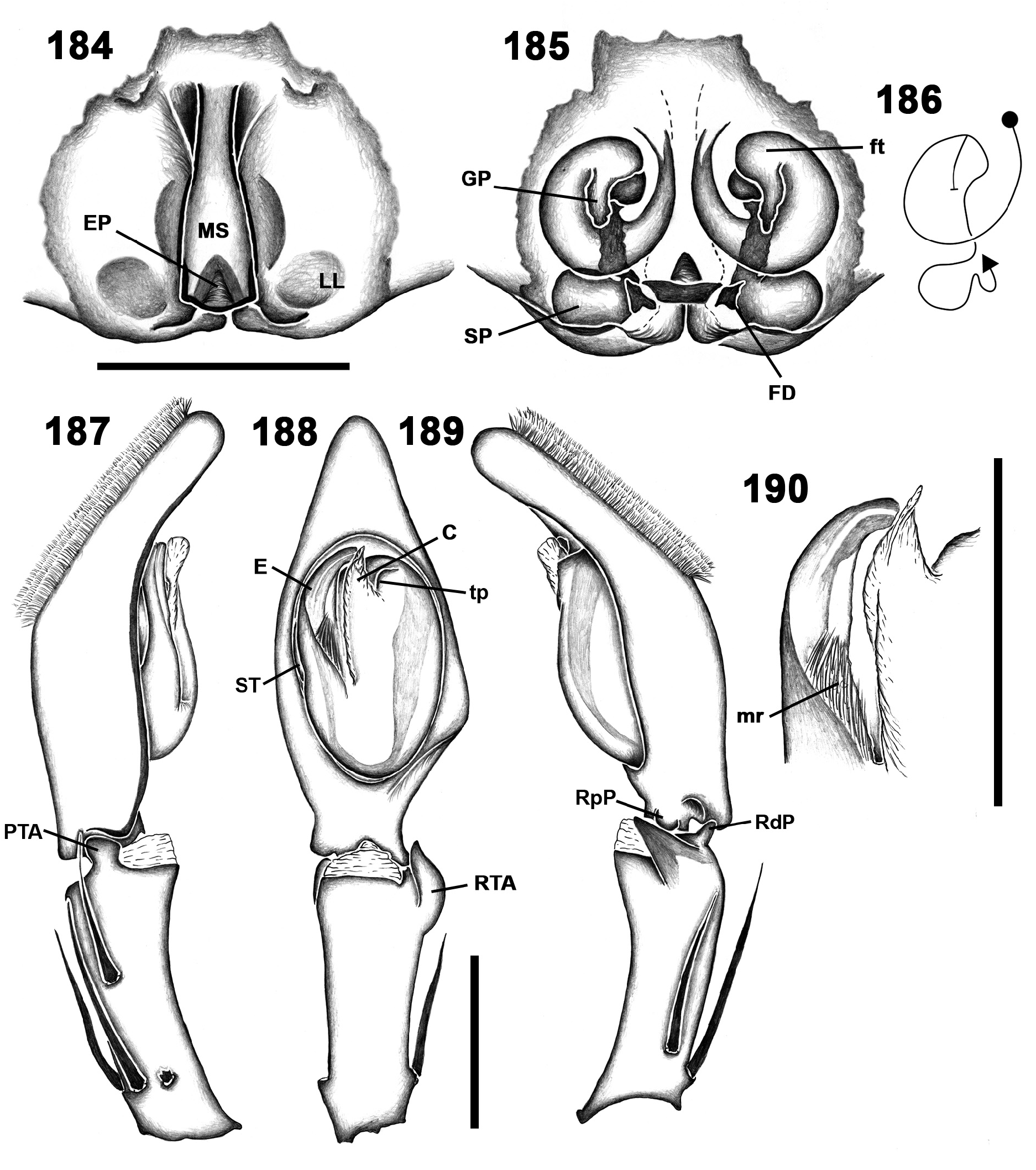
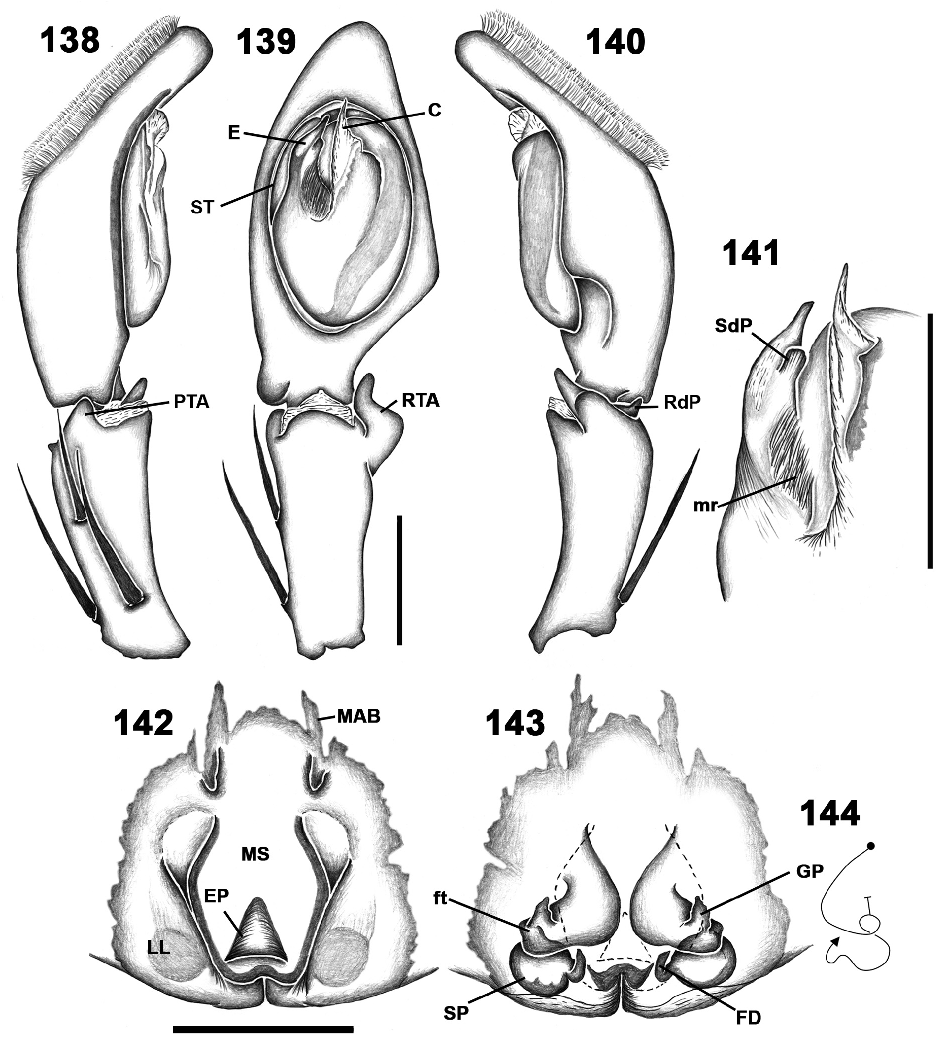
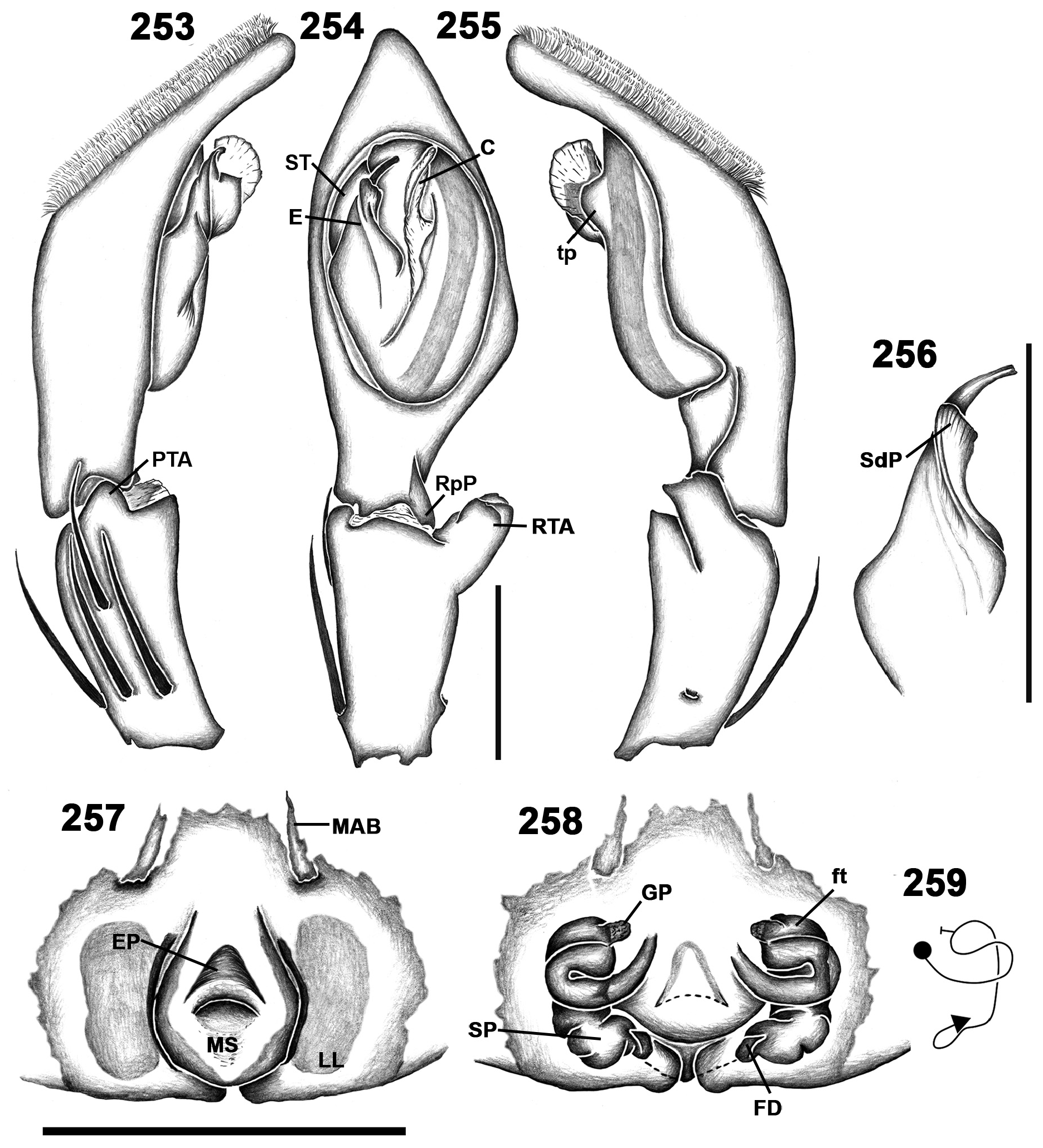
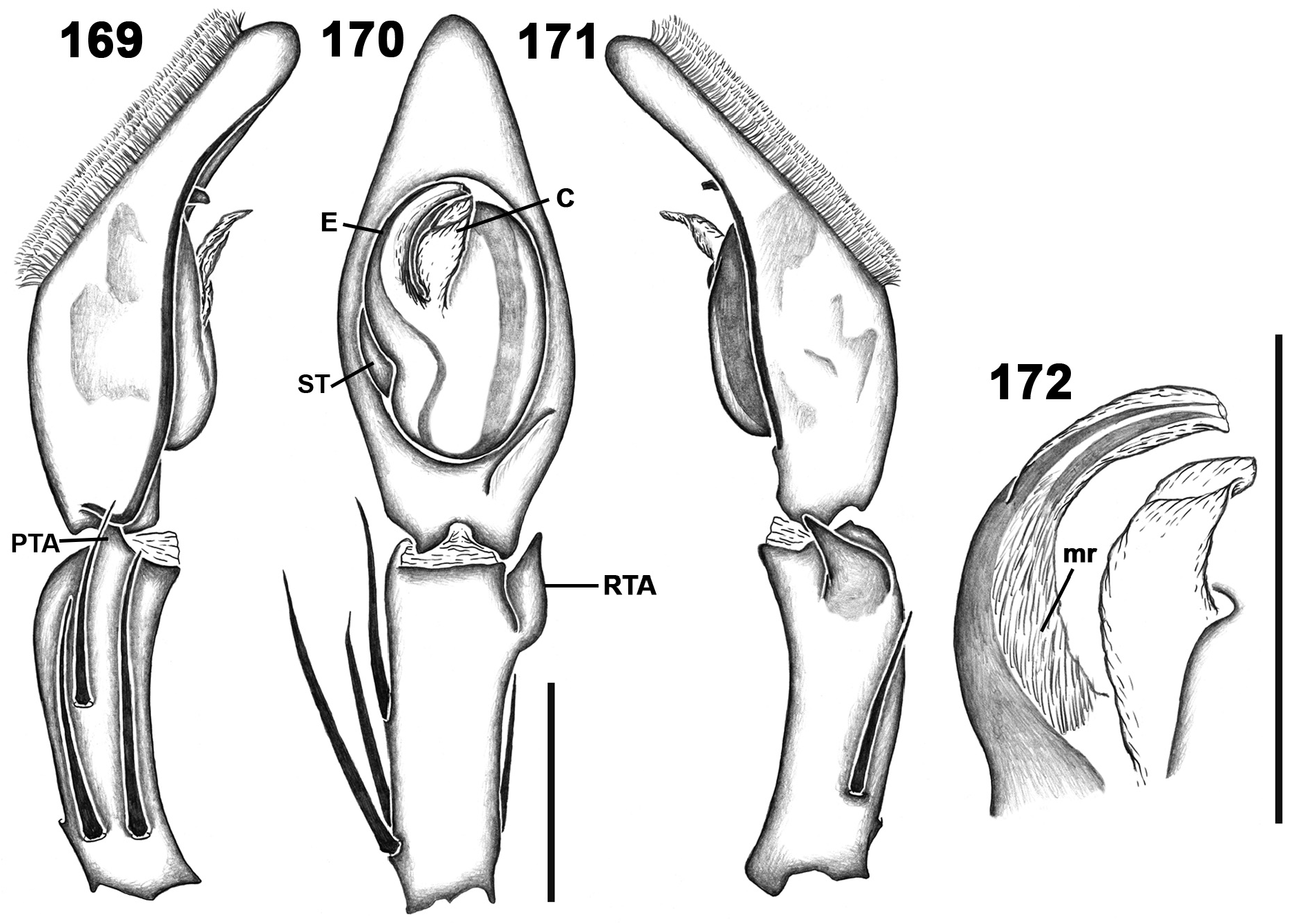
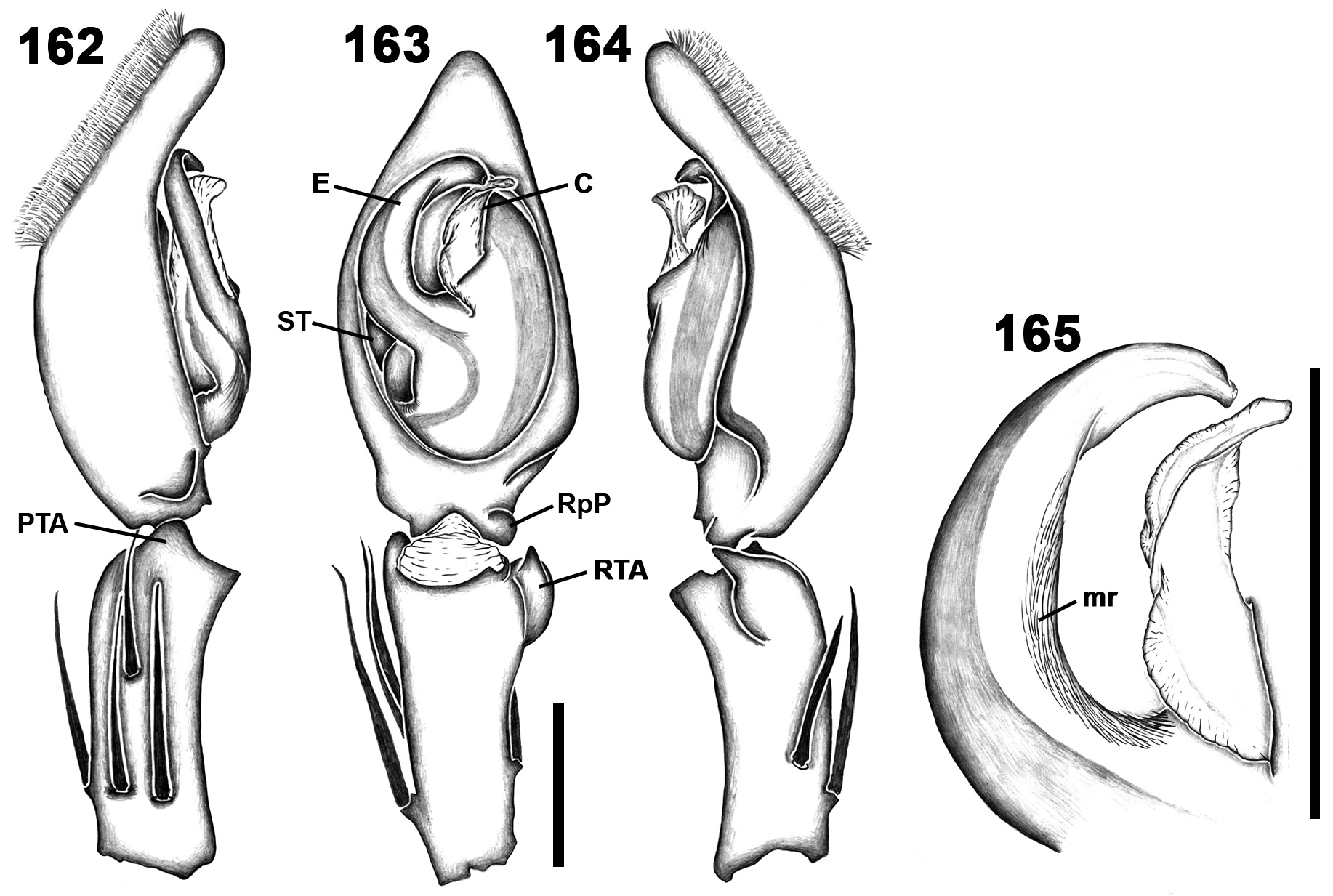
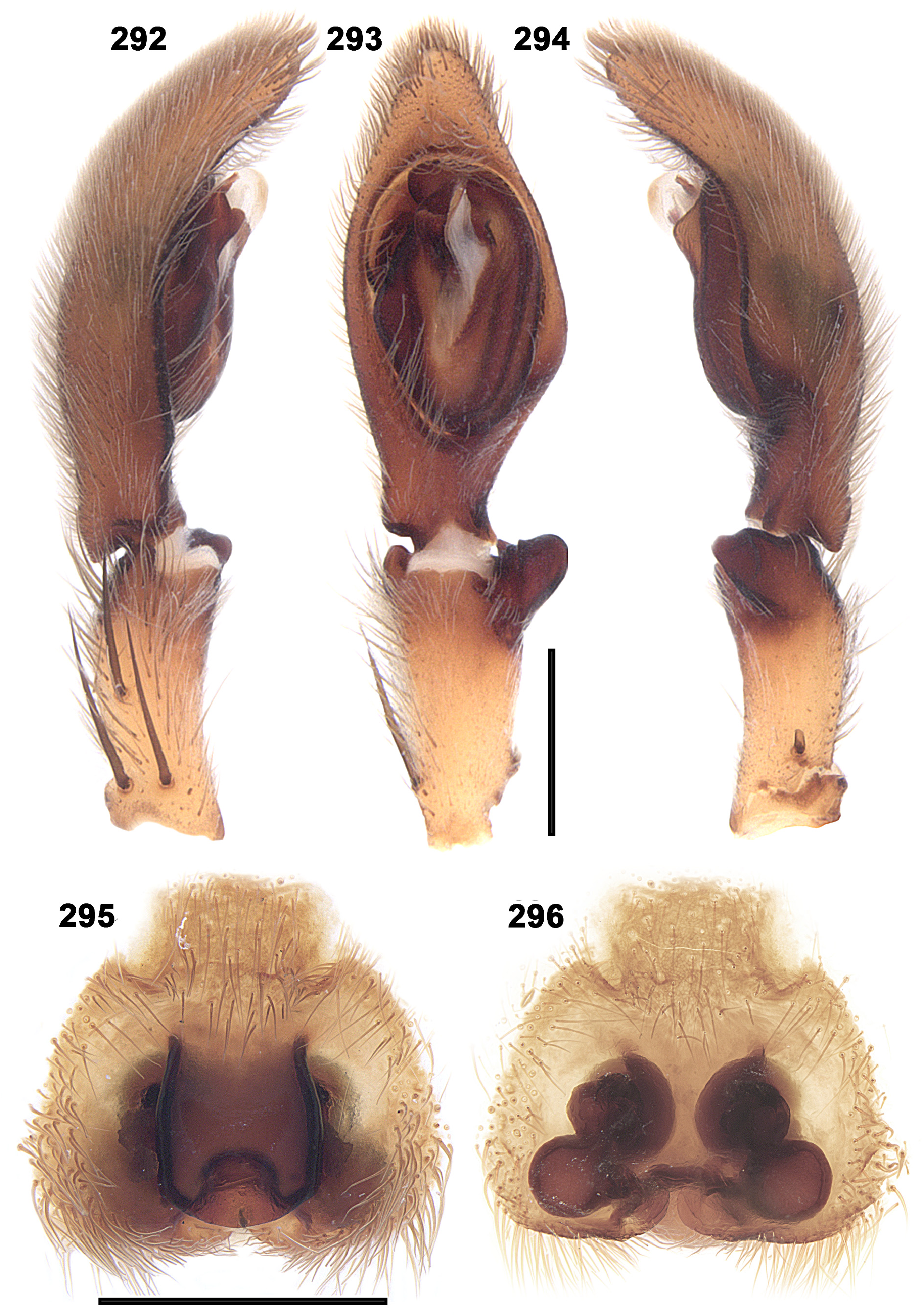
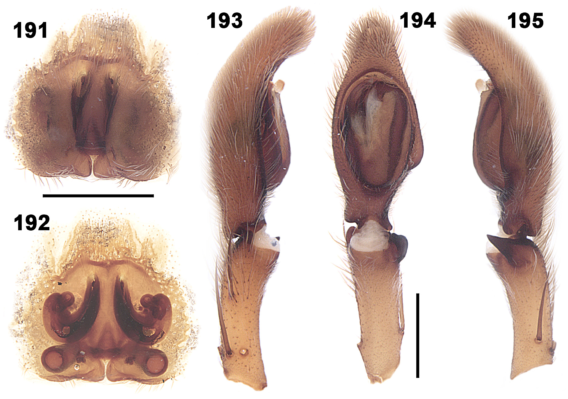
Description. Coloration pattern ranging from orange brown to brown; prosoma with faint darker reticulated pattern of thin lines mostly on cephalic region; opisthosoma with heart mark bearing dark margins and two pairs of oval marks on either side and median chevron or triangular marks down posterior half; legs with darker metatarsi and tarsi ( Figs 91–94 View FIGURES 91–94 ). Total length of males 9.2–15.1, of females 10.8–20.8. Dorsal shield of prosoma generally longer than wide, can be as wide as long. Cephalic region slightly higher than thoracic region, flattening posteriorly. Fovea conspicuous on posterior third of prosoma. Eyes arranged in two straight rows; AME larger than ALE and more separated from each other than from ALE; PME smaller than PLE mostly equidistant, but can be slightly closer or further apart from each other than from PLE. Clypeus low, less than AME diameter. Chelicerae longer than wide with three promarginal teeth, the median one largest, and 3–5 retromarginal teeth, three subequal and the others smaller. Between 15 and 25 intermarginal denticles present at the base of the groove, in front of promarginal teeth. Internal margin with 5–10 escort setae ( Fig. 95 View FIGURES 95–98 ). Labium rebordered as wide as long. Endites slightly convergent, longer than wide, with dense scopula on internal margin. Serrula with single row of denticles ( Fig. 96 View FIGURES 95–98 ). Sternum longer than wide, slightly projected between coxae IV. Female palp with single pectinate claw with 5–7 short teeth ( Fig. 97 View FIGURES 95–98 ); sensory setae long, distally curved, with barbules along the entire setae and with distal region bearing a large rounded pore and a single filiform extension, scattered dorsally along tarsus ( Fig. 102 View FIGURES 99–102 ). Legs laterigrade (2143). Spination pattern in males: femora I–III: p1-1-1, d0-1-1, r1-1-1; IV: p1-1-1, d0-1-1, r0-0-1; patellae I–III: p1, r1; IV: p0, r0; tibiae I–II: p1-0-1, d1-1-1, r1-0-1, v2-2-2; III–IV: p1-0-1, d0-0-1, r1-0-1, v2-2-2; metatarsi I–III: p1-1-0, r1-1-0, v2-2-0; IV: p1-1-2, r1-1-1, v2-2-0; palp: femur p0-0-1, d0-1-2, r0-0-1; patella: p1, r1; tibia: p2-1-0, d1-0-0, r1-0-0; in females: femora I–III: p1-1-1, d0-1-1, r1-1-1; IV: p1-1-1, d0-1-1, r0-0-1; patellae I–III: p0, r1; IV: p0, r0; tibiae I–VI: p1-0-1, r1-0-1, v2-2-2; metatarsi I–III: p1-1-0, r1-1-0, v2-2-0; IV: p1-1-2, r1-1-1, v2-2-0; palp: femur: p0-0-1, d0-1-2, r0-0-1; patella: p1, r1; tibia: p2-1-0, d1-0-0, r1-1-0; tarsus p2-1-0, r2-1-0. Trochanter deeply notched. Metatarsi I–IV distally with dorsal trilobate membrane with median hook as large as lateral projections ( Fig. 98 View FIGURES 95–98 ). Tarsi and antertior half of metatarsi scopulate. Trichobothria present on dorsal tibiae, metatarsi and tarsi, arranged in several rows on tarsi, converging to a single row on metatarsi. Dorsal plate of bothria with one or two transversal grooves, projecting over a smooth proximal plate. Tarsal organ capsulate, with oval opening, located dorsally, at distal end of tarsi. Tarsi with pair of pectinate claws with 20–23 short teeth and claw tufts. Opisthosoma oval, longer than wide. Male epiandrium with small groups of epiandrous spigots ( Fig. 99 View FIGURES 99–102 ). Six spinnerets: anterior lateral spinnerets contiguous, conical and bi-segmented. Basal segment elongated and cylindrical, distal segment short and truncated. Posterior median spinnerets conical and short. Posterior lateral spinnerets conical and bi-segmented. Basal segment elongated and cylindrical, distal segment short and truncated. Male palp: tibia generally shorter than cymbium; PTA short, of various shapes (e.g., Figs 103 View FIGURES 103–106 , 150 View FIGURES 150–156 , 187 View FIGURES 184–190 , 297 View FIGURES 297–303 ); RTA arising distally, of various shapes, single (e. g. Figs 104 View FIGURES 103–106 , 170 View FIGURES 169–172 , 254 View FIGURES 253–259 ) or with branches (e.g., Figs 222 View FIGURES 221–224 , 240 View FIGURES 238–244 , 298 View FIGURES 297–303 ); cymbium with large oval alveolus, dorsal oval scopula, bearing sensory setae, as in female, and, in some species, small retro proximal projection (RpP, e.g., Figs 163 View FIGURES 162–165 , 266 View FIGURES 265–268 ); subtegulum ring-shaped; tegulum oval, smooth (e.g., Figs 127 View FIGURES 126–132 , 170 View FIGURES 169–172 , 188 View FIGURES 184–190 ) or with depressions and/or protrusions, usually close to conductor (e.g., Figs 151 View FIGURES 150–156 , 188 View FIGURES 184–190 , 254 View FIGURES 253–259 ); embolus short, arising from tegulum mostly at 9 o’clock position, smooth (e.g., Fig. 129 View FIGURES 126–132 ), with membranous area short (not surpassing half embolus length) (e.g., Figs 141 View FIGURES 138–144 , 163 View FIGURES 162–165 , 190 View FIGURES 184–190 ), long (surpassing half embolus length (e.g., Figs 165 View FIGURES 162–165 , 172 View FIGURES 169–172 ), bearing long needlelike extensions ( Fig. 100 View FIGURES 99–102 ) or barely conspicuous, not visible with light microscopy, only SEM (e.g., Figs 241 View FIGURES 238–244 , 256 View FIGURES 253–259 , 268 View FIGURES 265–268 ), bearing short needle-like extensions ( Fig. 101 View FIGURES 99–102 ); conductor hyaline, laminar, arising centrally from tegulum.
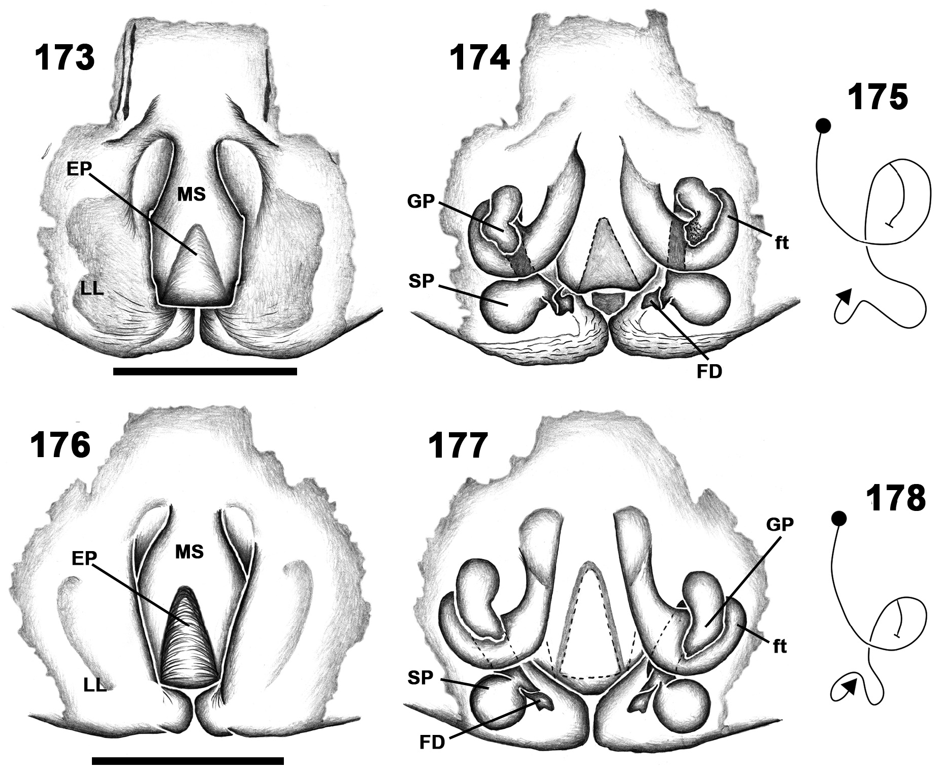
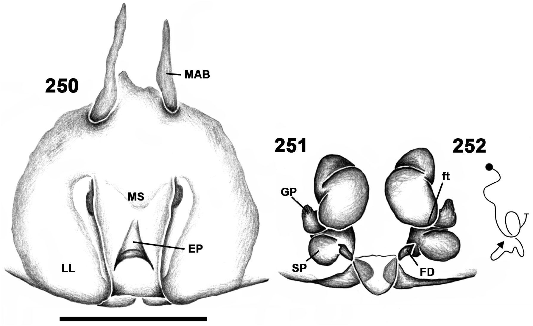
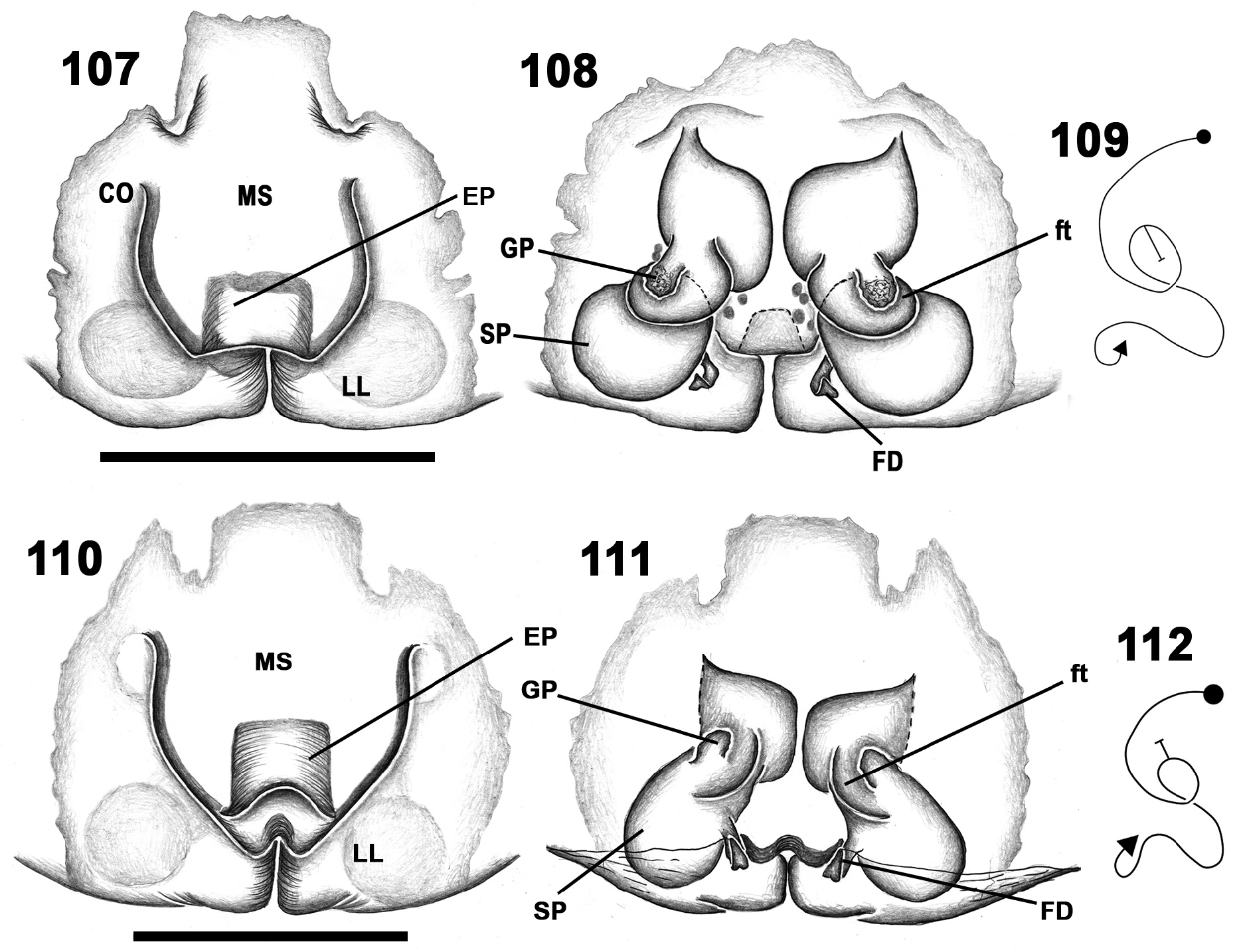
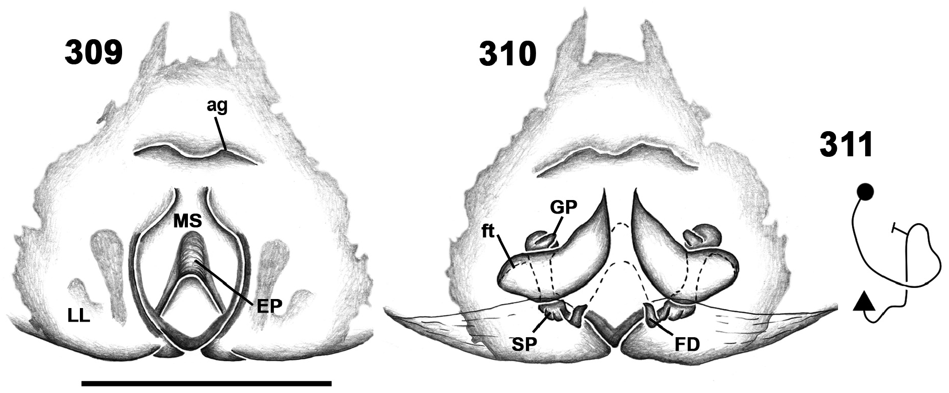
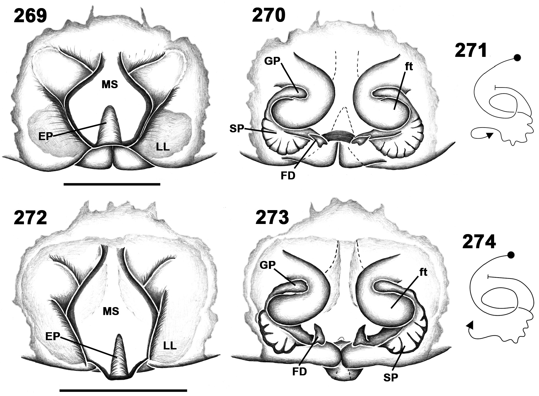
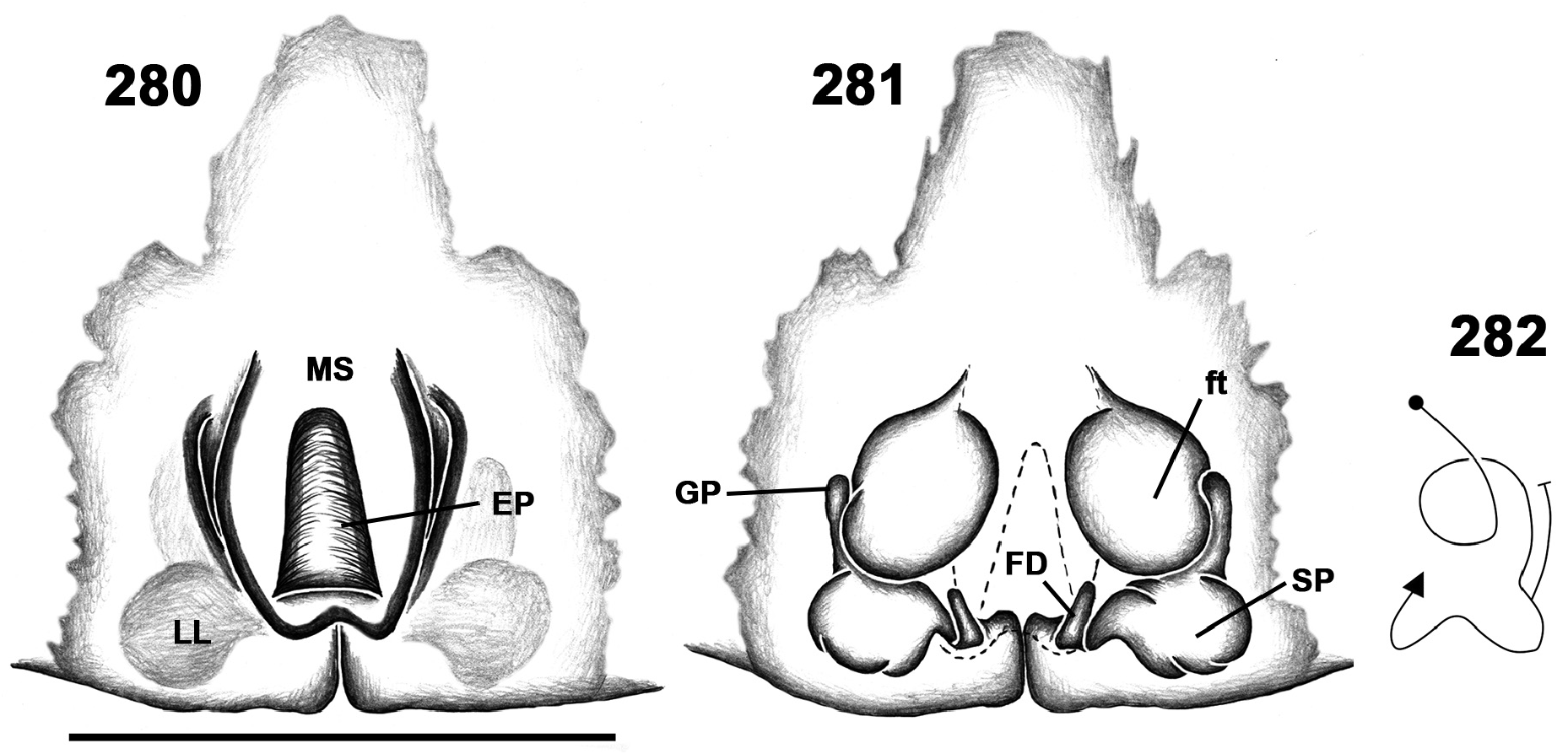
Female epigyne: EF of variable shapes; MAB usually present (e.g., Figs 130 View FIGURES 126–132 , 142 View FIGURES 138–144 , 250 View FIGURES 250–252 ), can be imbedded in EF (e.g., Figs 182 View FIGURES 179–183 , 206 View FIGURES 203–207 , 313 View FIGURES 313–314 ) or inconspicuous (e.g., Figs 208 View FIGURES 208–216 , 234 View FIGURES 234–237 , 295 View FIGURES 292–296 ); LL without projections, touching posteriorly (e.g., Figs 107 View FIGURES 107–112 , 118 View FIGURES 118–123 , 154 View FIGURES 150–156 ) or separated and posteriorly covered by MS (e. g. Figs 225 View FIGURES 225–230 , 289 View FIGURES 285–291 , 301 View FIGURES 297–303 ); MS of variable shapes, bearing EP; EP of various shapes, opening medially (e.g., Figs 250 View FIGURES 250–252 , 257 View FIGURES 253–259 , 309 View FIGURES 309–311 ) or posteriorly (e.g., Figs 173 View FIGURES 173–178 , 200 View FIGURES 196–202 , 269 View FIGURES 269–274 ) on MS. Vulva: internal ducts with one turn (e.g., Figs 112 View FIGURES 107–112 , 156 View FIGURES 150–156 , 178 View FIGURES 173–178 ); GP present, arising from ducts at first turn or close to SP (e.g., Figs 201 View FIGURES 196–202 , 270 View FIGURES 269–274 , 281 View FIGURES 280–282 ); SP of various shapes; FD short, hook-shaped (e.g., Figs 212 View FIGURES 208–216 , 290 View FIGURES 285–291 , 310 View FIGURES 309–311 ).
Composition. Twenty-five species: Meri abuna spec. nov.; M. aparia spec. nov.; M. arraijan spec. nov.; M. carabobo spec. nov.; M. conduri spec. nov.; M. formosus (Banks) comb. nov.; M. guri spec. nov.; M. jaraua spec. nov.; M. kaieteur spec. nov.; M. manaos spec. nov.; M. martinique spec. nov.; M. mathani (Simon) comb. nov.; M. munduruku spec. nov.; M. paiaia spec. nov.; M. pictitarsis (Simon) comb. nov.; M. quinari spec. nov.; M. rivai spec. nov.; M. santivincenti (Simon) comb. nov.; M. tambor spec. nov.; M. tapirapeco spec. nov.; M. trinitatis (Strand) comb. nov.; M. tumatumari spec. nov.; M. vanini spec. nov.; M. yaciba spec. nov.; M. zeteki spec. nov.
Distribution. North and northwestern South America, Panama and the Lesser Antilles ( Figs 313–320 View FIGURES 313–314 View FIGURES 315–318 View FIGURES 319–320 ).
Key to species of Meri View in CoL gen. nov.
1 Males .............................................................................................. 2
- Females........................................................................................... 15
2(1) Subdistal embolic projection absent ( Figs 129 View FIGURES 126–132 , 165 View FIGURES 162–165 , 190 View FIGURES 184–190 )..................................................... 3
- Subdistal embolic projection present (SdP, Figs 141 View FIGURES 138–144 , 224 View FIGURES 221–224 , 288 View FIGURES 285–291 )................................................. 7
3(2) RTA tapering towards pointed tip ( Figs 152 View FIGURES 150–156 , 171 View FIGURES 169–172 , 189 View FIGURES 184–190 ); embolus curved, with conspicuous membranous region bearing long needle-like extensions (mr, Figs 100–101 View FIGURES 99–102 , 165 View FIGURES 162–165 , 190 View FIGURES 184–190 )......................................................... 4
- RTA distally widened and blunt ( Figs 127–128 View FIGURES 126–132 ); embolus mostly straight, without membranous area or needle-like extensions ( Fig. 129 View FIGURES 126–132 )........................................................................ M. carabobo View in CoL spec. nov.
4(3) Tegulum strongly indented at base of embolus ( Figs 163 View FIGURES 162–165 , 170 View FIGURES 169–172 ); embolus with membranous region along more than half its length (mr, Figs 165 View FIGURES 162–165 , 172 View FIGURES 169–172 ).............................................................................. 5
- Tegulum not strongly indented at embolus base ( Figs 151 View FIGURES 150–156 , 188 View FIGURES 184–190 ); embolus with membranous area along less than half its length (mr, Figs 153 View FIGURES 150–156 , 190 View FIGURES 184–190 )................................................................................... 6
5(4) RTA abruptly narrowed medially; cymbium with small retroproximal projection; tegulum slightly protruding over conductor base (RpP, Figs 163–164 View FIGURES 162–165 )................................................................ M. guri View in CoL spec. nov.
- RTA gradually tapering; cymbium without retroproximal projection; tegulum not protruding over base of conductor ( Figs 170–171 View FIGURES 169–172 )........................................................................... M. jaraua View in CoL spec. nov.
6(4) RTA gradually tapering; tibia with retro dorsal projection; PTA anvil-shaped; tegulum not protruding over conductor base (RdP, Figs 188–189 View FIGURES 184–190 )...................................................................... M. manaos View in CoL spec. nov.
- RTA abruptly narrowed medially; tibia without retro dorsal projection; PTA triangular; tegulum protruding over conductor base ( Figs 151–152 View FIGURES 150–156 )............................................................................. M. formosus View in CoL
7(2) RTA with ventral branch (vb, Figs 222 View FIGURES 221–224 , 298 View FIGURES 297–303 )............................................................... 8
- RTA without ventral branch ( Figs 104 View FIGURES 103–106 , 239 View FIGURES 238–244 , 266 View FIGURES 265–268 )............................................................ 9
8(7) RTA distally blunt with ventral branch roughly triangular, slightly longer than wide ( Fig. 222 View FIGURES 221–224 ); embolus with subdistal projection long and laminar (SdP, Fig. 224 View FIGURES 221–224 )........................................................... M. pictitarsis
- RTA distally pointed with ventral branch slightly curved, roughly three times longer than wide ( Fig. 298 View FIGURES 297–303 ); embolus with subdistal projection hook-shaped (SdP, Fig. 300 View FIGURES 297–303 )............................................... M. yaciba View in CoL spec. nov.
9(7) Embolus with large, conspicuous membranous region, bearing long needle-like extensions (mr, Fig. 100 View FIGURES 99–102 ).............. 10
- Embolus with barely conspicuous membranous region (generally not distinguishable under light microscopy, only SEM), bearing short needle-like extensions ( Fig. 101 View FIGURES 99–102 )................................................................ 12
10(9) Tibia without retro dorsal projection ( Figs 105 View FIGURES 103–106 , 198 View FIGURES 196–202 ); embolus with subdistal projection as a small protuberance, not laminar (SdP, Figs 106 View FIGURES 103–106 , 199 View FIGURES 196–202 ).................................................................................. 11
- Tibia with retro dorsal projection (RdP, Fig. 140 View FIGURES 138–144 ); embolus with subdistal projection laminar (SdP, Fig. 141 View FIGURES 138–144 )............................................................................................... M. conduri View in CoL spec. nov.
11(9) PTA distally rounded ( Fig. 103 View FIGURES 103–106 ); RTA gradually tapering ( Fig. 105 View FIGURES 103–106 ); embolus bulging prolaterally at base ( Fig. 104 View FIGURES 103–106 ).......................................................................................... M. abuna View in CoL spec. nov.
- PTA triangular ( Fig. 196 View FIGURES 196–202 ); RTA abruptly narrowed medially ( Fig. 186 View FIGURES 184–190 ); embolus not bulging prolaterally at base ( Fig. 197 View FIGURES 196–202 )................................................................................. M. martinique View in CoL spec. nov.
12(9) RTA tapering towards tip ( Figs 240 View FIGURES 238–244 , 267 View FIGURES 265–268 ); embolus not bulging prolaterally at base ( Figs 239 View FIGURES 238–244 , 266 View FIGURES 265–268 )................... 13
- RTA distally blunt ( Figs 255 View FIGURES 253–259 , 287 View FIGURES 285–291 ); embolus bulging prolaterally at base ( Figs 254 View FIGURES 253–259 , 286 View FIGURES 285–291 )........................... 14
13(12) RTA gradually tapering, with small dorsal branch at base (db, Fig. 240 View FIGURES 238–244 ); embolus with subdistal projection directed antero-mediad (SdP, Fig. 239, 241 View FIGURES 238–244 ).............................................................. M. quinari View in CoL spec. nov.
- RTA abruptly narrowed medially ( Fig. 267 View FIGURES 265–268 ); embolus with subdistal projection directed laterad (SdP, Figs 266, 268 View FIGURES 265–268 )................................................................................................. M. trinitatis View in CoL
14(12) PTA triangular ( Fig. 253 View FIGURES 253–259 ); RTA longer than wide ( Figs 254–255 View FIGURES 253–259 ); embolus bulging retrolaterally at base ( Fig. 256 View FIGURES 253–259 ).......................................................................................... M. tambor View in CoL spec. nov.
- PTA distally rounded ( Fig. 285 View FIGURES 285–291 ); RTA wider than long ( Figs 286–287 View FIGURES 285–291 ); embolus not bulging retrolaterally ( Fig. 288 View FIGURES 285–291 ).......................................................................................... M. vanini View in CoL spec. nov.
15(1) EP opening medially on MS ( Figs 250 View FIGURES 250–252 , 257 View FIGURES 253–259 , 309 View FIGURES 309–311 ).......................................................... 16
- EP opening posteriorly on MS ( Figs 107 View FIGURES 107–112 , 200 View FIGURES 196–202 , 285 View FIGURES 285–291 );........................................................ 18
16(15) MS widest medially, with anterior margins converging ( Figs 257 View FIGURES 253–259 , 309 View FIGURES 309–311 ); SP elongated, with margins bearing few folds ( Figs 258 View FIGURES 253–259 , 310 View FIGURES 309–311 ).............................................................................................. 17
- MS widest anteriorly, with anterior margins diverging ( Fig. 250 View FIGURES 250–252 ) SP irregularly rounded, smooth ( Fig. 251 View FIGURES 250–252 ) M. sanctivincenti View in CoL
17(16) EF without anterior groove; MS with anterior margins gradually converging ( Fig. 257 View FIGURES 253–259 ); internal ducts with FW C-shaped, laterad ( Figs 259–259 View FIGURES 253–259 )............................................................... M. tapirapeco View in CoL spec. nov.
- EF with transversal anterior groove; MS with anterior margins constricted (eg, Fig. 302 View FIGURES 297–303 ); internal ducts with FW slightly reniform, postero-laterad ( Figs 310–311 View FIGURES 309–311 )..................................................... M. zeteki View in CoL spec. nov.
18(15) Internal ducts with GP arising close to SP, not at first turn ( Figs 201 View FIGURES 196–202 , 270 View FIGURES 269–274 , 281 View FIGURES 280–282 ).................................. 19
- Internal ducts with GP arising at first turn ( Figs 122 View FIGURES 118–123 , 177 View FIGURES 173–178 , 243 View FIGURES 238–244 )................................................ 21
19(18) MS with anterior margins gently converging anteriorly ( Figs 200 View FIGURES 196–202 , 280 View FIGURES 280–282 ); SP margins with few folds ( Figs 201 View FIGURES 196–202 , 281 View FIGURES 280–282 )...... 20
- MS with anterior margin constricted ( Figs 269, 272 View FIGURES 269–274 ); SP with convoluted margins ( Figs 270, 273 View FIGURES 269–274 )............ M. trinitatis View in CoL
20(19) MS medially constricted with EP slightly longer than wide ( Fig. 194 View FIGURES 191–195 )....................... M. martinique View in CoL spec. nov.
- MS widest medially with EP almost two times longer than wide ( Fig. 273 View FIGURES 269–274 ).................. M. tumatumari View in CoL spec. nov.
21(18) EP subrectangular or trapezoid ( Figs 118 View FIGURES 118–123 , 289 View FIGURES 285–291 )............................................................ 22
- EP triangular ( Figs 121 View FIGURES 118–123 , 142 View FIGURES 138–144 , 208 View FIGURES 208–216 )...................................................................... 25
22(21) EP wider than long or as long as wide with anterior margin not surpassing half MS length ( Figs 107 View FIGURES 107–112 , 154 View FIGURES 150–156 , 289 View FIGURES 285–291 )......... 23
- EP longer than wide with anterior margin surpassing MS length, along most of MS length ( Fig. 112 View FIGURES 107–112 )... M. aparia View in CoL spec. nov.
23(22) MS with lateral margins not constricted anteriorly and not covering LL posteriorly ( Figs 107 View FIGURES 107–112 , 154 View FIGURES 150–156 )................... 24
- MS with lateral margins constricted anteriorly, covering LL posteriorly ( Fig. 289 View FIGURES 285–291 ).................. M. vanini View in CoL spec. nov.
24(23) MS with lateral margins slightly curved ( Fig. 107, 110 View FIGURES 107–112 ); GP slightly longer than wide; SP ovoid ( Fig. 108, 111 View FIGURES 107–112 ).............................................................................................. M. abuna View in CoL spec. nov.
- MS with lateral margins straight ( Fig. 154 View FIGURES 150–156 ); GP three times longer than wide; SP with constrictions ( Fig. 155 View FIGURES 150–156 )..................................................................................................... M. formosus View in CoL
25(21) MS with conspicuous transversal anterior groove (ag, Figs 121 View FIGURES 118–123 , 130 View FIGURES 126–132 )........................................... 26
- MS without anterior groove ( Figs 142 View FIGURES 138–144 , 208 View FIGURES 208–216 , 242 View FIGURES 238–244 )........................................................... 28
26(25) MS widest anteriorly; EP with anterior margin not surpassing half MS length ( Fig. 121 View FIGURES 118–123 ); internal ducts with GP mediad ( Figs 122–123 View FIGURES 118–123 )......................................................................... M. arraijan View in CoL spec. nov.
- MS widest medially; EP with anterior margin surpassing half MS length ( Fig. 130 View FIGURES 126–132 ); internal ducts with GP antero-laterad ( Figs 131–132 View FIGURES 126–132 )........................................................................ M. carabobo View in CoL spec. nov.
28(26) MS widest anteriorly ( Figs 208, 211 View FIGURES 208–216 , 225 View FIGURES 225–230 )................................................................ 29
- MS widest medially or posteriorly ( Figs 142 View FIGURES 138–144 , 184 View FIGURES 184–190 , 242 View FIGURES 238–244 )...................................................... 32
29(28) MS covering LL posteriorly ( Figs 225 View FIGURES 225–230 , 301 View FIGURES 297–303 )............................................................... 30
- MS not covering LL posteriorly ( Figs 208, 211 View FIGURES 208–216 )............................................................ 31
30(29) HP 1.5 times longer than wide ( Fig. 225 View FIGURES 225–230 ); GP posterior to CO; FD antero laterad or laterad ( Figs 226–227 View FIGURES 225–230 ).... M. pictitarsis
- HP 2 times longer than wide ( Fig. 301 View FIGURES 297–303 ); GP in line with CO; FD antero mediad ( Figs 302–303 View FIGURES 297–303 )....... M. yaciba View in CoL spec. nov.
31(29) MS with lateral margins diverging anteriorly; EP more than two times longer than wide with anterior margin reaching anterior margin of MS ( Fig. 208 View FIGURES 208–216 ); internal ducts with FW not dilated ( Fig. 209 View FIGURES 208–216 )................................. M. mathani View in CoL
- MS with lateral margins running roughly parallel anteriorly; EP slightly longer than wide with anterior margin not surpassing half MS length ( Fig. 211 View FIGURES 208–216 ); internal ducts with FW strongly dilated ( Fig. 212 View FIGURES 208–216 )................ M. munduruku View in CoL spec. nov.
32(28) MS rhombus shaped with EP opening slightly anterior to posterior margin ( Fig. 142 View FIGURES 138–144 ); internal ducts with FW medio-posteriad and GP anteriad ( Figs 143–144 View FIGURES 138–144 )........................................................ M. conduri View in CoL spec. nov.
- MS of different shapes with EP opening at posterior margin ( Figs 173 View FIGURES 173–178 , 214 View FIGURES 208–216 , 242 View FIGURES 238–244 ); internal ducts with FW postero laterad or antero-laterad and GP mediad or posteriad ( Figs 174–175 View FIGURES 173–178 , 242–244 View FIGURES 238–244 )........................................... 33
33(32) MS with anterior margins strongly bottlenecked ( Figs 214 View FIGURES 208–216 , 242 View FIGURES 238–244 ); SP elongate ( Figs 215 View FIGURES 208–216 , 243 View FIGURES 238–244 )....................... 34
- MS with anterior margins slightly converging ( Figs 173, 176 View FIGURES 173–178 , 184 View FIGURES 184–190 ); SP ovoid ( Figs 174, 177 View FIGURES 173–178 , 185 View FIGURES 184–190 )................... 35
34(33) MS shaped as an inverted cognac glass with EP wider than long ( Fig. 214 View FIGURES 208–216 ); internal ducts with GP posteriad, anterior to CO; SP with grooved margins ( Figs 215–216 View FIGURES 208–216 )..................................................... M. paiaia View in CoL spec. nov.
- MS Erlenmeyer-shaped with EP onger than wide ( Fig. 242 View FIGURES 238–244 ); internal ducts with GP mediad, posterior to CO; SP with smooth margins ( Figs 243–244 View FIGURES 238–244 )................................................................. M. rivai View in CoL spec. nov.
35(33) MS widest posteriorly, gently narrowing posteriorly, roughly three times longer than wide; EP with anterior margin not surpassing 0.2 MS length ( Fig. 184 View FIGURES 184–190 ).......................................................... M. kaieteur View in CoL spec. nov.
- MS widest medially almost two times longer than wide; EP with anterior margin reaching half MS length ( Figs 173, 176 View FIGURES 173–178 )... M. jaraua View in CoL spec. nov.


No known copyright restrictions apply. See Agosti, D., Egloff, W., 2009. Taxonomic information exchange and copyright: the Plazi approach. BMC Research Notes 2009, 2:53 for further explanation.