
Atractides (Atractides) svislocensis, Cichocka, Maria & Biesiadka, Eugeniusz, 2013
|
publication ID |
https://doi.org/ 10.11646/zootaxa.3608.4.3 |
|
publication LSID |
lsid:zoobank.org:pub:E81A5018-222E-43C1-A9C5-9506F3EDAC0F |
|
DOI |
https://doi.org/10.5281/zenodo.5625820 |
|
persistent identifier |
https://treatment.plazi.org/id/039A8780-FF8A-FF9F-3AF4-EF296872D9D4 |
|
treatment provided by |
Plazi |
|
scientific name |
Atractides (Atractides) svislocensis |
| status |
sp. nov. |
Atractides (Atractides) svislocensis sp. nov.
( Figs. 15–27 View FIGURES 15 – 17 View FIGURES 18 – 21 View FIGURES 22 – 27 )
Type material. Holotype: male dissected and slide mounted in Hoyer’s fluid. Belarus, river Svisloč, Suchaja Dolina (N: 53 0 47’459; E: 23 0 97’78), 12.06.1998, leg. E. Biesiadka. Paratypes: 8 females, same data as holotype, 10 larvae reared in the laboratory from one female captured on 12.06.1998, same data as holotype, oviposition 30.06., hatching after 7 days.
Other material. Male and 3 females. Belarus, river Dzitva, Hančary (N: 53 0 43’964; E: 25 0 37’84), 15. 0 7.1997, leg. E. Biesiadka.
Diagnosis. In both genders integument smooth without microsculpture. Vgl 4 situated on nearly the same level as Vgl 2 and Vgl 3. Whip-like seta situated on the mediodistal margin of the I-L-5, bipartite in the proximal part. Genital organ of males with few hairs (10–13), maximum width in posterior half. P-3 with straight ventral margin. Sword seta on P-4 situated nearly ventrodistal seta. Female genital plates narrow, with distinct notches between the acetabula. The diameter of Vgl 4 twice the diameter of Vgl 3. P-3 enlarged proximally. Sword seta on P-4 situated nearly ventroproximal seta. Larval dorsal plate elongated, in the anterior part rounded, in the central part parallelsided. Lp2 distinctly distant from the plate’s margin. Lh1 halfway between Mh1 and Mh2. C1 and C4 close to each other. Larval organ large and oval, with the falciform process slightly distant from its anterior margin. Excretory pore large, located in the centre of the excretory pore plate, between the posterior anal setae.
Description. Male. (measurements: n = 2, holotype, second specimen in parantheses) L of idiosoma ( Fig. 15 View FIGURES 15 – 17 ) 770 (778), W 695 (397). L of coxal field 449 (452), maximal W 625 (427), L of suture between Cx-1 193 (194), Cx-1 W 316 (616), Cx-2 W 404 (408), Cx-3 W 536 (540). Distance between Cx-3+4 96 (98). Genital organ L 136 (136), W 185 (187), maximum W in posterior half. Ac oval or subtriangular in shape. Ac-1-3 L 44 (45)-57 (57)-75 (78). Genital organ with 10–13 hairs on each side. Excretory pore elongated, located posterior to Vgl-1; Vgl-1 separated from Vgl-2. Vgl-1, Vgl-2 and Vgl-3 situated nearly on the same level. Palp ( Fig. 16 View FIGURES 15 – 17 ) total dorsal L 410, dorsal L of individual segments: P-1, 37 (36); P-2, 92 (94); P-3, 97 (100); P-4, 132 (132); P-5, 52 (52); relative L of individual segments for holotype (in % of total L): P-1, 9.0; P-2, 22.4; P-3, 23.6; P-4, 32.2; P-5, 12.6; ventral margin of P-2 with a mediodistal protrusion, P-3 distinctly concave, P-4 slender, distance between ventral hairs 27, sword seta inserted halfway between ventral hairs, proximal and distal hairs of the same thickness.
I-L as given in Fig. 17 View FIGURES 15 – 17 , I-L-5 slightly enlarged in distal part, dorsal L 228 (132), ventral L 163 (165), S-1 L 136 (138), ratio S-1 L/ W 10.3, S-2 L 105 (108), ratio S-2 L/ W 7.52, distal, curved seta bipartite in the proximal part ( Fig. 17 View FIGURES 15 – 17 ). I-L-6 L 171.
Female (measurements: n = 5). L of idiosoma ( Fig. 18 View FIGURES 18 – 21 ) 990–1220, W 880– 980. L of coxal field 440–460, maximal W 750–765, L of suture between Cx-150–158, Cx-1 W 280–296, Cx-2 W 464–475, Cx-3 W 540–580. Distance between Cx-3+4 190–205. Genital organ L 240–258, W 242–256, genital plates arcuate, with distinct notches between the acetabula, genital plate L 158–163. Ac1-3 L (57–58)-(43–45)- (47–49). Location of excretory pore, Vgl-1, Vgl-2 and Vgl-3 as in male. Palp ( Fig. 19 View FIGURES 18 – 21 ) total dorsal L 453–462, dorsal L of individual segments: P- 1, 39–40; P-2, 95–98; P-3, 127–129; P-4, 148–150; P-5, 44–45; relative L of individual segments for one specimen (in % of total length): P-1, 8.6; P-2, 21.2; P-3, 27.9; P-4, 32.4; P-5, 9.7.
I-L-5 as in males ( Fig. 20 View FIGURES 18 – 21 ), dorsal L 295–300, ventral L 218–224, S-1 L 147–149, ratio S-1 L/ W 17.0, S-2 L 122–123, ratio S-2 L/ W 9.3. I-L-6 L 236–139. IV-L slender, distal end of IV-L-4 with five unequal setae—three significantly longer than two others, on distal end of IV-L-5 four setae—one longer than three others ( Fig. 21 View FIGURES 18 – 21 ). External claw denticle slightly longer than internal denticle.
Larva (n = 10). Idiosoma elongated, 234–247 long and 153–169 wide on average. Dorsal plate ( Fig. 22 View FIGURES 22 – 27 ) elongated, L 226–239 and W 127–143, rounded in its anterior part. Microrelief on the plates in the shape of polygons. Lp1 and Lp2 long, equal in length, Lp1 distant from the plate’s margin, located closer to Mp2. Lh1 setae halfway between mediopropodosomal setae Mh1 and Mh2. Coxal plates occupying a larger part of the ventral side of the idiosoma ( Fig. 23 View FIGURES 22 – 27 ), maximum L 213–231 medial L 150–163. Urstigma the processes which accompany this structure situated slightly in front of the organ. Coxal setae close to one another, especially C4 and C1. C1 relatively short. Posterior margin of the coxal plates with a transverse thickening. Anal plate ( Fig. 23 View FIGURES 22 – 27 ) large, 80–91 wide and 31–41 long, with slightly folded anterior margin. Excretory pore small and round. L of posterior ventral setae 187–197. Gnathosoma quite elongated. Capitulary bay narrow and deep, 44–52 long and 39–46 wide. Chelicerae ( Fig. 24 View FIGURES 22 – 27 ) quite big, 67–78 long and 31–41 wide at the base. Legs ( Figs 25–27 View FIGURES 22 – 27 ) quite long, especially the first and second pair ( Table 2 View TABLE 2 ), with rather long, thick and stiletto-like setae in the distal parts of genu, tibia and tarsus.
Remarks. From most points of view, the new species is similar to Atractides albaruthenicus sp. nov. from which it differs in the male sex in the very low number of genital setae. Such strong reduction of genital setae is not encountered in other species of the genus. P-2 and P-4 are more slender than ones in second species, and sword seta in P-4 inserted more distally. In females P-2 and P- 3 in proximal part are more enlarged. The shape of genital plates in females is similar to that in A. ovalis Koen. 1883 , A. inflatus (Walter, 1925) and A. diastema (Szalay, 1935) . Differences between larvae of both species are most significantly (character states of larva A. albaruthenicus sp.nov. are indicated in parentheses). Dorsal plate elongated (oval), lateral margins parallel (rounded), anterior margin rounded (straight), larval organ small (visibly bigger), C4 long and thick (short and fine), excretory pore situated in the centre of anal plate (in the anterior third of anal plate).
Etymology. The name of the new species originates from the name of the river in which it was found.
Acknowledgements
The authors are thankful to Dr. P. Martin and anonymous referees for their work for valuable comments and helping to improve the language.
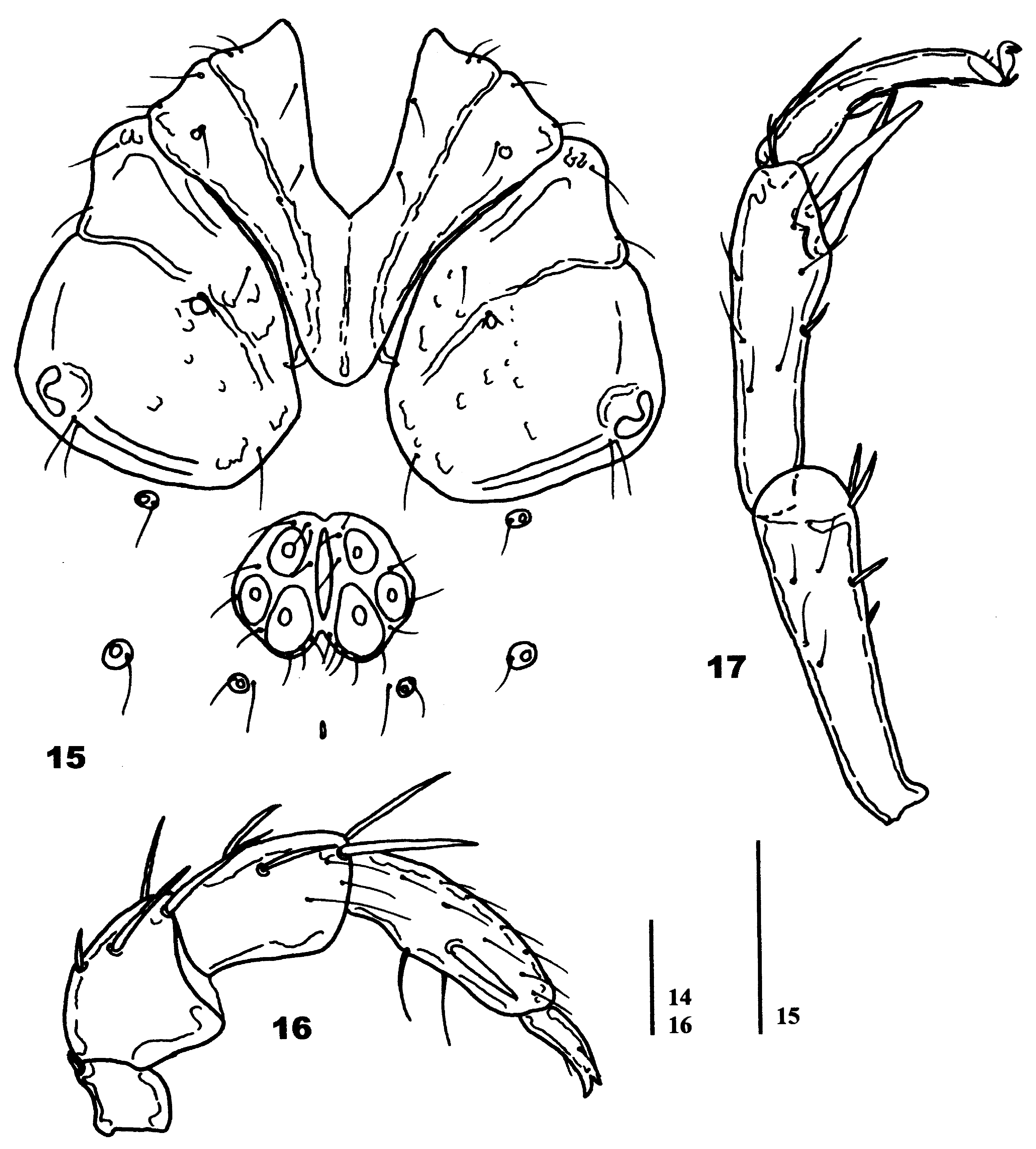
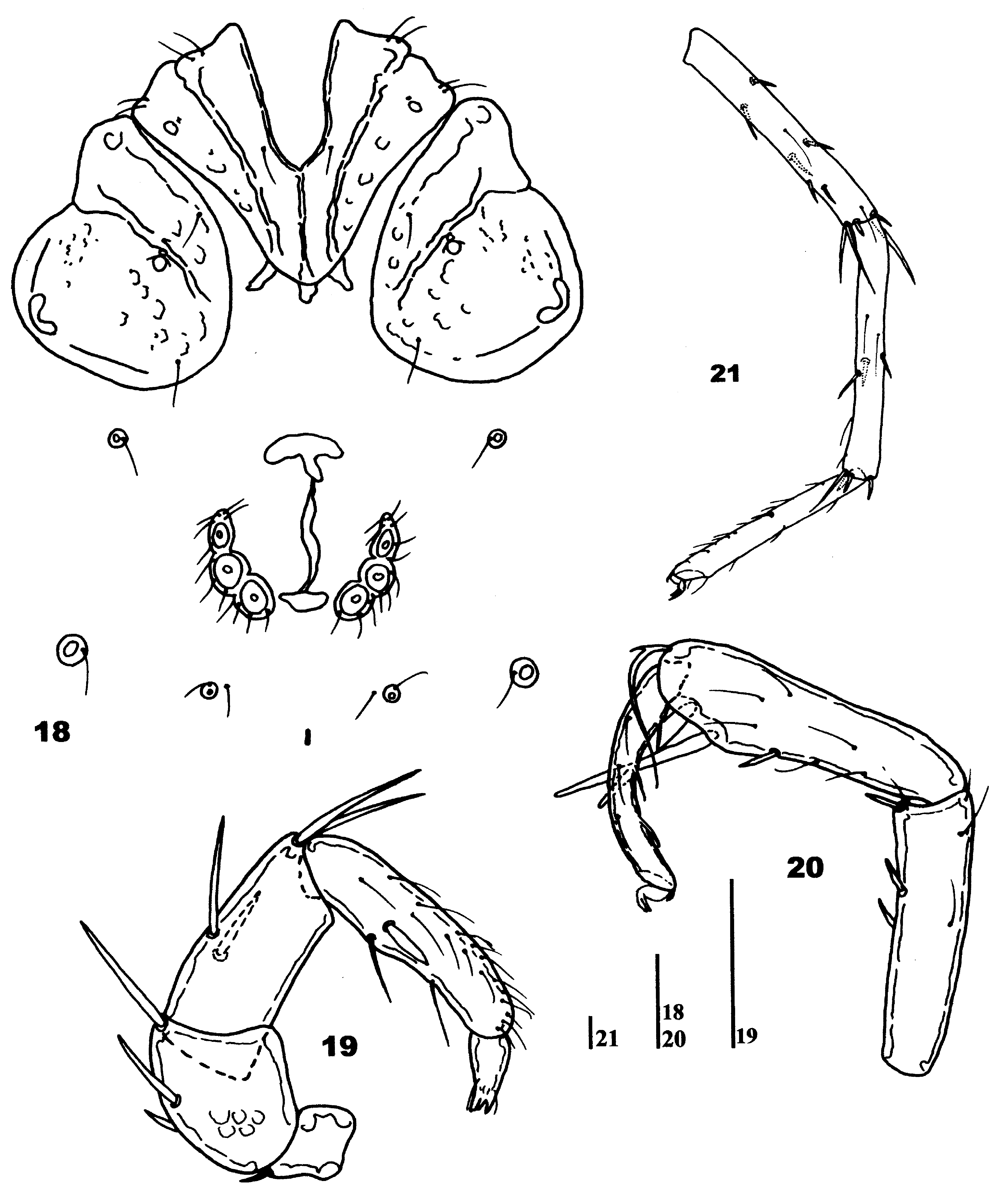
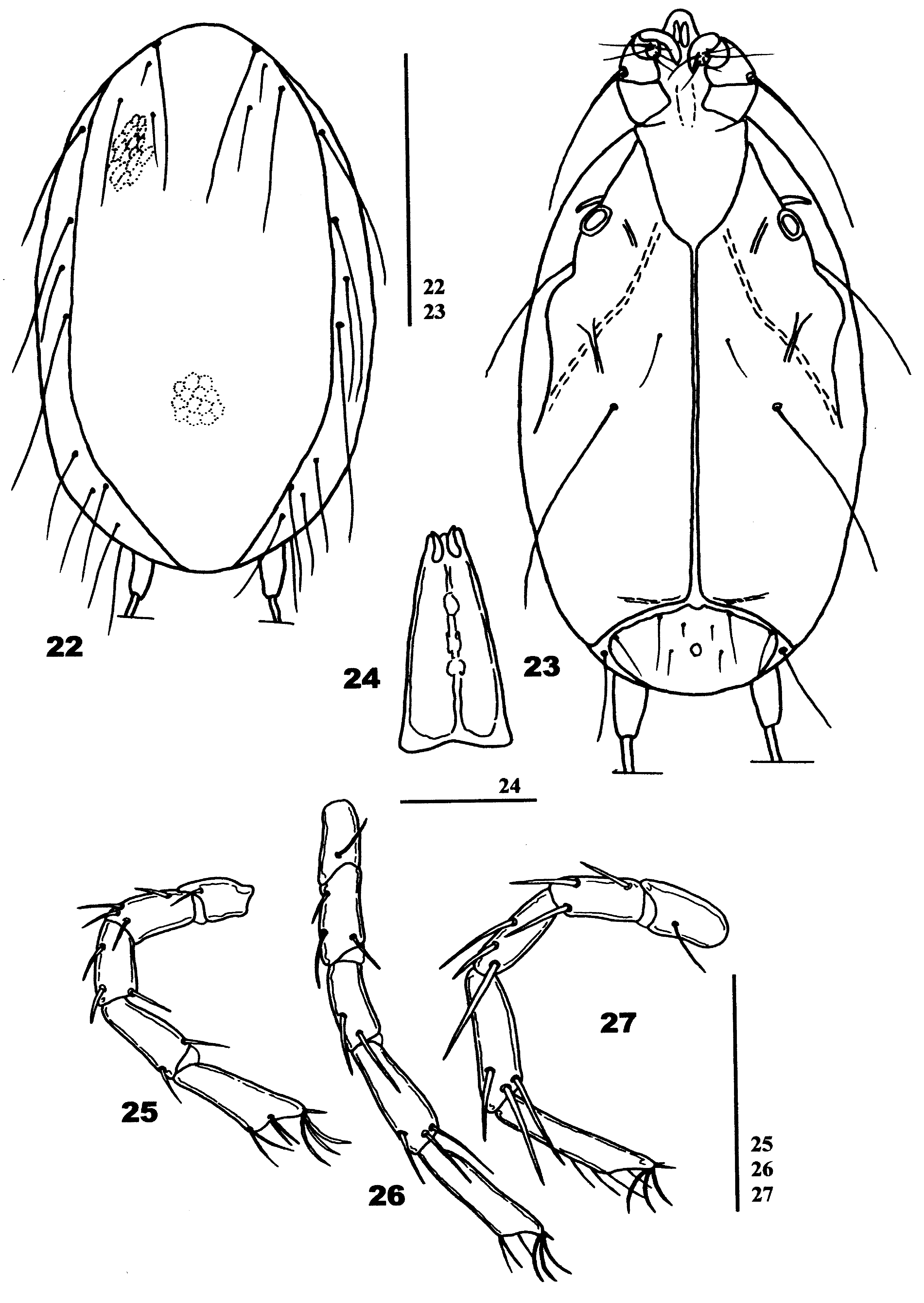
TABLE 2. Leg measurements (in μm) of larvae Atractides svisloczensis sp. nov.
| Measurements | Leg segments | Total length | |||
|---|---|---|---|---|---|
| trochanter femur | genu | tibia | tarsus | ||
| I–L min–max | 23.4–33.8 31.2–36.4 | 33.8–39.0 | 36.4–46.8 | 52.0–70.0 | 187.2–218.4 |
| mean | 27.8 34.3 | 35.9 | 41.4 | 55.4 | 194.8 |
| II–L min–max | 28.6–33.8 31.2–39.0 | 31.2–39.0 | 41.6–52.0 | 65.0–78.0 | 195.0–226.2 |
| mean | 30.9 34.6 | 35.4 | 48.4 | 68.4 | 217.6 |
| III–L min–max | 28.6–41.6 36.2–44.2 | 39.0–44.2 | 49.4–57.2 | 83.2–88.4 | 241.8–267.8 |
| mean | 33.0 38.7 | 40.3 | 53.0 | 86.1 | 251.1 |
No known copyright restrictions apply. See Agosti, D., Egloff, W., 2009. Taxonomic information exchange and copyright: the Plazi approach. BMC Research Notes 2009, 2:53 for further explanation.
|
Kingdom |
|
|
Phylum |
|
|
Class |
|
|
Order |
|
|
Family |
|
|
Genus |
|
|
SubGenus |
Atractides |