
minutus Roewer, 1919
|
publication ID |
https://doi.org/ 10.5281/zenodo.190591 |
|
DOI |
https://doi.org/10.5281/zenodo.6219188 |
|
persistent identifier |
https://treatment.plazi.org/id/039B87F0-FF88-EE79-F2B6-FC7F7B7FC623 |
|
treatment provided by |
Plazi |
|
scientific name |
minutus Roewer, 1919 |
| status |
|
Rivetinus minutus Roewer, 1919 , new family assignment
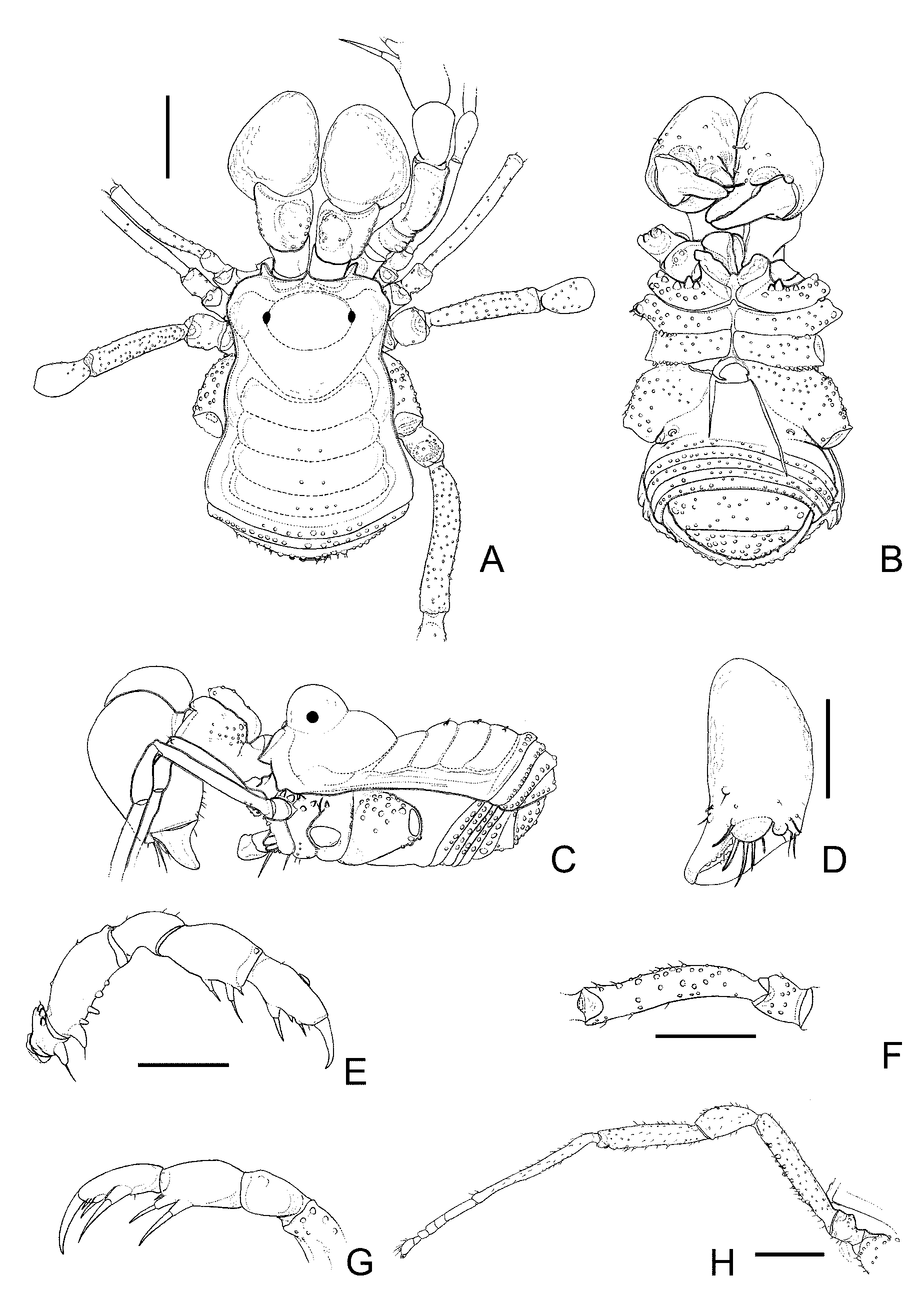
( Figs. 2 View FIGURE 2 and 4 View FIGURE 4 D,E)
Rivetinus minutus Roewer 1919:132 , pl. 13, Figs. 7 (dor hab), 7a–b (legI, IV), 1923:127 (rdesc), Fig. 133 (dor hab); Kury 2003: 28 (cat). (ma paratype; Ecuador, [Carchí], El Pelado, collected at 4150 m; without collector and date; SMF RI 311; examined). Note: Although not written in the label, according to the original description, the collector is Dr. Rivet, dated in i.1903.
Diagnosis: Zamorinae with unarmed ocularium, dorsal scutum mostly smooth, with mesotergal grooves I–IV backward, area I undivided, pedipalpal patella unarmed, coxae II–III without conspicuous armature, ventral plate of penis concave at the sides, with two distal roughly delimitated lobes, each one with acute and frontward corners.
Redescription: Male (holotype): Dorsum ( Fig. 2 View FIGURE 2 A,C): Measurements: DSL 3.0; OMW 2.3; PL 1.4; PW 1.8; Leg I 5.2; II 7.5; III 5.7; IV 7.6. Dorsal scutum mostly smooth, with constriction near groove I and between grooves II–III, widening from area II to posterior margin; mesotergal area I slightly wider. Prosoma high behind ocularium. Ocularium, large, high, domed. Mesotergal area I entire; II–III with a pair of small tubercles; IV with a short row of four small tubercles; grooves I–IV backward. Posterior margin and free tergites with one row of larger tubercles (on posterior margin shorter, placed in the middle). Anal operculum tuberculate.
Venter ( Fig. 2 View FIGURE 2 B): Coxae I–IV covered with tubercles, I with an anterior row of larger tubercles, two higher tubercles near coxapophysis.
Chelicera ( Fig. 2 View FIGURE 2 A,C,D): Segment I with several small dorsal tubercles, dorso-prolaterally swollen, bulla well-marked; II swollen, with a frontal-median projection between base of fingers ( Fig. 2 View FIGURE 2 D). Fixed and movable finger with five small teeth.
Pedipalpus ( Fig. 2 View FIGURE 2 E,G): Coxa with large ventral tubercle. Trochanter with two ventral tubercles (median larger). Femur with five ventral tubercles (basal larger). Patella unarmed. Tibia mesal IIi, ectal iIi. Tarsus mesal Iii, ectal Iii.
Legs ( Fig. 2 View FIGURE 2 F,H): Small tuberculate. Coxae I–II each with two dorsal tubercles; III with one tubercle directed to another lateral of II; IV ending at area II, tuberculate. Trochanter I with three ventral larger tubercles; IV inflated. Femur IV slightly curved inward, with two ventral rows of tubercles increasing in size subapically. Tarsal formula: 6(3), 11 (4), 6, 6.
Penis ( Fig. 4 View FIGURE 4 D,E): Truncus with three pairs of trifid long basal setae, two pairs of small, curved, unirramous setae on sides of stylus base and two unirramous pairs on ventral plate base. Ventral plate concave at the sides, with two distal roughly delimitated lobes, each one with acute and frontward corners. Stylus long, unique, without crest.
Coloration: Specimen discolored. Dorsal scute, chelicera and pedipalp dark yellow, legs light yellow. Female: unknown.

No known copyright restrictions apply. See Agosti, D., Egloff, W., 2009. Taxonomic information exchange and copyright: the Plazi approach. BMC Research Notes 2009, 2:53 for further explanation.
minutus Roewer, 1919
| Pinto-Da-Rocha, Ricardo & Hara, Marcos Ryotaro 2009 |
minutus
| Kury 2003: 28 |
| Roewer 1919: 132 |