
Coniocleonus nigrosuturatus (Goeze, 1777)
|
publication ID |
https://doi.org/ 10.5281/zenodo.5298722 |
|
publication LSID |
lsid:zoobank.org:pub:D1FF3534-A1C8-4B2B-ACDF-69F31ED12BC0 |
|
DOI |
https://doi.org/10.5281/zenodo.5306921 |
|
persistent identifier |
https://treatment.plazi.org/id/039C0A0F-FFFD-FFE1-FEB0-FB90EE40F9CB |
|
treatment provided by |
Marcus |
|
scientific name |
Coniocleonus nigrosuturatus (Goeze, 1777) |
| status |
|
Coniocleonus nigrosuturatus (Goeze, 1777) View in CoL
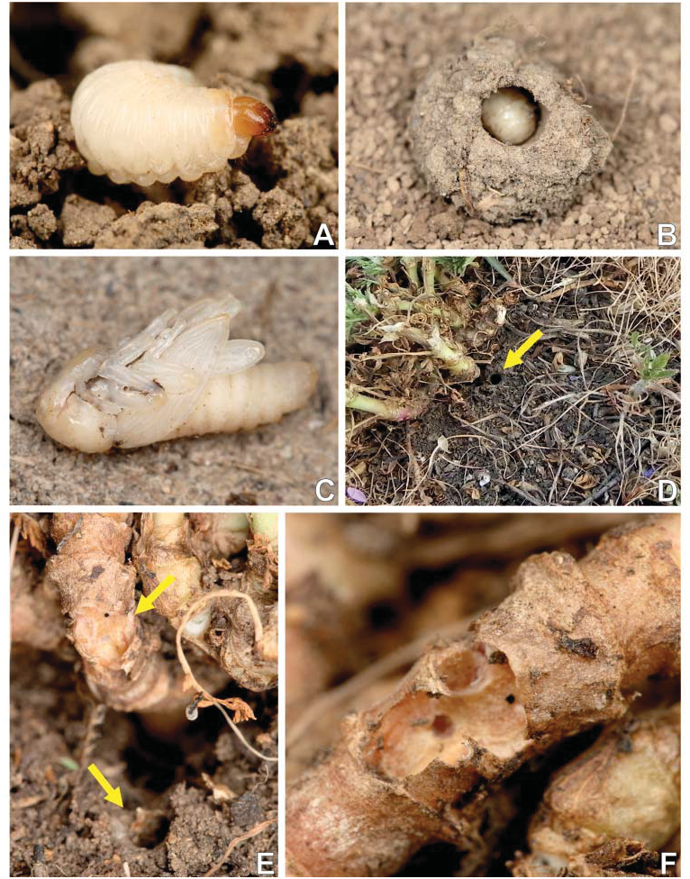
Material examined. ROMANIA: Sfânta Elena: 24.v.2013 (1 larva), 4.vii.2013 (1 larva, 1 pupa) (all F. Trnka leg.), 7.vi.2012 (1 larva), 10.vii.2012 (1 pupa reared, partly damaged), 19.v.2013 (2 larvae), 7.vii.2013 (1 pupa reared from larva from v.2013 (partly dried-up)) (all R. Stejskal leg.). SLOVAKIA: Dolné Zelenice, 16.vi.2013 (2 larvae), R. Stejskal leg.
Description of mature larva. Colouration. Head light brown or brown. All thoracic and abdominal segments white ( Fig. 4A View Fig ). Cuticle ¿nely spiculate.
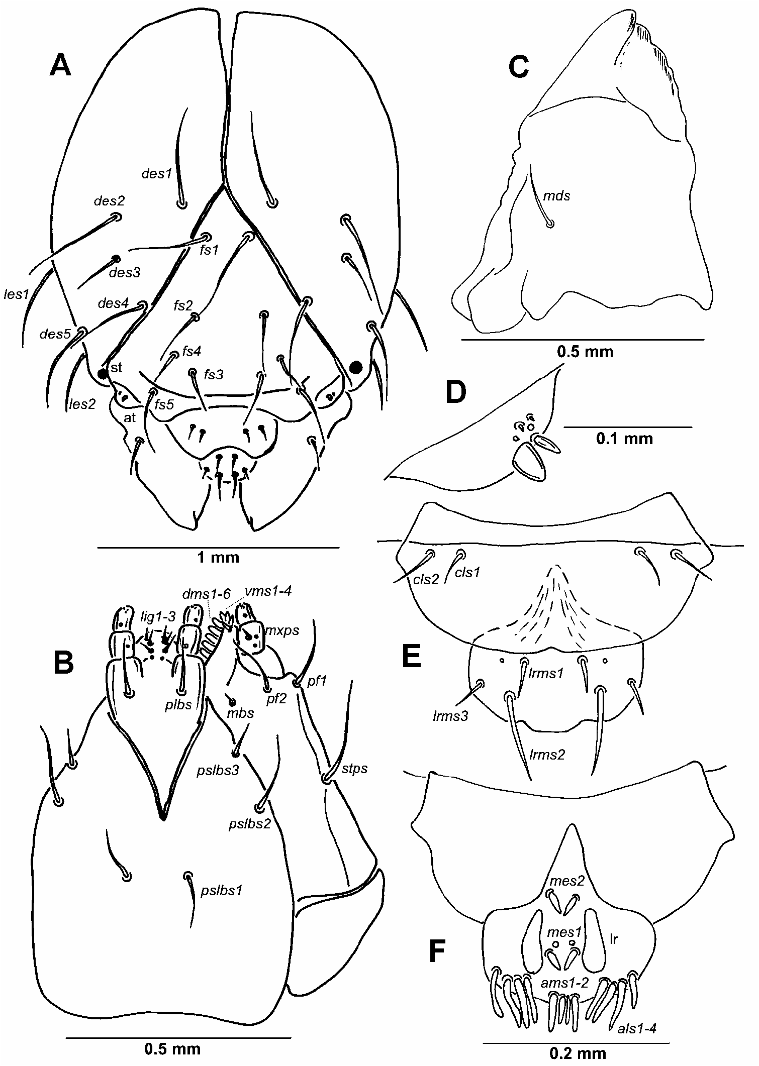
Head capsule and mouth parts. Head width: 1.5–1.7 mm (mean 1.6 mm), suboval, flattened laterally, endocarinal line absent. Frontal sutures on head distinct, Y-shaped, extended to stemmata. Single stemma (st), in the form of a dark pigmented spot, located on each side anterolaterlly. Des1 near to frontal suture, des2 and des3 located in the central part of epicranium, des4 located anterially near to frontal suture, des5 located anterolaterally; des1-2 and des4-5 long, equal in length, des3 distinctly shorter ( Fig. 1A View Fig ). Fs1 and fs2 placed medially, fs3 located anteriomedially, fs4 located anteriolaterally, and fs5 located laterally, close to the epistoma; all setae relatively long, fs3 and fs4 shorter than the remaining three setae ( Fig. 1A View Fig ). Les1–2 as long as des1; and ves1–2 as long as des3. Epicranial area without pores. Antennae located at the end of the frontal suture on each side, membranous and slightly convex basal article bearing conical triangular sensorium, relatively long; basal membranous article with 3 sensillae different in both shape and length ( Fig. 1D View Fig ).
Labrum ( Fig. 1E View Fig ) approximately 2 times as wide as long, with 3 pairs of hairform lrms, of different length; lrms3 and lrms1 distinctly shorter than lrms2; lrms1 placed close to the margin with clypeus, lrms2 located anteriomedially and lrms3 located anteriolaterally; anterior margin double sinuate. Clypeus ( Fig. 1E View Fig ) approx. 2.5 times as wide as long with 2 relatively short cls, unequal in length, localized posteriolaterally; anterior margin rounded to the inside; median part covered by thorn-shaped cuticular processes. Epipharynx ( Fig. 1F View Fig ) with 4 pairs of blunt, ¿nger-like als, of almost equal length; 2 pairs of ams, ams1 distinctly shorter than ams2; 2 pairs of short, blunt mes; labral rods (lr) elongated, converging anterially. Mandibles ( Fig. 1C View Fig ) relatively broad, bi¿d, tooth of unequal height; slightly truncate; mds relatively long, hairform. Maxilla ( Fig. 1B View Fig ) stipes with 1 stps, 2 pfs and 1 mbs, stps and pfs1-2 long, equal in length, mbs very short; mala with 6 bacilliform dms of different length; 4 vms short, almost equal in length; vms distinctly shorter than dms. Maxillary palpi with two palpomeres; basal palpomere with 1 mxps and two pores; length ratio of basal and distal palpomeres: 1:0.7; distal palpomere with one pore and a group of conical, cuticular apical processes. Praelabium ( Fig. 1B View Fig ) heart-shaped and distinctly elongated, with 1 pair of plbs; ligula with sinuate margin and 3 pairs of hairform micro ligs, equal in length; premental sclerite well visible. Labial palpi with two palpomeres; length ratio of basal and distal palpomeres: 1:0.6; distal palpomere with one pore and short, cuticular apical processes; basal palpomere with 1 dorsal pore. Postlabium ( Fig. 1B View Fig ) with 3 pslbs, pslbs1 located anterially, remaining two pairs laterally; different in length, pslbs3 distinctly shorter than pslbs1 and pslbs2.
Thorax and abdomen. Body length: 11.0– 13.5 mm (mean 12.0 mm) stocky, slightly curved, rounded in cross section ( Fig. 2A View Fig ). The widest place in the body (abdominal segments V–VI) measuring up to 4.5 mm. Prothorax distinctly smaller than meso- and metathorax. Metathorax and abdominal segments I–III of almost equal length, next abdominal segments increasing gradually to abdominal segments V–VI (the largest) and then decreasing to the terminal parts of the body. Abdominal segment X reduced to four anal lobes of unequal size, the dorsal being distinctly the largest, the lateral pair equal in size, and the ventral lobe very small. Anus located terminally. Spiracles (9 pairs) bicameral, the ¿rst placed between the pro- and mesothorax (see Material and methods), the abdominal spiracles located laterally, close to the anterior margin of abdominal segments I–VIII.
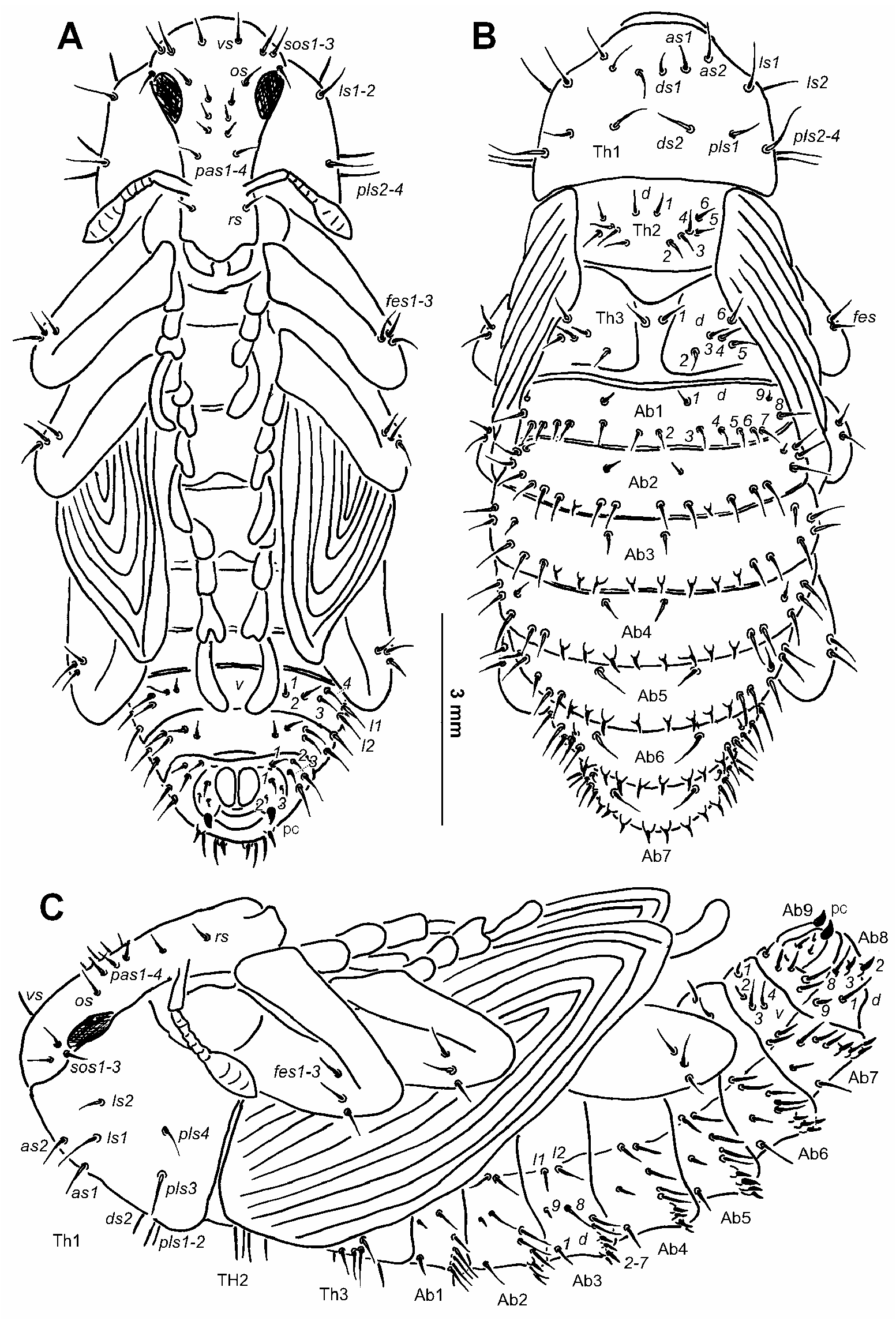
Chaetotaxy of mature larva. Slightly reduced, but in some parts with more than the most frequent number of setae in weevils (e.g., meso- and metathorax with 2(3) pds, and abdominal segments I-VII with 5 pds; the state in C. nigrosuturatus see below). Setae thin, long, light yellow or orange. Thorax. Prothorax ( Fig. 2B View Fig ) with 10 prns unequal in length, 2 vpls and 1 msts. Mesothorax ( Fig. 2B View Fig ) with 1 prs, 4 pds (pds2 distinctly shorter than the three remaining setae), 1 very short as, 2 short ss, 1 dpls, 2 vpls of different length and 1 short msts. Chaetotaxy of metathorax ( Fig. 2B View Fig ) identical to mesothoracal. Each pedal area of thoracic segments well separated, with 6-8 pda unequal in length. Abdomen. Abdominal segments I-VII ( Figs 2C–D View Fig ) with 1 prs, 6 pds (pds1, pds3 and pds5 very short), 1 dls, 1 very short ss, 2 dpls of different length, 2 vpls, 1 very short lsts and 1 micro msts. Abdominal segment VIII ( Fig. 2D View Fig ) with 1 prs, 3 very short pds, 1 very short ss, 2 dpls of different length, 2 short vpls, 1 very short lsts and 1 micro msts. Abdominal segment IX ( Fig. 2D View Fig ) with 3 ds (ds2 relatively long, ds1 and ds3 microsetae); 1 short ls and 2 sts of different length (sts1 very short, sts2 microsetae). Each anal lobe on abdominal segment X ( Fig. 2D View Fig ) with 1 microseta (ts).
Description of pupa. Morphology ( Figs 3A–C View Fig , 4C View Fig ). Body length: 11.0– 11.6 mm, at the widest region: 4.5–5.3 mm. The widest place in the body is commonly between the apex of the meso- or metafemora. Body stocky, elongated, white or yellowish. Cuticle smooth. Rostrum relatively short, approximately 1.8 times as long as wide, extended beyond procoxae. Antennae relatively long and stout. Pronotum almost 1.7 times as wide as long. Mesonotum and metanotum of almost equal length. Abdominal segments I–V of almost equal length; abdominal segment VI semicircular, next abdominal segments diminish gradually to the end of the body. Abdominal segments VII–IX distinctly smaller than other abdominal segments. Gonotheca (abdominal segment IX) of all three specimens divided. Sexual dimorphism in weevils is visible mainly in the length of rostrum and in the structure of abdominal segment IX: gonotheca of ƃ undivided, of Ƃ divided ( GOSIK & SPRICK 2012a,b, 2013; GOSIK & WANAT 2014).
Chaetotaxy ( Figs 3A–C View Fig ). Setae relatively long, unequal in length, light yellow or orange, some setae on abdominal segments II–VIII distinctly get stronger and located on protuberances. Setae well visible. Head capsule includes 1 vs, 3 sos, 1 os and 4 pas. Rostrum with 1 rs, located on the anterior margin. Setae on head capsule and rostrum straight, rs and pas1–3 distinctly shorter than the remaining setae on head, thoracic and abdominal segments.
Pronotum with 2 as, 2 ds, 2 ls and 4 pls. Dorsal parts of mesothorax with 1 pair of setae located posteromedially and 5 pairs located along its anterior margin. Chaetotaxy of metathorax identical to mesothoracal. Each apex of femora with groups of 3 fes. Dorsal parts of abdominal segments I–VIII each with 2 pairs of setae located posteriorly (d1, d9) and 7 pairs (d2-8) located along theirs anterior margins. Seta d4 (on abdominal segment II) and setae d2-5 (on abdominal segments III–VII) short, thorn-like, located on protuberances. Remaining setae long, hair-like. Abdominal segments I–VII with groups of 2 lateral setae and 4 pairs of ventral setae. Dorsal part of abdominal segment VIII with 2 pairs of setae located posteriorlly (d1, d9) and 3 pairs (d2, d3 and d8) located along its anterior margin; d2-–3 thorn-like, located on protuberances; remaining setae elongated. Abdominal segment VIII with groups of 2 lateral setae and 3 pairs of short ventral setae. Abdominal segment IX with 2 pairs of ventral microsetae and 1 pair of short, thin setae. Pseudocerci short, triangular.
Comparison with larvae of other Cleonini . Larvae of three cleonine taxa have been described so far ( SCHERF 1964): Cleonis pigra (Scopoli, 1763) , Cyphocleonus dealbatus (Gmelin, 1790) (as Cyphocleonus tigrinus (Panzer, 1789)) , and? Pachycerus segnis (Germar, 1824) (as Pachycerus scabrosus Brullé, 1832 , but identi¿cation is probaly incorrect, see below). The comparison of the larva of Coniocleonus nigrosuturatus with those decribed by SCHERF (1964) is somewhat problematic due to the use of differing terminology for morphology and chaetotaxy and/or an absence of good quality drawings. Despite these problems, we were able to compare the morphology of all four taxa ( Table 1). However, the identity of the larva described as Pachycerus segnis by SCHERF (1964) is unclear and it likely represents a genus other than Pachycerus . According to extensive surveys by BRUN et al. (1993) conducted in France, Greece and Turkey, larvae of P. segnis were always found within earthen cells attached to the taproot, but not in gall-like swellings as reported by SCHERF (1964). Descriptions by SCHERF (1964) perfectly ¿t the larval development of some species of the cleonine genus Rhabdorrhnychus, which takes place in a root gall-like swelling ( DIECKMANN 1983, STEJSKAL & TRNKA, unpubl. data).
MAY (1993) considered the increased number of pds on meso- and metathorax and abdominal segments I–VII and the increased number of epipharyngeal lining setae (als) (i.e. higher than the most frequent number of setae in weevils [for details see Chaetotaxy of mature larva of C. nigrosuturatus ]) as diagnostic for the mature larva of the subfamily Lixinae . Descriptions of mature larvae from the tribe Lixini ( Larinus species : GOSIK & SKUHROVEC 2011; Lixus species : SCHERF 1964, MAY 1994, NIKULINA 2001, 2007, NIKULINA & GÜLTEKIN 2011, GOSIK & WANAT 2014; Rhinocyllus conicus: MAY 1994 ) ¿t this diagnosis, as do all known species from the tribe Cleonini (see Table 1). The comparison of both tribes is recently not possible because our knowledge of immature stages in Cleonini is very scarce.

| R |
Departamento de Geologia, Universidad de Chile |
No known copyright restrictions apply. See Agosti, D., Egloff, W., 2009. Taxonomic information exchange and copyright: the Plazi approach. BMC Research Notes 2009, 2:53 for further explanation.