
Drakensbergena
|
publication ID |
https://doi.org/ 10.5281/zenodo.188410 |
|
DOI |
https://doi.org/10.5281/zenodo.6217097 |
|
persistent identifier |
https://treatment.plazi.org/id/039CE125-FF9E-FFCC-FF2C-FF3FCDCD0E32 |
|
treatment provided by |
Plazi |
|
scientific name |
Drakensbergena |
| status |
|
Key to species of Drakensbergena View in CoL males
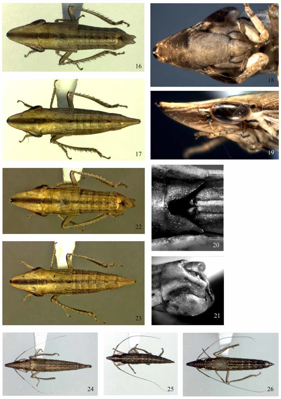
1. Antenna very long, 0.7–0.8 times as long as body length ( Figs 24–26 View FIGURES 16 – 26 , 37 View FIGURES 37 – 46 )................................................................. 2
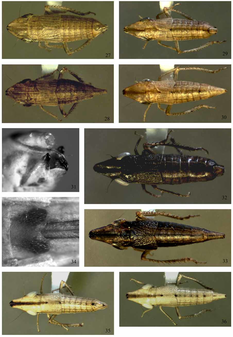
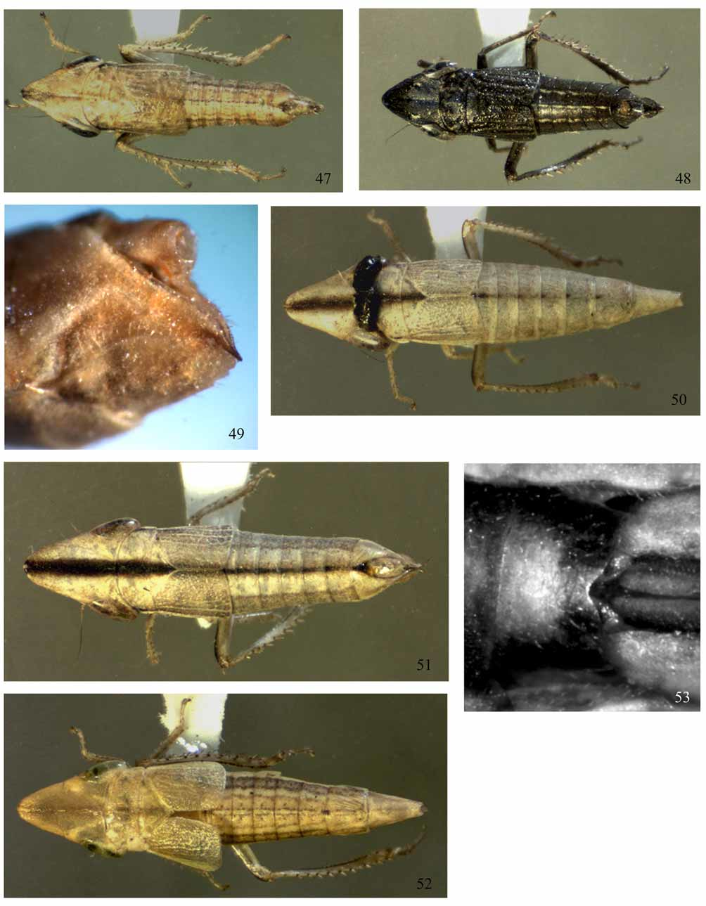
– Antenna short, 0.1–0.4 times as long as body length (e.g. Figs 2, 5 View FIGURES 1 – 15 , 17 View FIGURES 16 – 26 , 30, 35 View FIGURES 27 – 36 , 42 View FIGURES 37 – 46 , 47 View FIGURES 47 – 53 ) ............................................. 3
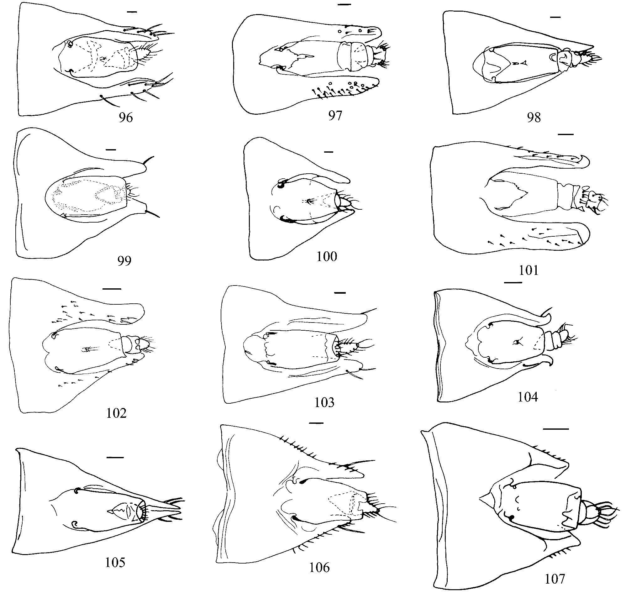
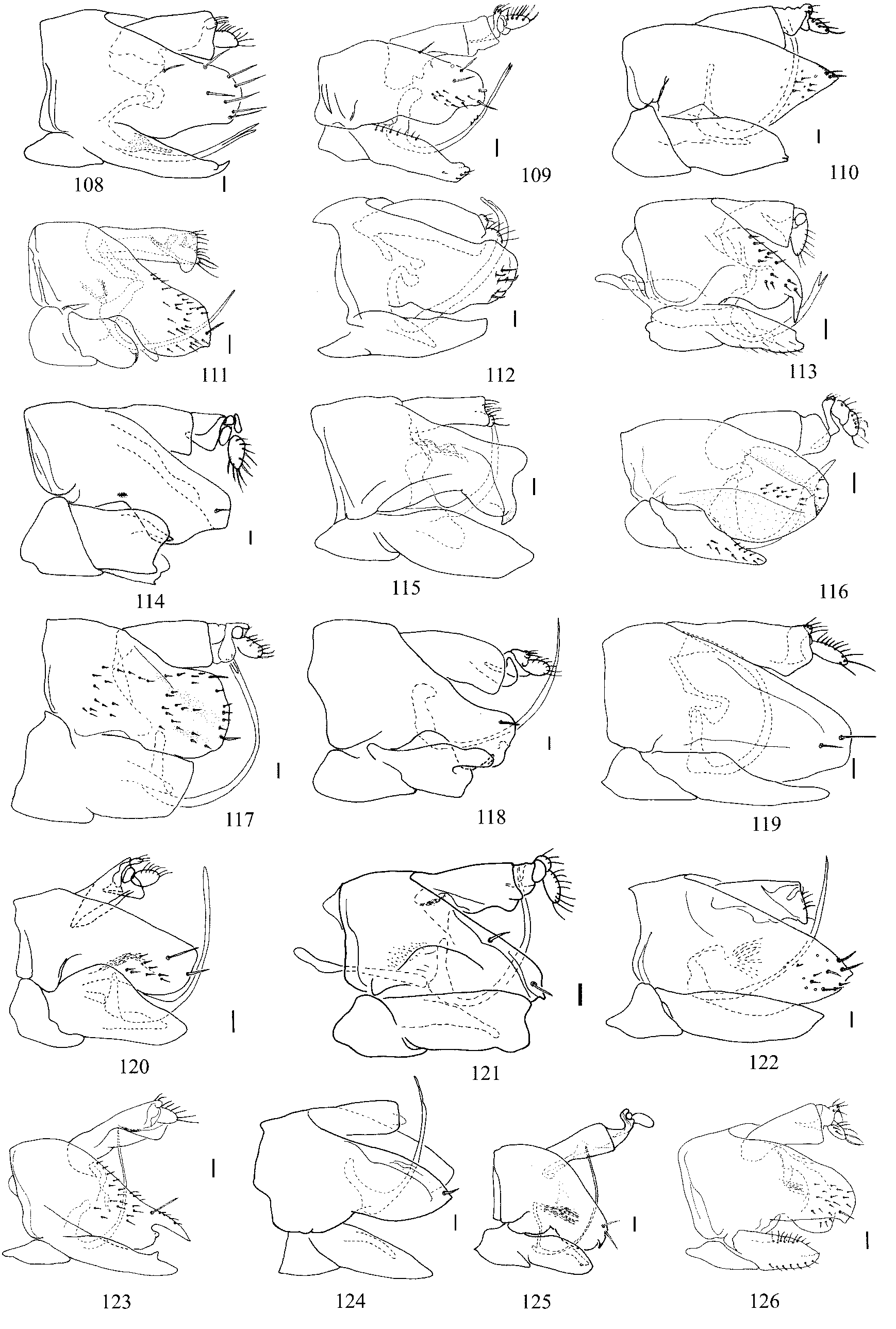
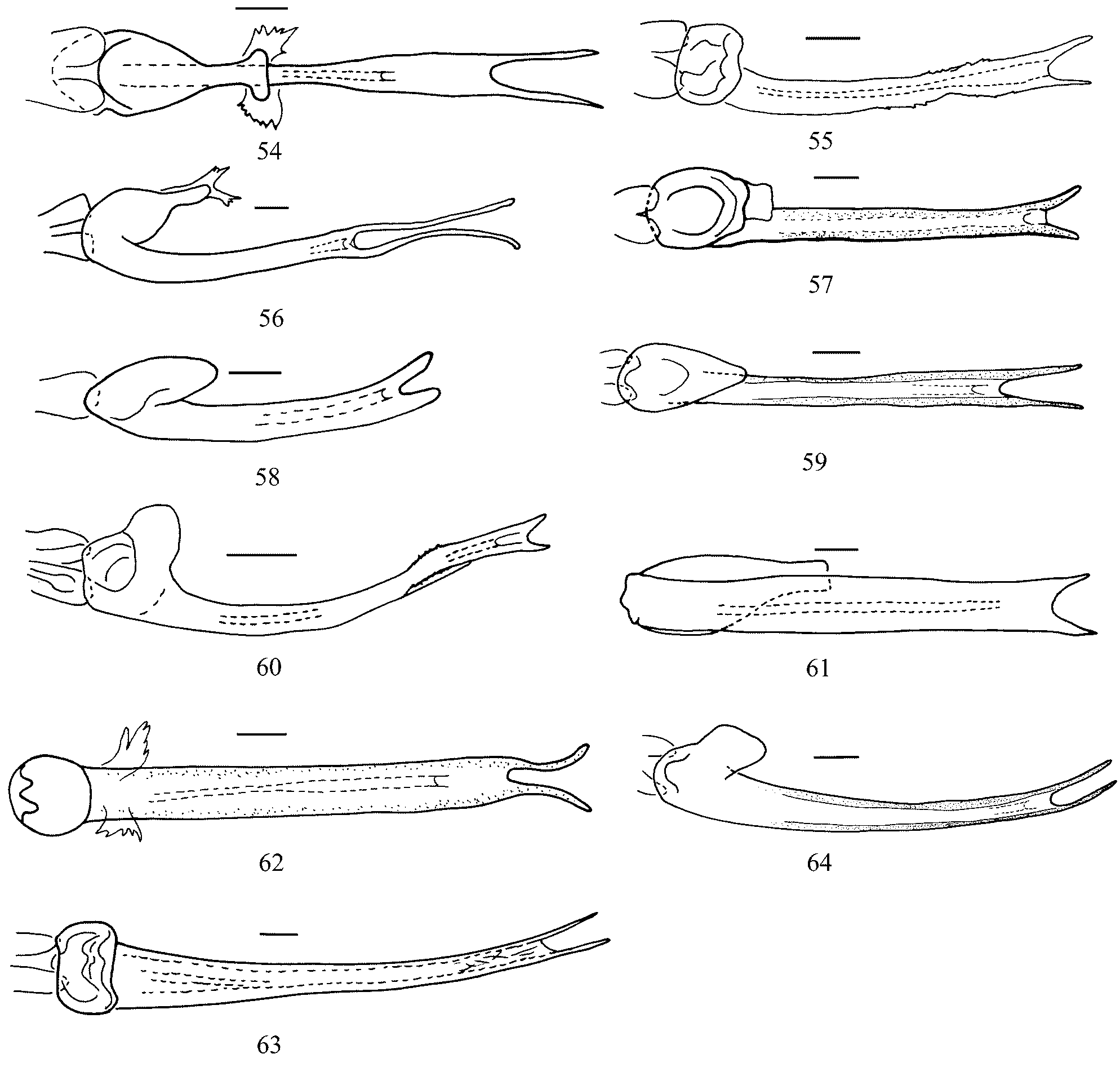
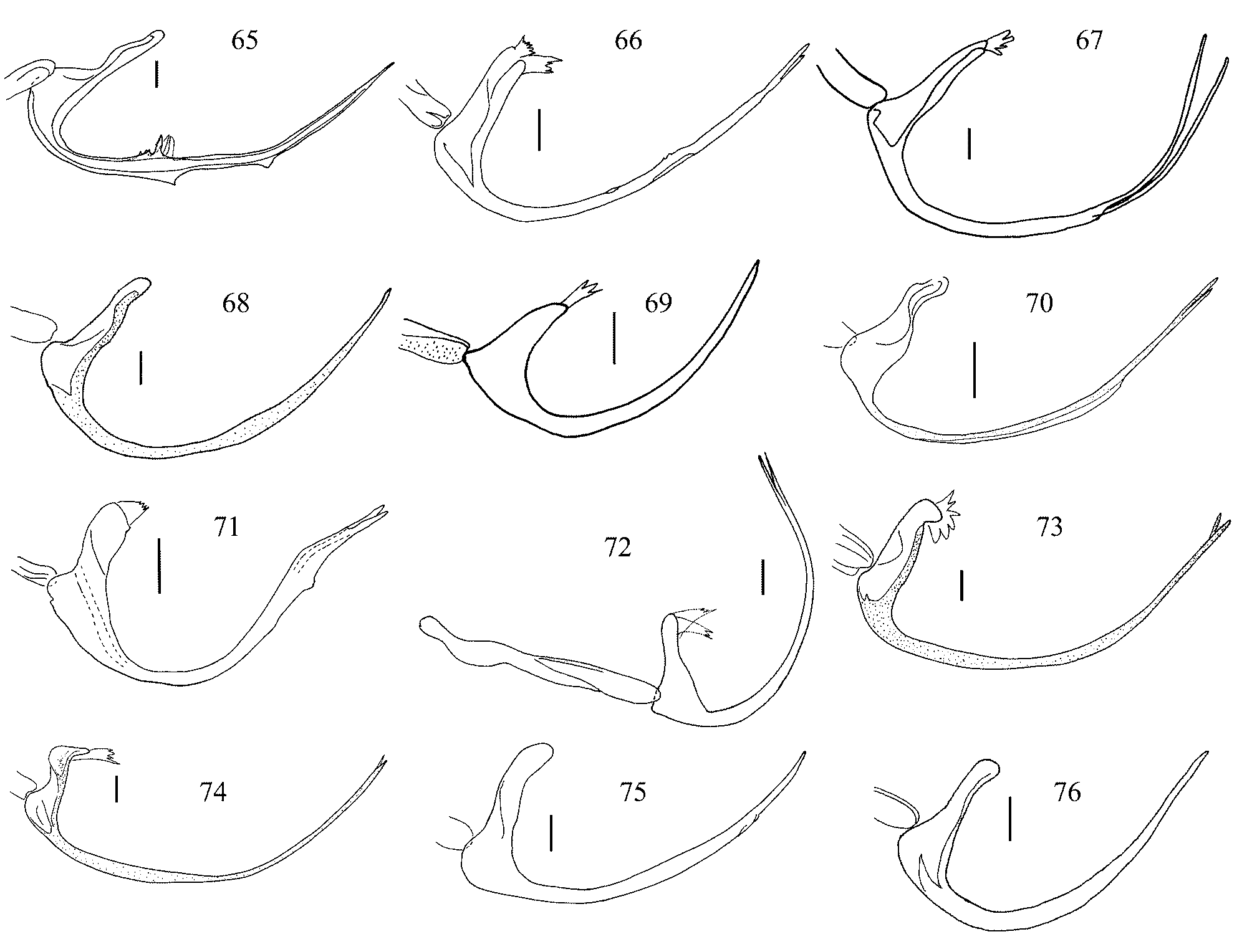
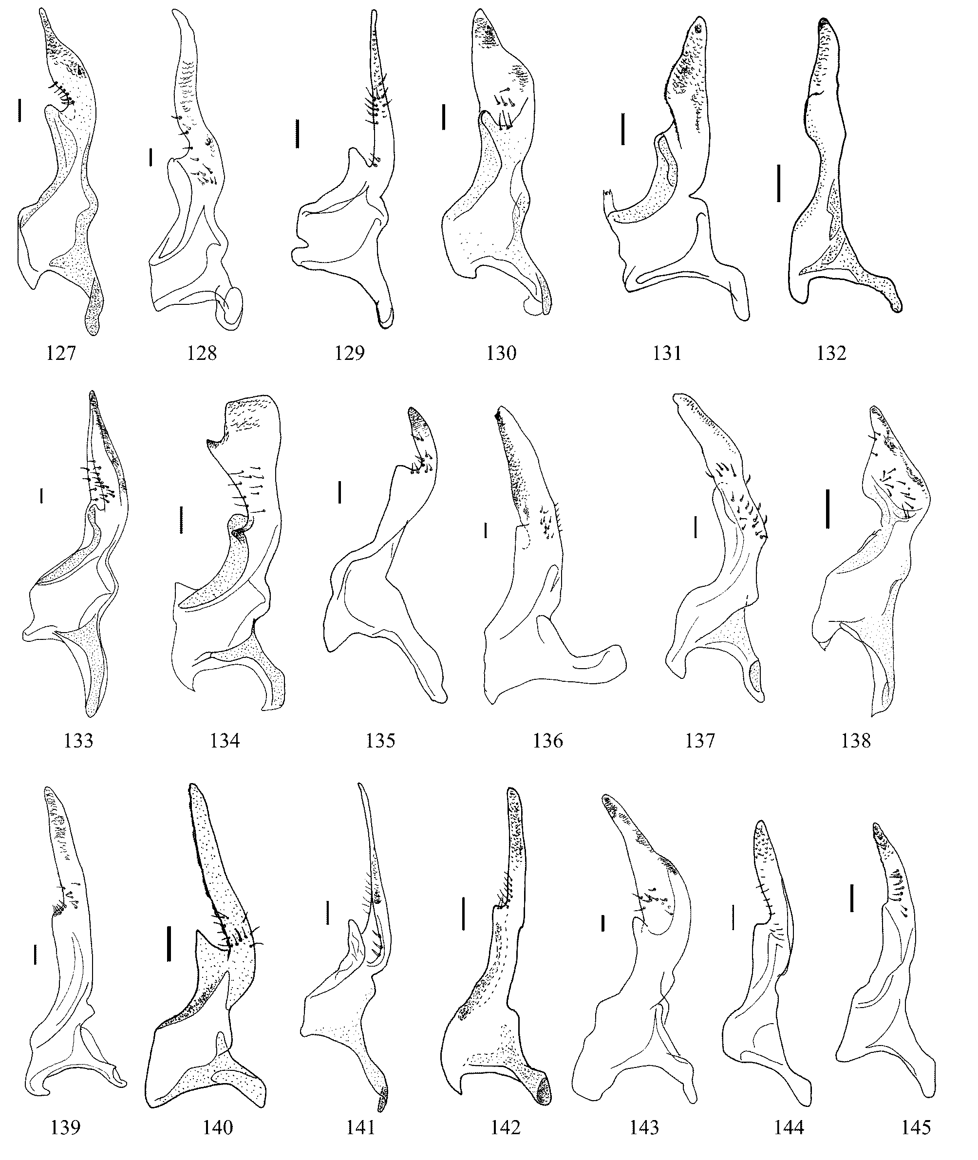
2. Vertex elongate (median length of vertex 1.5–1.6 mm) ( Figs 24–26 View FIGURES 16 – 26 ); pygofer lobe with concealed, membranous ventral tooth; pygofer lobe with fine setae, no macrosetae ( Figs 101 View FIGURES 96 – 107 , 116 View FIGURES 108 – 126 ); aedeagal shaft with subapical lateral margin denticulated and subapical ventral tooth ( Figs 60 View FIGURES 54 – 64 , 71 View FIGURES 65 – 76 ); style with anterior medial arm elongate, posterior apophysis short, digitate, curved laterad ( Fig. 135 View FIGURES 127 – 145 ) ......................................................................................... D. gigascutica View in CoL sp.n.
– Vertex short (median length of vertex 0.9–1.0 mm); pygofer lobe truncate, without tooth, with two macrosetae ( Fig. 120 View FIGURES 108 – 126 ); aedeagal shaft with margin entire; style with anterior medial arm short, posterior apophysis long, linear, directed posteriad ( Fig. 139 View FIGURES 127 – 145 ) ..................................................................................................... D. ochracea View in CoL Linnavuori
3. Aedeagal shaft bearing teeth dorsally and ventrally ( Figs 54 View FIGURES 54 – 64 , 65 View FIGURES 65 – 76 ) or lateral subapical margin concave with marginal denticulation (Figs, 55, 66) ........................................................................................................................................... 4
– Aedeagal shaft without teeth or denticles, smooth (e.g. Figs 56, 57 View FIGURES 54 – 64 , 67, 68 View FIGURES 65 – 76 )............................................................... 5
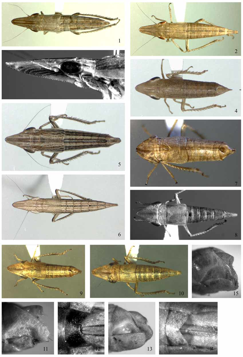
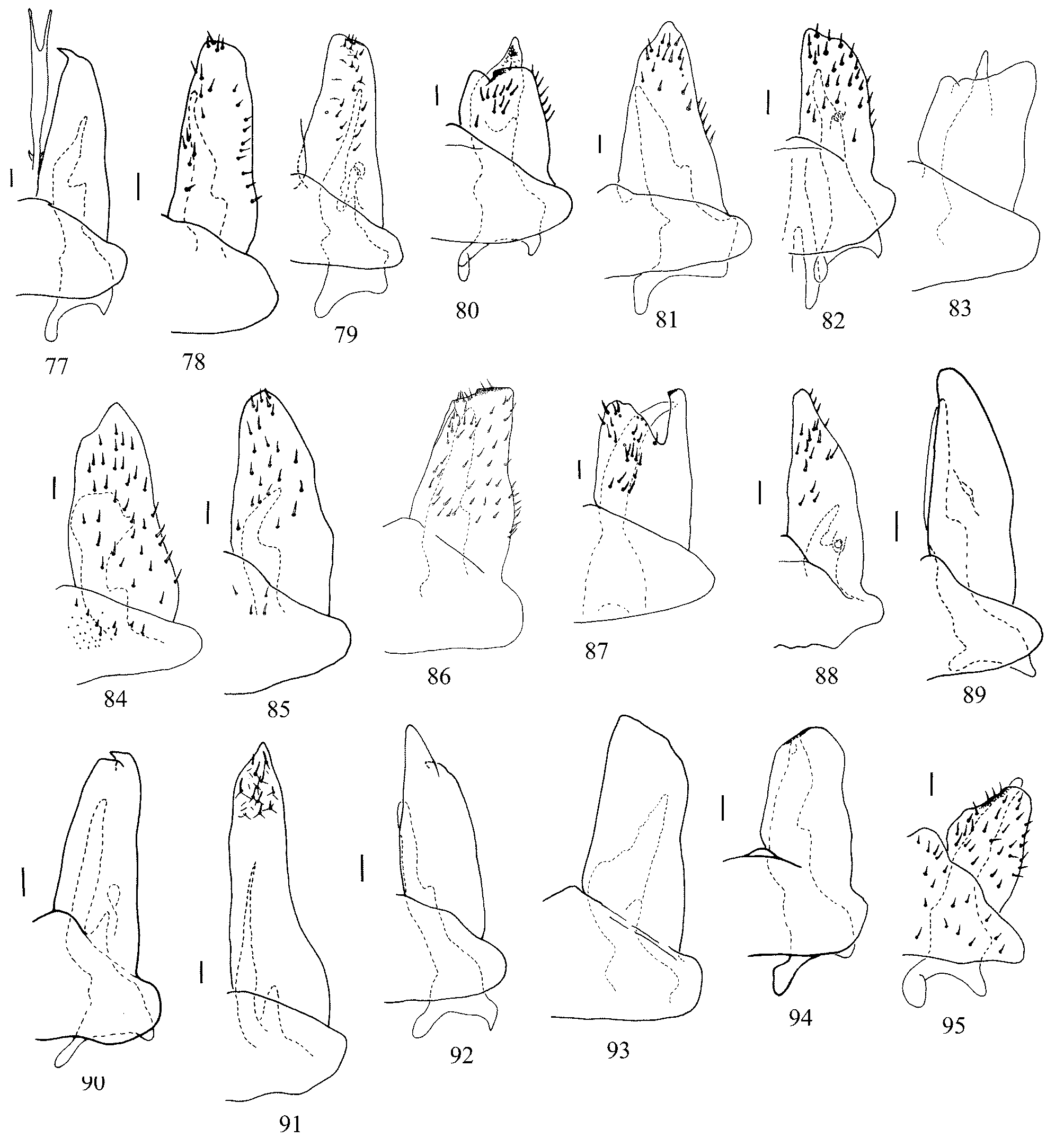
4. Aedeagal shaft medially with dorsal and ventral teeth ( Fig. 65 View FIGURES 65 – 76 ), dorsal view, apex of shaft wider than base ( Fig. 54 View FIGURES 54 – 64 ); pygofer lobe broadly rounded ( Fig. 108 View FIGURES 108 – 126 ); plate apex with pointed protrusion ( Fig. 77 View FIGURES 77 – 95 ); style posterior apophysis with apex narrow, tubular, base much wider than apex ( Fig. 127 View FIGURES 127 – 145 ); length of body 10.4 mm ( Figs 1, 2 View FIGURES 1 – 15 ) ............................... ......................................................................................................................................................... D. armstrongi View in CoL sp.n.
– Aedeagal shaft without dorsal and ventral teeth, with subapical lateral margin concave, with marginal denticulation ( Figs 55 View FIGURES 54 – 64 , 69 View FIGURES 65 – 76 ), dorsal view, parallel-sided ( Fig. 55 View FIGURES 54 – 64 ); pygofer lobe with apex narrow, rounded, base with rounded recess ( Fig. 109 View FIGURES 108 – 126 ); plate with apex rounded ( Fig. 78 View FIGURES 77 – 95 ); style with posterior apophysis tubular, S-shaped, dorsal view ( Fig. 128 View FIGURES 127 – 145 ); length of body 7.3–8.1 mm ( Figs 5, 6 View FIGURES 1 – 15 ) .............................................................................. D. austrina View in CoL sp.n.
5. Style apophysis projecting beyond posterior margin of plate ( Figs 11 View FIGURES 1 – 15 , 80 View FIGURES 77 – 95 , 111 View FIGURES 108 – 126 ), apex of apophysis visible in undissected specimen plate 0.6–0.9 times as long as wide ( Fig. 80 View FIGURES 77 – 95 ); .......................................................... D. breviata View in CoL sp.n.
– Style apophysis retracted into plate (e.g. Fig. 85 View FIGURES 77 – 95 ), sometimes reaching distal margin of plate (e.g. Fig. 94 View FIGURES 77 – 95 ); plate more than 1.0 times as long as wide ............................................................................................................................. 6
6. Plate posterior margin with deep notch extending two thirds into plate, forming lateral tubular process ( Figs 87 View FIGURES 77 – 95 , 118 View FIGURES 108 – 126 ) ............................................................................................................................................................ D. lobulata View in CoL sp.n.
– Plate with posterior margin entire, generally triangular, apex blunt (e.g. Fig. 79 View FIGURES 77 – 95 ) or truncate (e.g. Figs 83, 93 View FIGURES 77 – 95 ), sometimes with small tooth dorsally on posterior margin (e.g. Figs 90, 94 View FIGURES 77 – 95 ) or with sclerotized ridge (e.g. Fig. 86 View FIGURES 77 – 95 ) .......... 7
7. Posterior margin of plate truncate, with rounded lip-like sclerotized ridge, ridge at least half width of posterior margin ( Fig. 86 View FIGURES 77 – 95 ) ......................................................................................................................................... D. labeona View in CoL sp.n.
– Posterior margin of plate never with a long sclerotized ridge, 1–2 dorsally directed teeth that sometimes arise from a sclerotized base (e.g. Figs 92, 94, 95 View FIGURES 77 – 95 ).............................................................................................................................
8. Style apophysis flattened dorsoventrally, expanded, with small median tooth ( Fig. 134 View FIGURES 127 – 145 ) ... D. fuscovittata View in CoL Linnavuori
– Style apophysis never flattened, tubular, very thin (e.g. Figs 129, 141 View FIGURES 127 – 145 ), thick, digitate (e.g. Figs 131, 136, 142 View FIGURES 127 – 145 ), or triangular (e.g. Figs 130, 138, 143 View FIGURES 127 – 145 )............................................................................................................................... 9
9. Style apophysis thin, uniformly tapering to a fine point............................................................................................. 10
– Style apophysis digitate, base robust, apex blunt or abruptly pointed........................................................................ 11
10. Aedeagal shaft with deep cleft apically, length of cleft 0.4–0.5 times as long as length of shaft ( Figs 56 View FIGURES 54 – 64 , 67 View FIGURES 65 – 76 ); plate 1.9–2.1 times as long as wide, left and right sides similar in length, apex truncate ( Figs 79 View FIGURES 77 – 95 , 110 View FIGURES 108 – 126 ) ....... D. bisulca View in CoL sp.n.
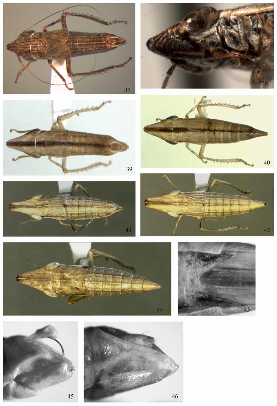
– Aedeagal shaft with shallow cleft apically ( Fig. 63 View FIGURES 54 – 64 ), length of cleft 0.1–0.2 times as long as length of shaft; plate 2.7–3.4 times as long as wide, left and right plates of unequal length, apex triangular ( Figs 91 View FIGURES 77 – 95 , 122 View FIGURES 108 – 126 ). D. prolixa View in CoL sp.n.
11. Pygofer lobe with prominent apical tooth (3.3–3.5 times as long as wide) ( Figs 113, 123 View FIGURES 108 – 126 ) ...................................... 12
– Pygofer lobe rounded, truncate, sometimes with minute or small weakly developed tooth or process (1.5 times as long as wide) (e.g. Figs 121, 124, 125, 126 View FIGURES 108 – 126 )............................................................................................................... 13
12. Pygofer process posteriad ( Fig. 123 View FIGURES 108 – 126 ); plate with subapical, lateral, dorsally directed tooth ( Figs 92 View FIGURES 77 – 95 , 123 View FIGURES 108 – 126 ), plates 2.0–2.4 times as long as wide .......................................................................................................... D. retrospina View in CoL sp.n.
– Pygofer process ventrad ( Fig. 113 View FIGURES 108 – 126 ); plate apex with or without lateral triangular process, variably sclerotized ( Figs 82 View FIGURES 77 – 95 , 113 View FIGURES 108 – 126 ), plates 1.3–1.6 times as long as wide .............................................................................. D. deorsuspina View in CoL sp.n.
13. Style apophysis with wide base, length up to 3 times as long as greatest width, generally short, triangular or elongate, digitate ( Figs 131, 138, 143 View FIGURES 127 – 145 ) ................................................................................................................................ 4
– Style apophysis with narrow base, length more than 3 times as long as greatest width, generally more elongate ( Figs 133, 140, 144, 145 View FIGURES 127 – 145 ) ..................................................................................................................................................... 16
14. Style with apophysis elongate, narrowly triangular or digitate, up to 3 times as long as greatest width ( Figs 131, 138 View FIGURES 127 – 145 ) .................................................................................................................................................................................... 15
– Style with apophysis short and broadly triangular, apophysis of style about 1.2 times as long as greatest width ( Fig. 138 View FIGURES 127 – 145 ) ................................................................................................................................................... D. longinqua View in CoL sp.n.
15. Apophysis of style about 1.6 times as long as greatest width, medial margin, lateroventral view, denticulate ( Fig. 143 View FIGURES 127 – 145 ); plate 1.9–2.2 times as long as wide, with lateral and medial margins subparallel and posterior margin angled acutely ( Fig. 93 View FIGURES 77 – 95 ); pygofer lobe pointed, with dorsal and ventral margins arcuate, with 0–2 macrosetae ( Fig. 124 View FIGURES 108 – 126 ) ..... ............................................................................................................................................................ D. simulata View in CoL sp.n.
– Apophysis of style about 2.8 times as long as greatest width, lateral margin, lateroventral view, denticulate, with prominent tooth subapically ( Fig. 131 View FIGURES 127 – 145 ); plate 1.4–1.8 times as long as wide, with lateral and medial margins converging triangularly ( Fig. 81 View FIGURES 77 – 95 ); pygofer lobe broadly rounded, with about 6 macrosetae ( Fig. 112 View FIGURES 108 – 126 ) ... D. cuneifer View in CoL sp.n.
16. Plate long, 1.4–2.0 times as long as wide, posterior margin variable, sometimes with tooth, or notch flanked by rounded margins ( Fig. 90 View FIGURES 77 – 95 ); style apophysis digitate, about 5.6 times as long as greatest width ( Fig. 140 View FIGURES 127 – 145 ) ................... ................................................................................................................................................... D. phaeogramma View in CoL sp.n.
– Plate short, less than 1.4 times as long as wide, posterior medial margin obtusely angled, apex of plate reaching half way or two thirds into pygofer lobe ( Figs 114, 125, 126 View FIGURES 108 – 126 ); style apophysis less than 5.0 times as long as wide........ 17
17. Pygofer, lateral view, with apex of lobe rounded, with ventrally directed apical tooth that is of variable length, about as wide at base as long; pygofer lobe with two macrosetae ( Figs 125, 126 View FIGURES 108 – 126 ); plate with two dorsally directed teeth at margin, posterior margin narrowly or more broadly rounded, 0.8–1.4 times as long as wide ( Figs 94, 95 View FIGURES 77 – 95 ); style with apophysis at its base narrower than width across preapical lobe, 3.0–4.4 times as long as wide, digitate ( Figs 144, 145 View FIGURES 127 – 145 ) ....................................................................................................................................................... D. spinula View in CoL sp.n.
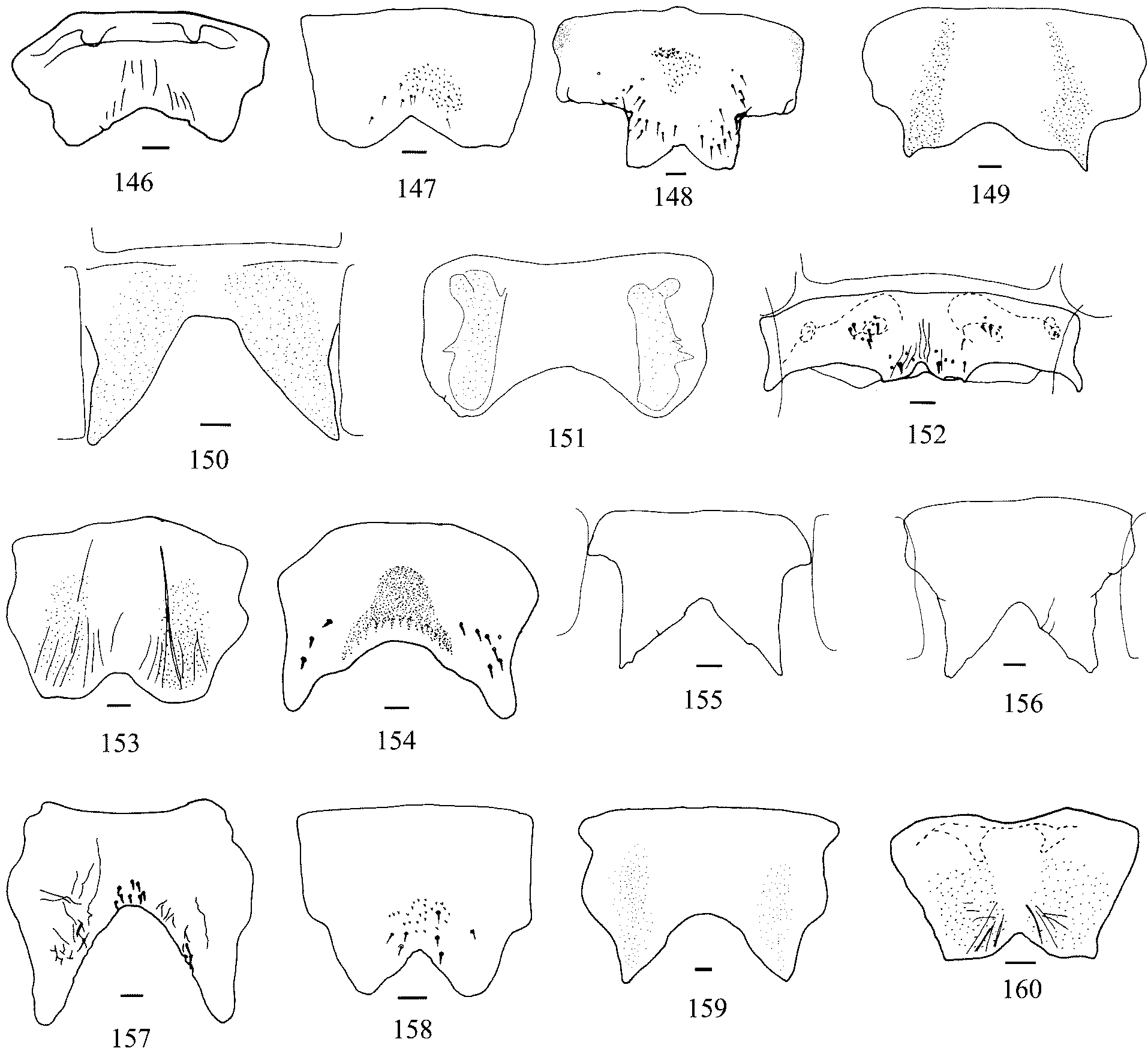
– Pygofer lobe, lateral view, with dorsal margin straight, posterior margin broadly triangular and ventral margin broadly rounded; pygofer lobe with few macrosetae (0–2) ( Fig. 114 View FIGURES 108 – 126 ); plate with posterior margin truncate, medially with small lobe, 1.1–1.3 times as long as wide ( Fig. 83 View FIGURES 77 – 95 ); style with apophysis at its base as wide as width across preapical lobe, 3 times as long as wide ( Fig. 153 View FIGURES 146 – 160 ) .......................................................................... D. festucacola View in CoL sp.n.
No known copyright restrictions apply. See Agosti, D., Egloff, W., 2009. Taxonomic information exchange and copyright: the Plazi approach. BMC Research Notes 2009, 2:53 for further explanation.
|
Kingdom |
|
|
Phylum |
|
|
Class |
|
|
Order |
|
|
Family |
|
|
Tribe |
Drakensbergeninae |