
Apoamphisiella foissneri, Paiva, Thiago Da Silva & Neto, Inácio Domingos Da Silva -, 2004
|
publication ID |
https://doi.org/ 10.5281/zenodo.157497 |
|
publication LSID |
lsid:zoobank.org:pub:D98FCACC-570E-41EA-98CE-75B85EE0CE16 |
|
DOI |
https://doi.org/10.5281/zenodo.5613529 |
|
persistent identifier |
https://treatment.plazi.org/id/039E0D1A-FF80-D245-315E-FD92FE0F5BC6 |
|
treatment provided by |
Plazi |
|
scientific name |
Apoamphisiella foissneri |
| status |
sp. nov. |
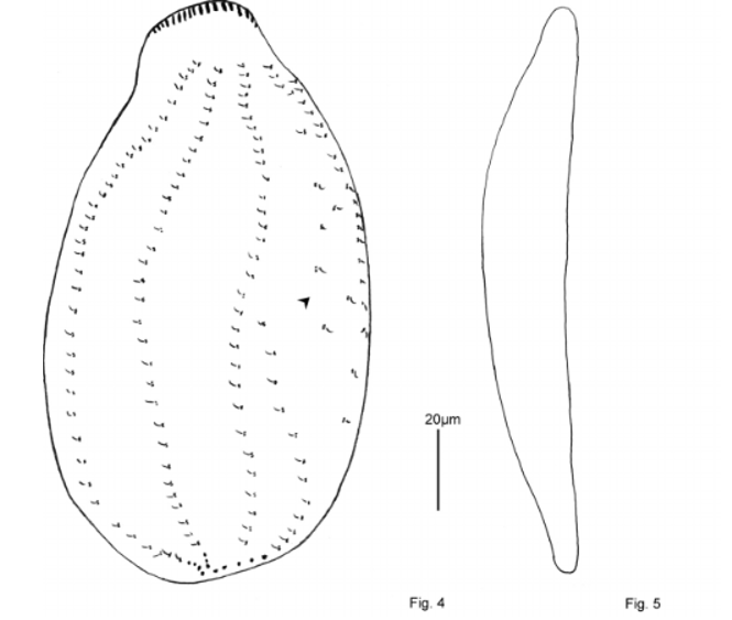
Characterization of Apoamphisiella foissneri sp. n. ( Figs. 2–5 View FIGURES 2, 3 View FIGURES 4, 5 , 17–21; Table 1)
Derivatio nominis
This species is dedicatied to Dr. Wilhelm Foissner (Salzburg, Austria) who made the first observation about a population of this genus from Brazil that closely matches our observations.
Locus typicus
Limoeiro River is located in the region of Além Paraíba, in the Minas Gerais State, Brazil. It is polluted by domestic and industrial wastewater, and is a very narrow affluent of Paraíba do Sul, a river of great economical importance. The samples were collected in July 2003, near Educandário Sérgio Ferreira, a local elementary school.
FIGURES 17–21:
phisiella foissneri . 17: view of a freely motile men under phase
showing body outline protruding transverse 18: protargol
specimen showing
ciliature and nuclear tus; 19: silver
specimen showing dorsal iature. Arrowhead
field of scattered
Dorsal kineties marked arabic numbers; 20: look of the ventral anterior gion of protargol
ed specimen, showing two postperistomial cirri; close look of the ventral terior region of protargol pregnated specimen
numerous transverse Legends: AFC =
frontal cirri; AZM = zone of membranelles; buccal cirrus; CC = cirri; eM = endoral brane; LMR = left
row; LVR = left ventral Ma = macronuclear Mi = micronucleus pM = paroral membrane; = postperistomial cirri; posterior frontal cirri; right marginal row; right ventral row; TC = verse cirri. Scale bars 10µm.
Diagnosis
Cells of Apoamphisiella foissneri sp. n. had a mean size of 150 x 70 µm in vivo, a flexible cell body, and a slightly oblique anterior end that was curved to the left. This new species also had two postperistomial cirri, two contractile vacuoles, and lacked cortical granules. Under dissection microscope, these cells had a green colouration, probably due to ingested algae. On average, this new species had 27 cirri in the left ventral row, 38 cirri in the right ventral row; 50 cirri in the left marginal row; 48 cirri in the right marginal row; 9 transverse cirri, and 11 caudal cirri.
Morphological characterization
Apoamphisiella foissneri sp. n. had two contractile vacuoles. Considered to be a rare feature in oxytrichid hypotrichs ( Berger, 1999), this feature was checked carefully in 10 additional free living (i.e. not squeezed under a coverslip) individuals. The vacuoles were located along the left margin of body. The anterior vacuole was located behind the middle region of the adoral zone of membranelles (AZM). It was difficult to readily observe it because of the delayed pulsation intervals in relation to the posterior vacuole, which was located at the equatorial region of body. The anterior vacuole diastolis usually occurred after three complete pulsations (systolis and diastolis) of the posterior. Sometimes, both were observed during simultaneous diastolic condition. Both vacuoles had very short, thin and inconspicuous collector ducts that were visible only during systolis. The cytoplasm showed “L” shaped crystals of greenyellowish colouration, which were heavily concentrated at the anterior end of body.
All specimens showed enlarged anterior frontal cirri that were arranged almost in a straight line, oblique to the body length axis. The leftmost cirrus of this set was located to the right of the anterior portion of the undulating membranes. The buccal cirrus was located to the right of the paroral membrane, above its virtual intersection with the endoral membrane. There were also two posterior frontal cirri, located to the inferiorright diagonal of the buccal cirrus, usually at level of the undulating membranes virtual intersection.
The AZM was conspicuous, occupied 43% of average cell length, and was composed of 62–80 membranelles. The distal region of the AZM was somewhat straight and angular. The paroral and endoral membranes virtually intercepted each other and were disposed like in cyrtohymenid hypotrichs. The infundibulum of A. foissner was the deepest of the three species characterized in this paper, and the peristomial lip engulfed at least the 5–8 proximal adoral membranelles. All specimens studied had two postperistomial cirri (n = 15). The anterior postperistomial cirrus was sometimes located just below the proximal end of the paroral membrane or across the outer surface of the infundibulum, which made it difficult to distinguish from the cirri in the left ventral cirral row. In contrast, the rear postperistomial cirrus was always located below the infundibulum vertex.
As in all characterized species of this genus, all specimens showed two ventral cirral rows. The left ventral row started below the rightmost posterior frontal cirri, and to the right of equatorial region of the posterior (preintersection) part of the endoral membrane.
In some specimens, the distance between the first and the second cirri was larger than the usual distance between each of the remaining cirri on this row, which was softly curved at the cytostome, ending in the transverse cirri. The right ventral row started below the distal end of the AZM. This row extended through the body almost parallel to its longitudinal axis, ending in the rightmost transverse cirri. The left marginal row started behind the AZM and extended along the left body margin, bending to the opposite margin at the posterior region and ending past the transverse cirri, after the posterior end of the right marginal row. The right marginal row started at the level of the second cirrus of the right ventral row and extended almost parallel to the longitudinal axis through the right margin. After the equatorial region, it bent slightly inwards, ending near to the rightmost transverse cirrus, above the last two or three cirri of the left marginal row. Both marginal rows did not merge. In addition, the terminal cirri were not as thick as the remaining cirri on these rows.
The transverse cirri were disposed in an oblique row behind the ventral rows. Their number was variable and ranged from 6–11 in the studied specimens. Also, some of the leftmost cirri of this set (usually 3 or more) were displaced to the left of the ventral rows. The rightmost transverse cirri protruded about 15–20µm (n = 10) beyond the anterior end of cell, were considerably thicker than the cirri in the ventral and marginal rows, and could easily be seen in living specimens ( Figs. 2 View FIGURES 2, 3 , 17). Due to their size, they may be misidentified as caudal cirri, but their placement can be elucidated as the organism swims, rotates across its longitudinal axis, and is observed laterally.
On the dorsal surface, we observed three long kineties next to a fourth shortened kinety, which began near the equatorial region. There was also a field of scattered dikinetids near the right margin. One or two dorsomarginal kineties were present close to the right margin of cell body in 2 of the 14 specimens studied. A set of caudal cirri was present, located in the posterior region of body. In total, there are 9–14 caudal cirri at the end of dorsal kineties 1, 2 and 4.
The nuclear apparatus was composed of two ellipsoid macronuclear nodules and 1–4 globulous micronuclei were disposed close to the macronuclei, along the left side of the body.
Typification
Slides containing the holotype (IBZUFRJ 00101) and paratypes (IBZUFRJ 00102; 1 slide) of Apoamphisiella foissneri were deposited in the collection of Laboratório de Protistologia, Dept. de Zoologia, Inst. de Biologia, CCS, Universidade Federal do Rio de Janeiro (UFRJ).

No known copyright restrictions apply. See Agosti, D., Egloff, W., 2009. Taxonomic information exchange and copyright: the Plazi approach. BMC Research Notes 2009, 2:53 for further explanation.