
Dactylosoma sp.
|
publication ID |
https://doi.org/ 10.1016/j.ijppaw.2019.12.006 |
|
publication LSID |
lsid:zoobank.org:pub:F5CBE7E7-D57F-4854-A98B-A6D6ED6FD342 |
|
DOI |
https://doi.org/10.5281/zenodo.11085332 |
|
persistent identifier |
https://treatment.plazi.org/id/039F87C1-FF9E-FF94-954E-FC902657FD12 |
|
treatment provided by |
Felipe |
|
scientific name |
Dactylosoma sp. |
| status |
|
3.1.2. Dactylosoma sp. from Belgium
Host: Pelophylax lessonae (Camerano, 1882) ( Anura : Ranidae ).
Site in host: Peripheral blood.
Vector: Unknown.
Localities: Haacht , Belgium (N50.979434 ̊, E4.659686 ̊) GoogleMaps .
Voucher material – 1 × blood smear with a parasitaemia of 0.3% from Pel. kl. esculentus deposited in the protozoan collection of the National Museum, Bloemfontein, South Africa, under accession number [ NMB P 538].
Representative DNA sequences: The sequence data specifically associated with Dactylosoma sp. ex Pel. lessonae have been submitted to GenBank and are as follows: Nuclear 18S rDNA (nu 18S) partial sequence: MN 879399.
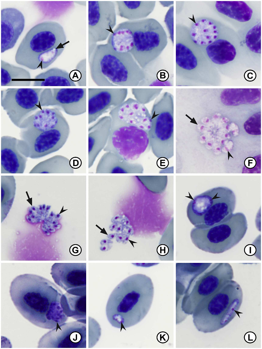
3.1.2.1. Description. Two distinct stages of merogony present in erythrocytes of Pel. lessonae ( Fig. 3 A–L View Fig ). The first stage characterised by large dactylate meronts producing up to approximately 14 clear merozoites. The second stage meronts seemingly smaller, although no clear merozoites were observed. The cytoplasm has a hyaline appearance. Parasitaemia of infected individual (n = 1) was 0.3%.
Primary merogony: trophozoites ( Fig. 3 A View Fig ) elongated to oval, usually tapering towards one end, vacuoles in some cases present ( Fig. 3 A View Fig arrow). Small round dense nuclei located at blunt end ( Fig. 3 A View Fig arrowhead), chromatin staining deep magenta. Trophozoites measure 6.3 ± 0.9 (5.3–7.4) long × 3.8 ± 0.8 (2.6–4.9) wide (n = 10).
Young primary meronts ( Fig. 3 B View Fig ) round to ovoid causing slight displacement of the host cell nucleus. Multinucleate, nuclear division located peripherally, chromatin staining bright pink to dark purple ( Fig. 3 B View Fig arrowhead). Young meronts measure 9.4 ± 1.1 (8.0–10.9) long × 8.0 ± 0.5 (7.0–8.6) wide (n = 7).
Primary meronts ( Fig. 3 C–G View Fig ) vary in form, round to pear shape, in some cases causing slight distortion or displacement of the host cell or nucleus, cytoplasm staining whitish-purple. Mature forms have dactylate appearance ( Fig. 3 F–G View Fig ). Multinucleate, normally showing more than 10 nuclei ( Fig. 3 View Fig C-E arrowhead), staining deep magenta. Meronts measure 11.3 ± 2.8 (8.4–16.5) long × 9.1 ± 1.9 (7.1–12.6) wide (n = 10).
Primary merozoites ( Fig. 3 F–H View Fig ) elongate to ovoid, hyaline cytoplasm staining white to bluish-purple. Dense nuclei chromatin centrally located, staining deep magenta ( Fig. 3 F–H View Fig , arrowhead). Merozoites ( Fig. 3 F–H View Fig , arrow) measure 4.0 ± 0.6 (3.1–5.0) long × 1.9 ± 0.6 (1.1–3.1) wide (n = 20).
Secondary merogony: young secondary meront ( Fig. 3 I View Fig ) irregular ovoid shape, cytoplasm staining bluish-purple, occasionally causing displacement of the host cell nucleus. Multinucleate, nuclei located peripherally, staining deep magenta ( Fig. 3 I View Fig , arrowhead). Young meronts measure 6.2 ± 1.1 (4.5–7.5) long × 4.5 ± 1.5 (2.7–7.3) wide (n = 7).
Secondary meronts ( Fig. 3 J View Fig ) ovoid shape, cytoplasm stains dark bluish-purple. Multinucleate, up to 6 nuclei located peripherally, chromatin staining dark purple ( Fig. 3 J View Fig , arrowhead). Secondary meronts measure 7.7 ± 0.9 (5.9–8.9) long × 6.6 ± 1.1 (5.8–8.5) wide (n = 10).
Secondary merozoites ( Fig. 3 K View Fig ) rare, short crescent shape, hyaline cytoplasm staining purple. Small round dense nuclei located peripherally, staining deep magenta ( Fig. 3 K View Fig arrowhead). Secondary merozoites measure 4.1 long × 2.3 wide (n = 1).
Gamonts ( Fig. 3 L View Fig ) elongate and slender shape, hyaline appearance, nuclei chromatin visible slightly off centre ( Fig. 3 L View Fig arrowhead). Extracellular forms often elongate with slight curvature, nucleoplasm visible slightly off centre, staining dark purplish-pink. Gamonts measure 9.8 ± 1.8 (7.9–12.1) long × 2.3 ± 0.1 (2.2–2.5) wide (n = 5).
3.1.2.2. Remarks. The species of Dactylosoma under study possesses several phenotypic characteristics mentioned in the original description of Dactylosoma splendens , later synonymised with D. ranarum , such as the round shape of the meronts and the number of primary and secondary merozoites produced (ranging approximately between 12 and five). However, this synonymisation happened over 100 years ago when knowledge of the close resemblance between different species was unknown. Unfortunately, no precise measurement data are available from the original description of D. splendens , which was described from Pel. kl. esculentus in Paris, France ( Labbé, 1894). However, based on the information of this species of Dactylosoma provided above, the parasite cannot, based on the data collected in the current study, be identified to species level.
Furthermore, this species of Dactylosoma can be characterised by its round young meront stages, the dactylate appearance of the mature meront stages, and the hyaline appearance of the cytoplasm. Although the trophozoite and gamont stages of Dactylosoma kermiti n. sp. are superficially similar in morphology and morphometrics, primary and secondary meront stages are wider (primary meront: 5.1–8.0 vs 7.1–12.6; secondary meront: 4.4–6.9 vs 5.8–8.5) and secondary merozoites stages slightly shorter (4.2–5.5 vs 4.1) in the species of Dactylosoma from Pel. lessonae . This species also conforms closely to D. sylvatica , however, the species from the current study produces more than eight merozoites in primary merogony and less than eight in secondary merogony as compared to D. sylvatica . Also the secondary meront stages are wider as compared to D. sylvatica (secondary meront: 5.9–8.9 × 5.8–8.5 vs 5.2–4.0).
| NMB |
Naturhistorishes Museum |
| MN |
Museu Nacional, Universidade Federal do Rio de Janeiro |
No known copyright restrictions apply. See Agosti, D., Egloff, W., 2009. Taxonomic information exchange and copyright: the Plazi approach. BMC Research Notes 2009, 2:53 for further explanation.