
Aspidiophorus gullmarsfjordensis, Kånneby, Tobias & Todaro, M. Antonio, 2017
|
publication ID |
https://doi.org/10.11646/zootaxa.4290.2.11 |
|
publication LSID |
lsid:zoobank.org:pub:A6C626EA-6A18-4E3F-9556-5635C21FA0BB |
|
DOI |
https://doi.org/10.5281/zenodo.6041717 |
|
persistent identifier |
https://treatment.plazi.org/id/03A14E50-482E-FFED-30C5-F9263E08FD43 |
|
treatment provided by |
Plazi |
|
scientific name |
Aspidiophorus gullmarsfjordensis |
| status |
sp. nov. |
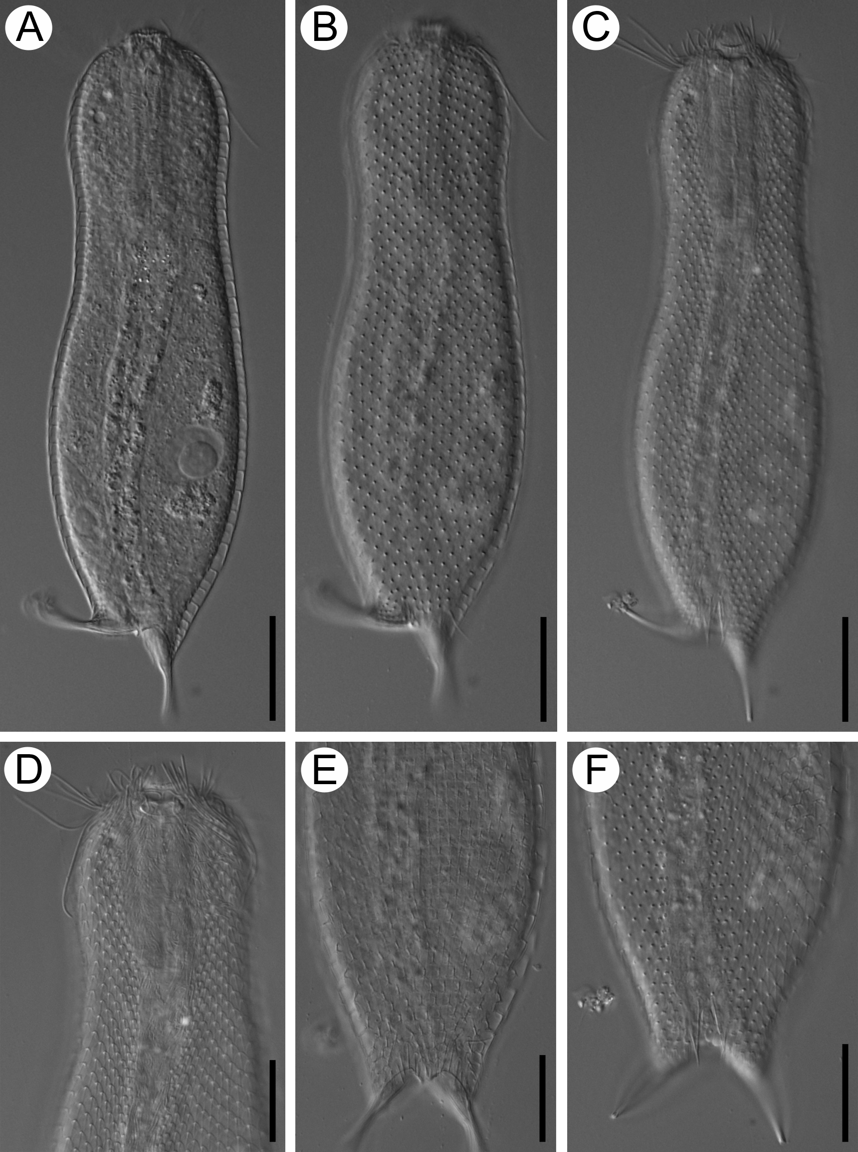
Aspidiophorus gullmarsfjordensis View in CoL n. sp. ( Figs. 1–2 View FIGURE 1 View FIGURE 2 )
(Zoobank F9A6D262-69EC-415D-9FC3-1F5F0AD2572E)
Type locality. In medium sand, Klubban, Östersidan, Sweden ( N 58° 15.10’, E 11° 27.93’). Sediments collected by GoogleMaps
snorkelling 0.5 to 2.5 m depths on 17 and 21 July, 2009.
Type material. Photographs of two specimens, available at the Swedish Museum of Natural History , Stockholm,
Sweden. Accession numbers: Holotype, SMNH Type-8880 ; Paratype, SMNH Type-8881. Etymology. This species is named after the Gullmarsfiord where it was first found. Diagnosis. A medium sized Aspidiophorus , 186–190 µm in length. Head rounded to three-lobed with small
cephalion and pleuria, slightly separated from trunk by neck constriction. Hypostomium present. Furca straight, 23–25 µm in length. Anterior and posterior pair of sensory bristles present. Body covered by pedunculate keeled rounded to rhomboidal scales. Four dorsal pairs of non-pedunculate keeled elliptical scales present just above furcal branches. Ventral interciliary area naked, except two pairs of keeled spined scales. Ventral ciliation in two separate longitudinal bands. Pharynx with slight terminal swellings, 40–44 µm in length.
Description. A medium sized Aspidiophorus , 186–190 µm in total body length. Head rounded to three-lobed with a pair of small pleuria. Cephalion small, 12 µm in width. Hypostomium concave, 4 in length and 10 µm in width, with two small hook-like projections on each side. One pair of ill-defined sensory ciliary tufts, the longest cilia of each tuft 20–25 µm in length. Ocellar granules absent.
Body width variable, 41–42 µm at head (U15–16), 37–39 µm at neck (U26–28), 51–55 µm at trunk (U59–61), and 20–25 µm at base of furca (U88–92). Head slightly separated from trunk by neck constriction that progressively widens into the trunk, which reaches its greatest width approximately three fifths down the length of the body. The trunk then tapers into a straight furca, 23–25 µm in length, with 13–15 µm long adhesive tubes. Anterior and posterior pair of dorsal sensory bristles present, anterior pair inserted at U22–23 and posterior pair inserted at U83–84.
Dorsal body surface covered by 21–22 columns of 35–38 pedunculate keeled scales, oval to round rhomboidal in shape. The total number of columns is 42–46. The median column of scales is straight, while the columns on either side slowly approach parallelism with the lateral body outline. Scales increase slightly in size from anterior to posterior end of body, scales of head, neck and trunk measure 3– 4 x 3, 4– 5 x 4 and 4– 5 x 4 µm respectively. The posteriormost pedunculate scales smaller than rest of trunk scales. Peduncle 1-2 µm in height. On either side of the caudal incision, just above each furcal branch, four pairs of non-pedunculate keeled elliptical scales are present. Proximal part of furcal branches covered by small rounded pedunculate keeled scales.
Ventrolateral scales of same type as dorsal scales, but decrease in size towards the locomotory cilia. Ventral interciliary area naked except for two pairs of terminal keeled spined scales. The anterior pair measure 3 x 2 µm and each bear a 10 µm long spine. The posterior pair of terminal scales are hard to discern; however each of them bear a spine 8 µm in length. All four spines reach beyond the caudal incision and can be seen between the furcal branches from the dorsal side. Ventral ciliation distributed in two separate longitudinal bands.
Mouth terminal, 8–9 µm in diameter. Pharynx 40–44 µm long with slight anterior and posterior swellings. Pharyngeal intestinal junction at U27–28. Intestine straight. Anus at U83–84.
The two specimens observed were in parthenogenetic phase. One individual showed two developed eggs on either side of the intestine.

| SMNH |
Saskatchewan Museum of Natural History |
No known copyright restrictions apply. See Agosti, D., Egloff, W., 2009. Taxonomic information exchange and copyright: the Plazi approach. BMC Research Notes 2009, 2:53 for further explanation.
|
Kingdom |
|
|
Phylum |
|
|
Order |
|
|
SubOrder |
Paucitubulatina |
|
Family |
|
|
SubFamily |
Chaetonotinae |
|
Genus |