
Fissiphallius tucupi, Tourinho, Ana Lúcia & González, Abel Pérez, 2006
|
publication ID |
https://doi.org/10.5281/zenodo.174070 |
|
DOI |
https://doi.org/10.5281/zenodo.6494533 |
|
persistent identifier |
https://treatment.plazi.org/id/03A2CA19-765A-FFD1-FEF4-24232CEFFCBF |
|
treatment provided by |
Plazi |
|
scientific name |
Fissiphallius tucupi |
| status |
sp. nov. |
Fissiphallius tucupi n. sp.
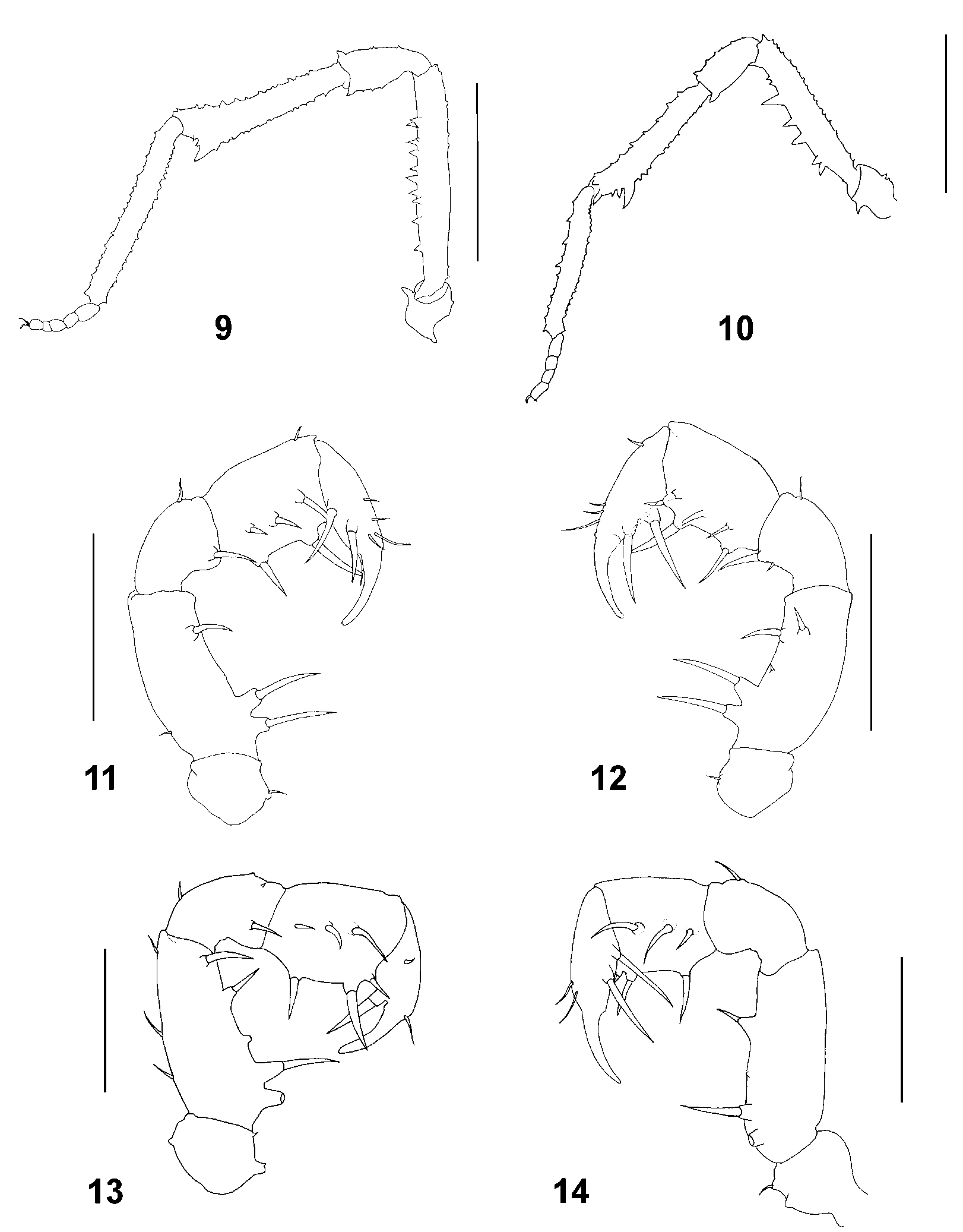
Figs 2, 4, 6, 8 View FIGURES 1 – 8 , 10, 13–14 View FIGURES 9 – 14 , 19–22 View FIGURES 16 – 21 View FIGURE 22
Type material. Male holotype ( INPA 907), Brazil, Amazonas State, Castanho municipality, between km 81 and 83 of the BR319 highway, 3.60665ºS, 06.19784ºW, 10–16.X.2005, Ana Tourinho, Rodrigo Dias & Sergio Marques de Souza leg. Paratypes: 7 males, 5 females ( INPA 908), 5 males, 4 females, 2 juveniles ( INPA 909), 8 males, 6 females ( INPA 910), 1 male, 11 females ( INPA 911), 1 male ( INPA 912), Ana Tourinho leg., 11 females ( INPA 913), 3 males, 2 females ( MNRJ 17815), with the same data as the holotype.
Etymology. Noun in apposition. Tucupi is a Tupi word, used for a regional manioc soup, which is prepared with duck or fish and which is widely consumed in the Amazon region.
Diagnosis. Eye mound armed with a pair of long and sharp spiniform median aphophyses very close to each other ( Figs 2, 4 View FIGURES 1 – 8 ). Clearly distinguished from congeneric species by its male genitalia: rutrum with slightly bifid apex, truncus ventrally with a rounded protuberance proximally of pergula ( Figs 19–21 View FIGURES 16 – 21 ).
Description of male holotype. Measurements. Total length: 2.8. Carapace region of scutum: 1.3 long, 1.1 wide. Scutum: 1.5 long, 2.0 maximum width. Pedipalp: 2.4 long. Legs: 7.1/4.9/6.3/4.0 long.
Dorsal view. Anterior border of prosoma with a pair of very small tubercles on each side ( Fig. 2 View FIGURES 1 – 8 ). Scutal areas I–IV each with a row of very small tubercles (almost indiscernible). Lateral margin of dorsal scutum armed with 16 sharppointed tubercles on each side, these increasing in length posteriorly (variation: 12–17/13–17), posterior margin armed with 15 small tubercles, the median one largest ( Figs 2, 4, 8 View FIGURES 1 – 8 ). Free tergites I–III armed with sharppointed tubercles: free tergite I with 15, II with 13 and III with 11, the median one always larger than others (variation: 11– 15 /11–13/7–11).
Ventral view. Posterior margin and sternites armed with tubercles increasing in length laterally. Sternites IV and V with two pairs of larger median tubercles ( Fig. 6 View FIGURES 1 – 8 ). Anal operculum with two rows of tubercles, anterior row with four sharppointed tubercles, posterior row with five tubercles, the median one largest ( Figs 6, 8 View FIGURES 1 – 8 ).
Chelicerae. Without remarkable armature. Bulla short and well marked. Fingers without teeth.
Pedipalps. Coxa with one ventral tubercle and two small dorsal protuberances. Femur ventrally with ventral tubercles, two basal and one distal setiferous tubercle, one distal setiferous tubercle in mesal region. Patella mesally with one setiferous tubercle. Tibia mesally and ectally with one row of three setiferous tubercles. Tarsus with two setiferous tubercles on each side; small setae scattered. Tarsal claw long and strong ( Figs 13–14 View FIGURES 9 – 14 ).
Legs. I–IV tuberculate. Posterior margin of coxae II–III armed with ventral tubercles ( Fig. 6 View FIGURES 1 – 8 ). Coxa IV with sharppointed dorsal tubercles ( Fig. 2 View FIGURES 1 – 8 ). Trochanters I–IV with one retrolateral sharppointed tubercle ( Figs 6 View FIGURES 1 – 8 , 10 View FIGURES 9 – 14 ). Femur IV with two rows of seven sharppointed ventral tubercles larger than others. Patellae with sharppointed distal tubercle. Tibia with one larger tubercle ( Fig. 10 View FIGURES 9 – 14 ). Tarsi tuberculate, anterior and posterior margins with a row of tubercles. Tarsal formula: 4(2), 5(3), 6, 5.
Penis. Stragulum with only short distal cleft. Apical portion of stragulum in the shape of a “parrot bill” ( Figs 19–20 View FIGURES 16 – 21 ). Rutrum medially with slightly bifid apex and three pairs of setae ( Figs 19, 21 View FIGURES 16 – 21 ). Pergula laterally with two pairs of setae ( Figs 19, 21 View FIGURES 16 – 21 ). A pair of setae present proximally of pergula and a rounded protuberance at same level as stragulum basis ( Figs 20–21 View FIGURES 16 – 21 ).
Color (in 75% ethanol). Body yellowish, carapace region of scutum with reticulated brown stains and with yellow stripes behind eye mound ( Fig. 2 View FIGURES 1 – 8 ). Lateral borders and median region of scutal areas I–IV with brown stains. Posterior margin of dorsal scutum and free tergites and sternites with brown lateral stains. Anal operculum brown, with yellow stain in the middle. Pedipalps and chelicerae pale yellow. Trochanters of legs yellow, femora and tibiae brown. Coxae I–IV yellow, coxa IV with brown dorsal stains.
Description of the female paratype. Very similar to the male, different in the following features: legs considerably shorter, armature of free tergites and sternites shorter, tubercles on free sternites all of the same length.
Measurements. Total length: 2.1 Carapace region of scutum: 0.9 long, 0.8 wide. Scutum: 1.5 long, 2.1 maximum width. Pedipalp: 2.3 long. Legs: 5.1/3.9/5.2/3.0 long. Tarsal formula: 4(2), 5(3), 6, 5.
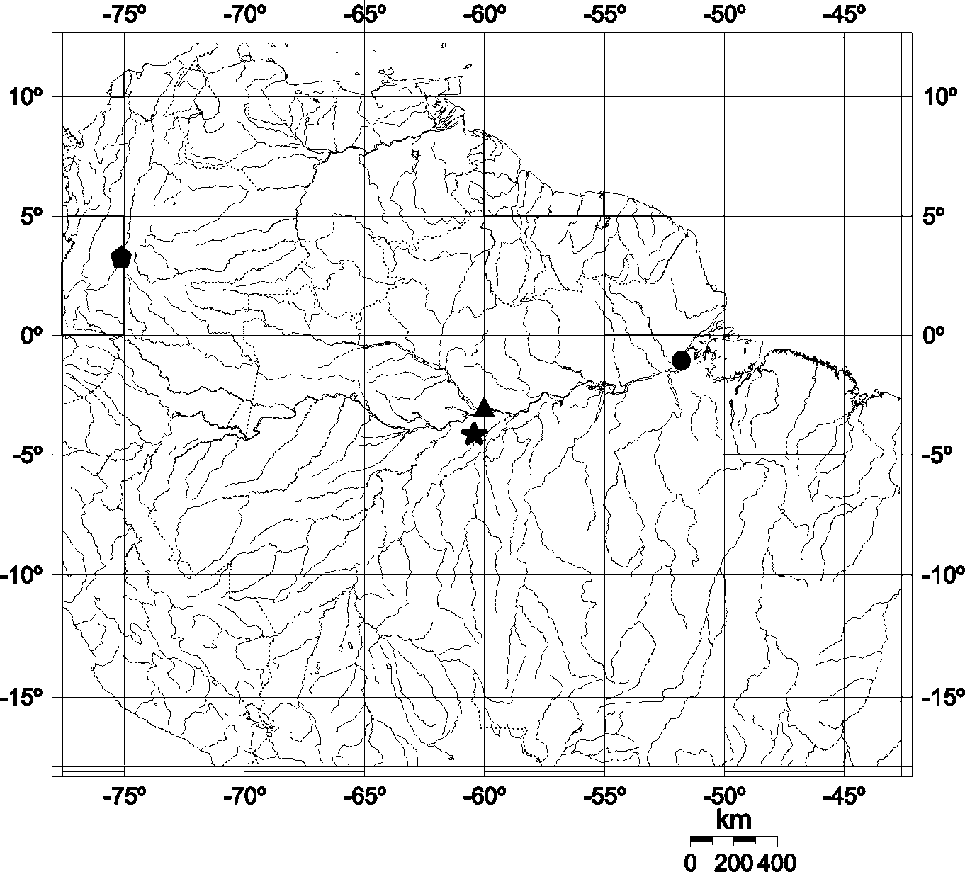
Distribution. Known only from the type locality ( Fig. 22 View FIGURE 22 ).


No known copyright restrictions apply. See Agosti, D., Egloff, W., 2009. Taxonomic information exchange and copyright: the Plazi approach. BMC Research Notes 2009, 2:53 for further explanation.
|
Kingdom |
|
|
Phylum |
|
|
Class |
|
|
Order |
|
|
SubOrder |
Laniatores |
|
Family |
|
|
Genus |