
Fissiphallius orube, Polydoro, Nathalia Sena & Pinto-Da-Rocha, Ricardo, 2012
|
publication ID |
https://doi.org/ 10.5281/zenodo.214946 |
|
DOI |
https://doi.org/10.5281/zenodo.6177685 |
|
persistent identifier |
https://treatment.plazi.org/id/03A41C0A-CF20-2D21-FF7A-FDB83FAE92C1 |
|
treatment provided by |
Plazi |
|
scientific name |
Fissiphallius orube |
| status |
sp. nov. |
Fissiphallius orube sp. nov.
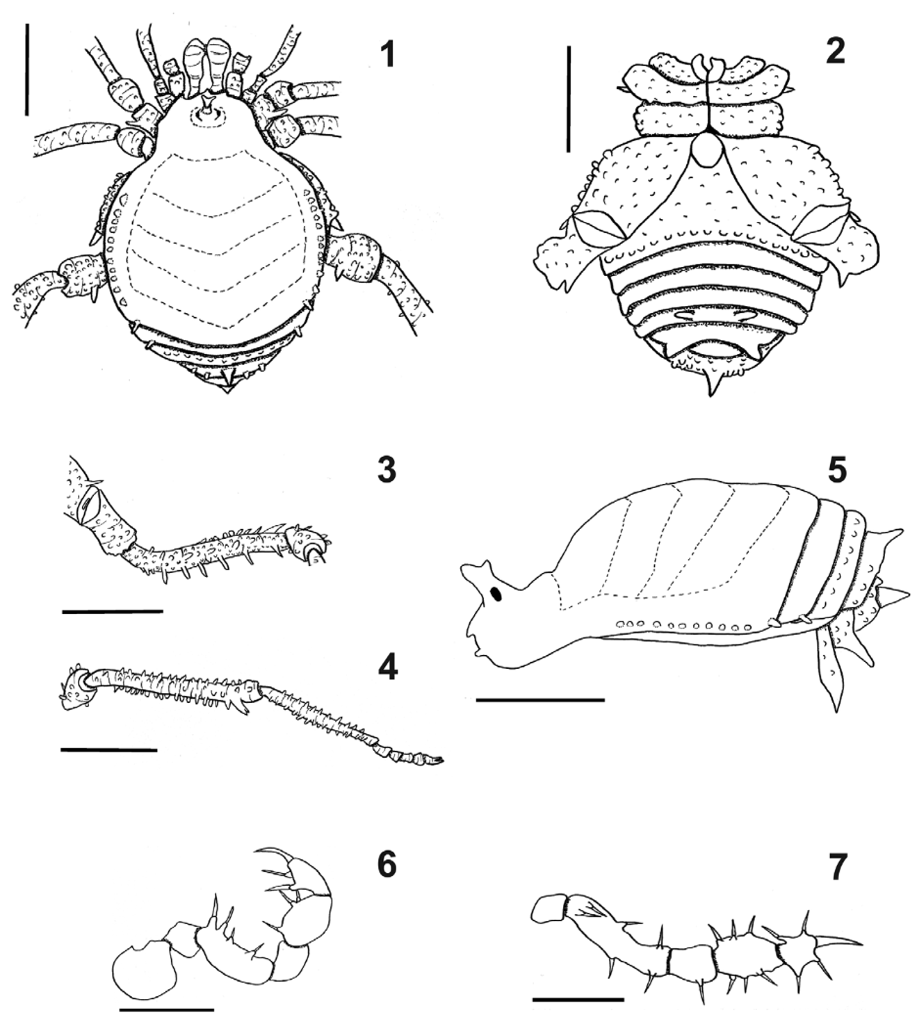
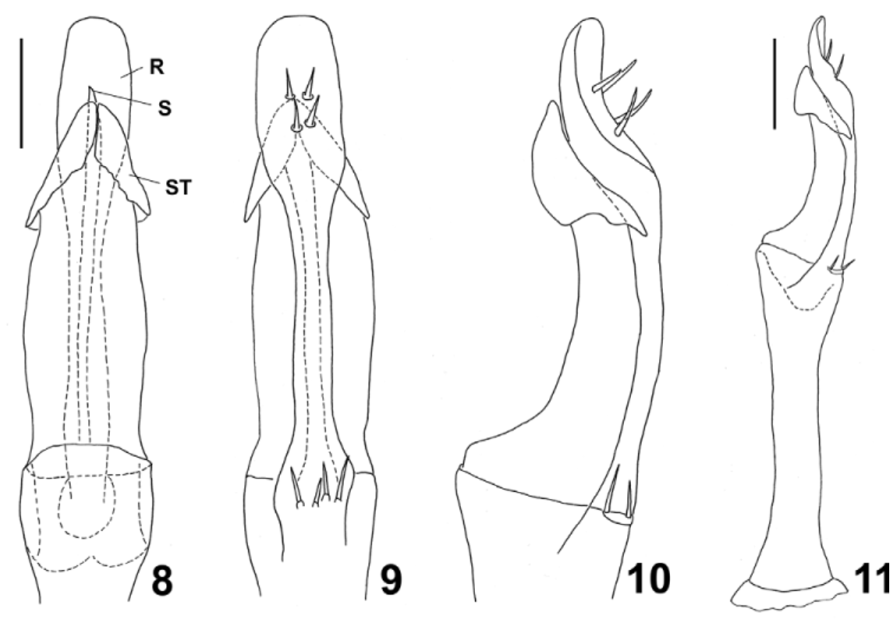
( Figs. 1–11 View FIGURES 1 – 7 View FIGURES 8 – 11 )
Type material. Male holotype (MZSP-15914), Brazil, Acre, Cruzeiro do Sul, near shore of river Moa, 7.6500°S, 72.6833°W, R. S. Vieira leg. Paratypes: 4 males and 7 females ( MZSP 36.498, IBSP, MNRJ), same collecting data as holotype.
Etymology. Orube is a noun in apposition taken from the main Brazilian indigenous language (Tupi), meaning “happiness”.
Diagnosis. Fissiphallius orube sp. nov. can be distinguished from other species of the family by the absence of a pergula (except F. s t u r m i Martens, 1988), the presence of four setae near encounter of stragulum with rutrum ( Fig. 9 View FIGURES 8 – 11 ), by the ocularium bearing a spiniform tubercle with apex single or divided in two ( Figs. 1, 5 View FIGURES 1 – 7 ) or three branches, by the stragulum with bifid apex (arrow-like, see Fig. 8 View FIGURES 8 – 11 ). It is similar to the Colombian F. s t u r m i Martens, 1988, in the genitalia, especially the rutrum, which is a narrow plate with rounded apex, and by the lack of a pergula (see Martens 1988).
Description (male holotype). Measurements. Total length: 2.65; prosoma length: 0.75; opisthosoma maximum width: 2.2, width of prosoma across ocularium 1.00; femur IV length: 1.75.
Dorsum. ( Figs. 1, 5 View FIGURES 1 – 7 ) Anterior margin of prosoma with three paracheliceral projections ( Fig. 1 View FIGURES 1 – 7 ). Ocularium with large central spiniform projection, with apex divided in three branches (other specimens have only one or two branches). Areas I–IV V-shaped, undivided and without tubercles. Lateral margin with 11 rounded similar sized tubercles from groove II to posterior margin. Posterior margin with one tubercle on each corner ( Fig. 5 View FIGURES 1 – 7 ). Free tergites covered by setae; free tergite I with one tubercle on each side; free tergite II with 12 tubercles; free tergite III with five tubercles, median larger than tergite length. Anal operculum with one large tubercle and several smaller tubercles scattered.
Venter. ( Fig. 2 View FIGURES 1 – 7 ) Coxae I–IV irregularly tuberculate. Genital operculum not enlarged, stigmatic area long. Posterior margin with one row of larger tubercles. Free sternites with a row of minute tubercles. Free sternites IV and V with two median, divergent, large tubercles (larger on sternite V).
Chelicera. Covered by setae. Bulla short, well marked, with one prolateral and one retrolateral tubercles. Movable finger (III) with four teeth, fixed finger smooth.
Pedipalp. ( Fig. 6–7 View FIGURES 1 – 7 ) Trochanter smooth. Femur dorsally smooth, ventrally with two basal, two median and two subapical (one ectal, one mesal) setiferous tubercles ( Fig. 7 View FIGURES 1 – 7 ). Patella with mesal setiferous tubercle. Tibia with three mesal (III) and three ectal (III) setae. Tarsus with two ectal (II), two mesal setae (II).
Legs. ( Figs. 3–4 View FIGURES 1 – 7 ) Legs I–IV covered by tubercles. Coxa II with apical retrolateral spiniform tubercle ( Fig. 1–2 View FIGURES 1 – 7 ). Coxa IV with dorsal apical large tubercle. Trochanter IV with sub-basal retrolateral spiniform tubercle. Femora II–IV with two ventral rows of large tubercles, larger on IV. Tibia IV with small tubercle on base, increasing in size distally, with subapical, ventral, large, bifid tubercle ( Fig. 4 View FIGURES 1 – 7 ). Metatarsus IV tuberculated. Tarsal formula: 3 (2), 5 (3), 5, 5.
Penis. ( Figs. 8–11 View FIGURES 8 – 11 ) Stragulum with central portion long and with an apical projection bifid ( Fig. 8 View FIGURES 8 – 11 ), apex arrow-like ( Fig. 8–9 View FIGURES 8 – 11 ). At 2/3rd length of rutrum, base is half the width of distal part, distal margin with two pairs of large setae, apex of distal margin rounded; base of rutrum (just below articulation of rutrum and truncus) with four large setae. Pergula (median ventral lobe) absent ( Fig. 10–11 View FIGURES 8 – 11 ).
Coloration. (in 70% ethanol) Body from pale yellow to orange, with small darker spots on lateral margin of dorsal scutum and median diamond shaped patch (lighter than body). Legs pale yellow.
Description of female paratype: Measurements. Total length 2.15; prosoma length 0.65; opisthosoma maximum width 1.75, width of prosoma at ocularium level 0.85; femur IV length 1.5. Very similar to male, differing in the following features: body slightly smaller, legs markedly shorter (in relation to body), ocularium less developed, with bifid apex, spiniform retrolateral projection of trochanter IV less developed, femur-tibia IV without evident tubercles. Tarsal formula: 3(2), 5(3), 5, 5.
Distribution: Recorded only from type locality.
No known copyright restrictions apply. See Agosti, D., Egloff, W., 2009. Taxonomic information exchange and copyright: the Plazi approach. BMC Research Notes 2009, 2:53 for further explanation.
|
Kingdom |
|
|
Phylum |
|
|
Class |
|
|
Order |
|
|
Family |
|
|
Genus |