
Scolelepis ( Scolelepis ) lefebvrei ( Gravier, 1905 )
|
publication ID |
https://doi.org/10.5281/zenodo.190434 |
|
DOI |
https://doi.org/10.5281/zenodo.6219684 |
|
persistent identifier |
https://treatment.plazi.org/id/03A43259-FF90-2B34-CAC6-FEAEFAA63FE4 |
|
treatment provided by |
Plazi |
|
scientific name |
Scolelepis ( Scolelepis ) lefebvrei ( Gravier, 1905 ) |
| status |
|
Scolelepis ( Scolelepis) lefebvrei ( Gravier, 1905) View in CoL
Figure 2 View FIGURE 2
Material examined. MBMCAS 198572 (2 spms), tidal flat of Dazuo, Fujian Province, sandy mud, 23 Apr 1963; MBMCAS 198603 (1 spm), 123°40΄ E, 35°15΄ N, Yellow Sea, silty mud, 77 m, 26 Jun 1976; MBMCAS 228653 (5 spms), tidal flat of Weizhou Is., Guangxi Province, sandy mud, 5 Apr 1978, coll. Ruiping Sun; MBMCAS 228911 (2 spms), 113°30΄ E, 21°45΄ N, South China Sea, sandy mud, 32 m, 12 Jul 1959, coll. Xiutong Ma; MBMCAS 228995 (2 spms), 111°45΄ E, 22°30΄ N, South China Sea, sandy mud, 18 m, 12 Jul 1959, coll. Xixing Liu.
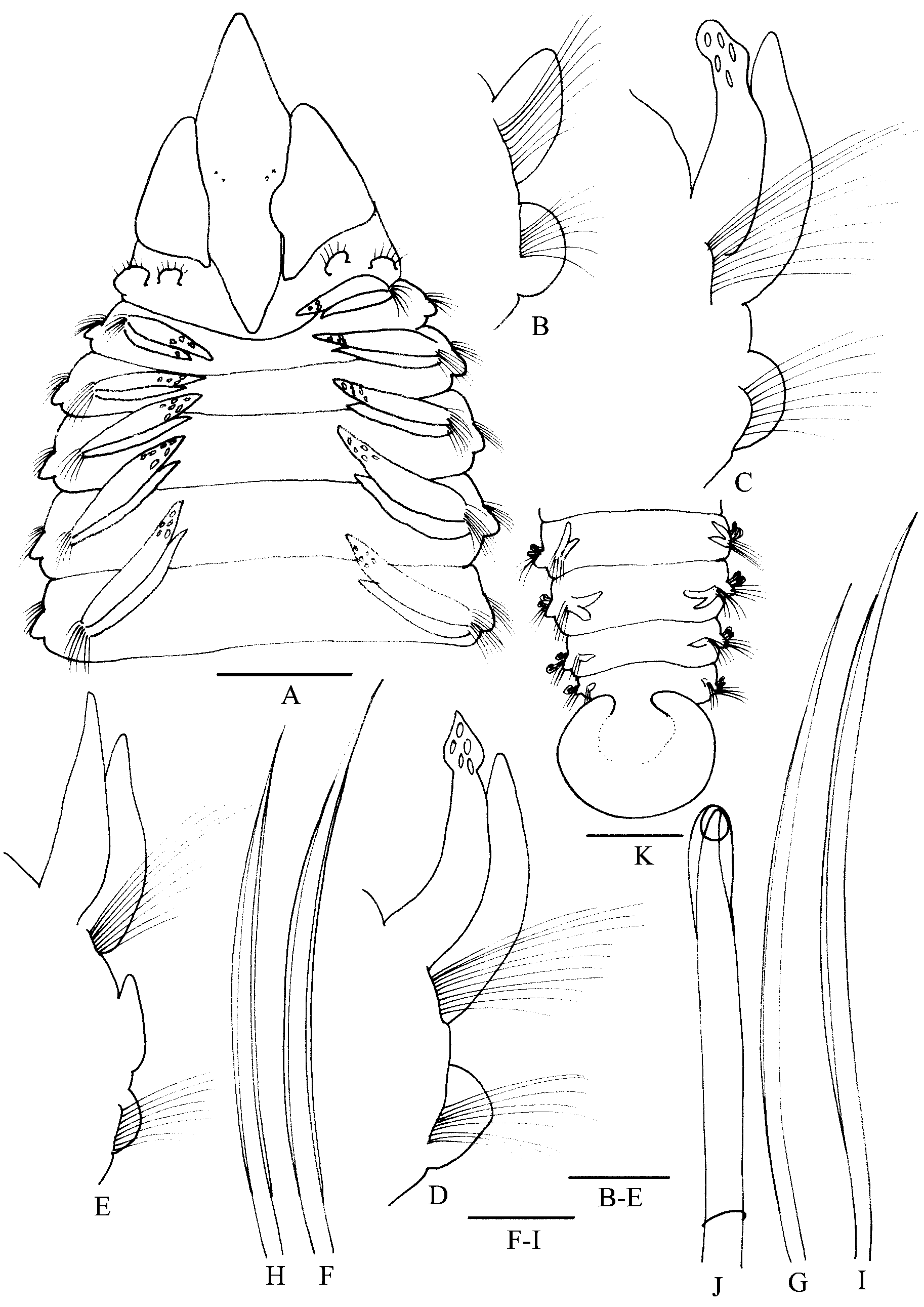
Description. Prostomium elongated, with pointed anterior margin. Caruncle extending posteriorly to setiger 1. Occipital tentacle absent. Two pairs of eyes, arranged in a transverse row. Peristomium distinct from setiger 1, forming well-developed lateral wings ( Fig. 2 View FIGURE 2 A).
Parapodia of setiger 1 well developed; notopodial postsetal lamellae large, subtriangular; neuropodial postsetal lamellae small; with capillary setae in both rami ( Fig. 2 View FIGURE 2 B). Branchiae present from setiger 2, continuing posteriorly to end of body; branchiae on anterior region of body with thick, glandular, subtriangular tips. Notopodial postsetal lamellae distally free from branchiae, long, narrow with ruffled edge in anterior setigers ( Fig. 2 View FIGURE 2 C–D). Posterior lamellae triangular with elongated pointed tip, nearly equal in length through posterior setigers. Notopodial presetal lamellae rounded. Neuropodial postsetal lamellae rounded anteriorly; slight notch present from setigers 26–30; lamellae divided into low, rounded interramal lobe and small triangular ventral lobe from setigers 45–49 ( Fig. 2 View FIGURE 2 E). Interramal lamellae in posterior setigers divided into 3–4 lobes. Neuropodial presetal lamellae thick and round, from setiger 2 through posterior setigers, smaller than corresponding postsetal lamellae.
All anterior setae sheathed capillaries, arranged in two rows on both rami. Anterior row thick, heavily granulated, posterior row thin capillaries without obvious sheaths ( Fig. 2 View FIGURE 2 F–I). Neuropodial hooded hooks from setiger 38, unidentate, with open hoods ( Fig. 2 View FIGURE 2 J), 7–9 in a series, accompanied by capillaries. Notopodial hooded hooks absent.
Pygidium with brownish, incised ventral cushion; anus opening dorsally ( Fig. 2 View FIGURE 2 K).
Remarks. The specimens correspond well with previous descriptions and illustrations. Glands in the distal part of branchiae can be easily detected in some of the samples; however, they are easily faded. The number of hooded hooks in posterior neuropodia in samples from China ( 7–9 in a series) differs slightly from those in the material from Japan (no more than five in a series). Scolelepis ( S.) lefebvrei ( Gravier, 1905) was previously recorded from the South China Sea by Wu et al. (1990). The distribution of the species is extended in the present paper; besides the South China Sea, it also inhabits the East China Sea.
Distribution. China (East China Sea, South China Sea, 0–45 m); Red Sea, Japan, Madagascar.
No known copyright restrictions apply. See Agosti, D., Egloff, W., 2009. Taxonomic information exchange and copyright: the Plazi approach. BMC Research Notes 2009, 2:53 for further explanation.
|
Kingdom |
|
|
Phylum |
|
|
Class |
|
|
Order |
|
|
Family |
|
|
Genus |