
Labyrinthus subplanatus ( Petit, 1843 )
|
publication ID |
https://doi.org/10.5281/zenodo.172563 |
|
DOI |
https://doi.org/10.5281/zenodo.6261407 |
|
persistent identifier |
https://treatment.plazi.org/id/03A4A163-FFCE-E36D-BA40-41659DCA4A31 |
|
treatment provided by |
Plazi |
|
scientific name |
Labyrinthus subplanatus ( Petit, 1843 ) |
| status |
|
Labyrinthus subplanatus ( Petit, 1843) View in CoL
Figures 13–22 View FIGURES 13 – 16 View FIGURES 17 – 22
Caracolla subplanatus Petit, 1843 , 238.
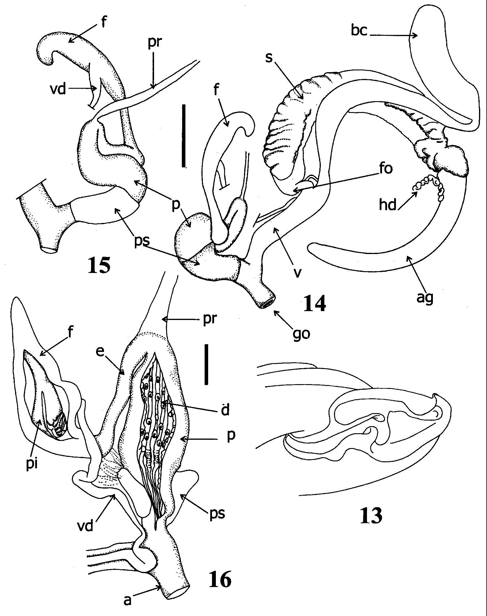
Description: Shell: Morphology as described by Solem (1966: 116), ( Fig. 13 View FIGURES 13 – 16 ).
Pallial Organs: Kidney long, thin, occupying about 60% of pulmonary roof length. Surface of pulmonary roof densely speckled with dark spots. Thin transversal veins abundant between kidney and rectum. Main pulmonary vein branched before reaching mantle collar. Pulmonary roof not expanded beyond top of kidney. Secondary ureter closed up to mantle collar. Ureteric interramus triangular, deeply excavated. Diaphragm thick, muscular.
Reproductive system ( Figs 14–16 View FIGURES 13 – 16 ): Ovotestis formed by elongated digitiform nonramified acini. Albumen gland yellowish, beanshaped. FPSC undifferentiated. Hermaphroditic duct reflexed over itself at base of albumen gland. Albumen gland continuous with spermoviduct. Folds of uterine portion transversal with respect to longitudinal axis of spermoviduct ( Fig. 14 View FIGURES 13 – 16 ). Free oviduct short, cylindrical, naturally forming angle with vagina. Prostatic duct of spermoviduct extending into proximal portion of free oviduct. Vagina as long as penis. Proximal portion of vagina with inner thick longitudinal ridges in zigzag pattern. Distally, ridges become thinner, straight. Hooked denticles absent in entire length of vagina. Duct of bursa copulatrix long, thick, parallel to spermoviduct up to junction with albumen gland. Bursa copulatrix sac globose, reflexed over duct ( Fig. 14 View FIGURES 13 – 16 ). Penial complex formed by flagellum, epiphallus, penis ( Fig. 15 View FIGURES 13 – 16 ). Diameter of flagellum progressively decreasing towards tip. Thick internal pilaster extending between flagellum and epiphallus. Pilaster forming papillalike structure transversally divided at junction between flagellum and epiphallus, point of insertion of vas deferens ( Fig. 16 View FIGURES 13 – 16 ). Penis sheath thick, muscular overlapping distal zone of penis. Epiphallus reflexed over penis, wrapped with muscular fibres and connective tissue. Distal zone of penis thicker than proximal. Penis retractor muscle thick, inserting at proximal portion of penis close to junction with epiphallus. Inner penis wall with thick folds, deeper in proximal zone. Dorsal surface of folds with white, oval, hooked pustules, regularly distributed as shown in Fig.16 View FIGURES 13 – 16 . Verge absent.
Digestive system: Jaw ( Figs 17–18 View FIGURES 17 – 22 ) arched, without vertical ribs ( Fig. 17 View FIGURES 17 – 22 ). Surface with thin transverse striation crossed by thin, shallow perpendicular lines giving appearance of reticulated surface ( Fig. 18 View FIGURES 17 – 22 ). Lower edge without medial protruded cutting edge.
Central tooth of radula unicuspid, triangular ( Fig. 19 View FIGURES 17 – 22 ). Fourteen lateral teeth unicuspid ( Figs 19, 20 View FIGURES 17 – 22 ). Marginal teeth tricuspid ( Fig. 21 View FIGURES 17 – 22 ). Outermost marginal teeth becoming wider but always tricuspid with more pronounced mesocones ( Fig. 22 View FIGURES 17 – 22 ). Buccal mass oval. Rest of digestive tract as in L. dunkeri .
Material examined: COLOMBIA: FMNH 163706, Choco Department, Choco, Caño Taparral, 20 Km to the north of Palestina, on San Juan River. November 25, 1968. B. Malkin Coll. FMNH 173866, Choco Department, San Juan River, Quebrada Docordo. June 5–8, 1969. B. Malkin Coll. FMNH 223561, Choco Department, San Juan River, 110 Km to the north of Palestine. January 20,1971. B. Malkin & P. Bouchard Coll.
Remarks: Labyrinthus subplanatus belongs to the L. otis species complex that was supported as a monophyletic clade within the genus ( Cuezzo 2003). The group of L. otis is composed by L. otis (Lightfoot, 1786) , L. subplanatus , L. marmatensis Pilsbry, 1910 , and L. plicatus . Labyrinthus subplanatus is similar to L. otis and both have been confused mostly because their distributional ranges are partly overlapping. However, L. otis differs from L. subplanatus mainly in having a deep supraperipheral groove and in being much larger ( L. otis shell diameter up to 58 mm, while L. subplanatus diameter up to 45 mm). Labyrinthus subplanatus also present a periphery acutely angulated with protruding keel, and the peristome forming a peripheral notch sometimes slightly twisted (see Solem 1966) which is absent in L. otis . Differences in the genitalia between these two species are difficult to evaluate because of the poor descriptions presently available of L. otis .

| FMNH |
Field Museum of Natural History |
No known copyright restrictions apply. See Agosti, D., Egloff, W., 2009. Taxonomic information exchange and copyright: the Plazi approach. BMC Research Notes 2009, 2:53 for further explanation.
|
Kingdom |
|
|
Phylum |
|
|
Class |
|
|
Order |
|
|
Family |
|
|
Genus |
Labyrinthus subplanatus ( Petit, 1843 )
| Cuezzo, Maria Gabriela 2006 |
Caracolla subplanatus
| Petit 1843 |