
Nousia Navas 1918
|
publication ID |
https://doi.org/10.11646/zootaxa.3754.4.9 |
|
publication LSID |
lsid:zoobank.org:pub:CECF329A-F13D-4E5F-A314-807211781D08 |
|
DOI |
https://doi.org/10.5281/zenodo.6141117 |
|
persistent identifier |
https://treatment.plazi.org/id/03A58780-FFCD-FFB3-FF41-F92E10B1F8E0 |
|
treatment provided by |
Plazi |
|
scientific name |
Nousia Navas 1918 |
| status |
|
Genus Nousia Navas 1918 View in CoL View at ENA
= Atalonella Needham & Murphy 1924 Type species: N. delicata Navas 1918 View in CoL .
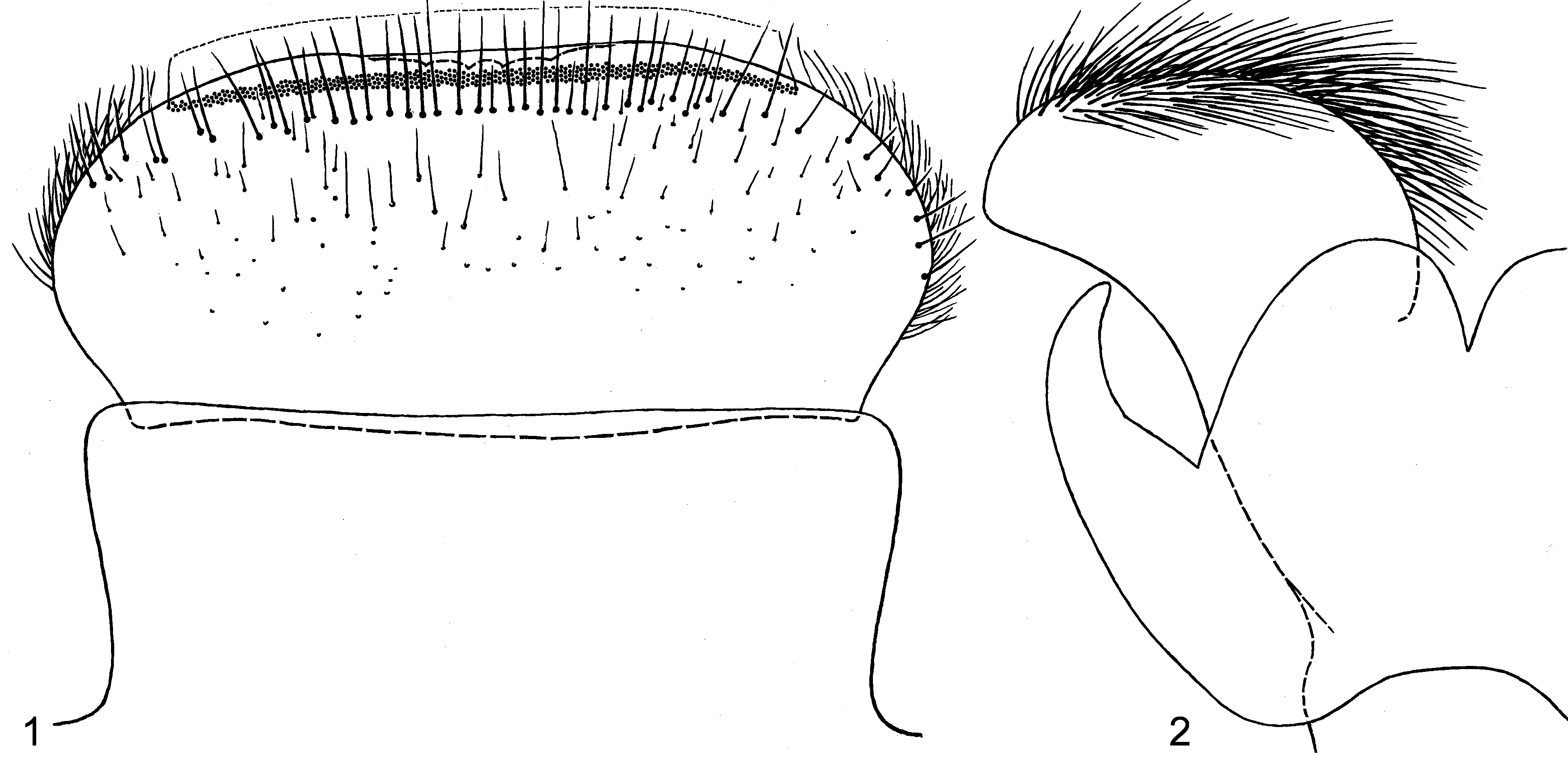
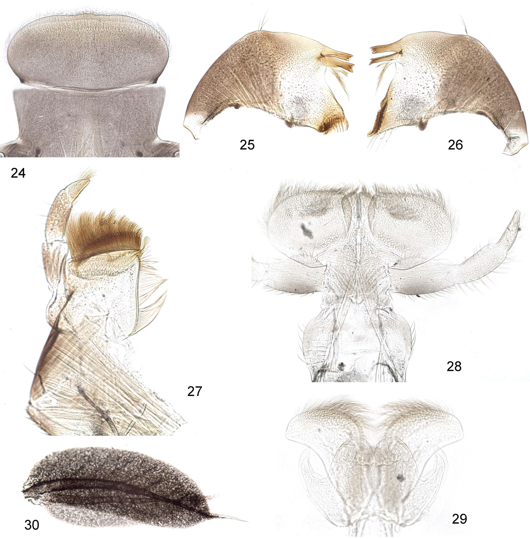
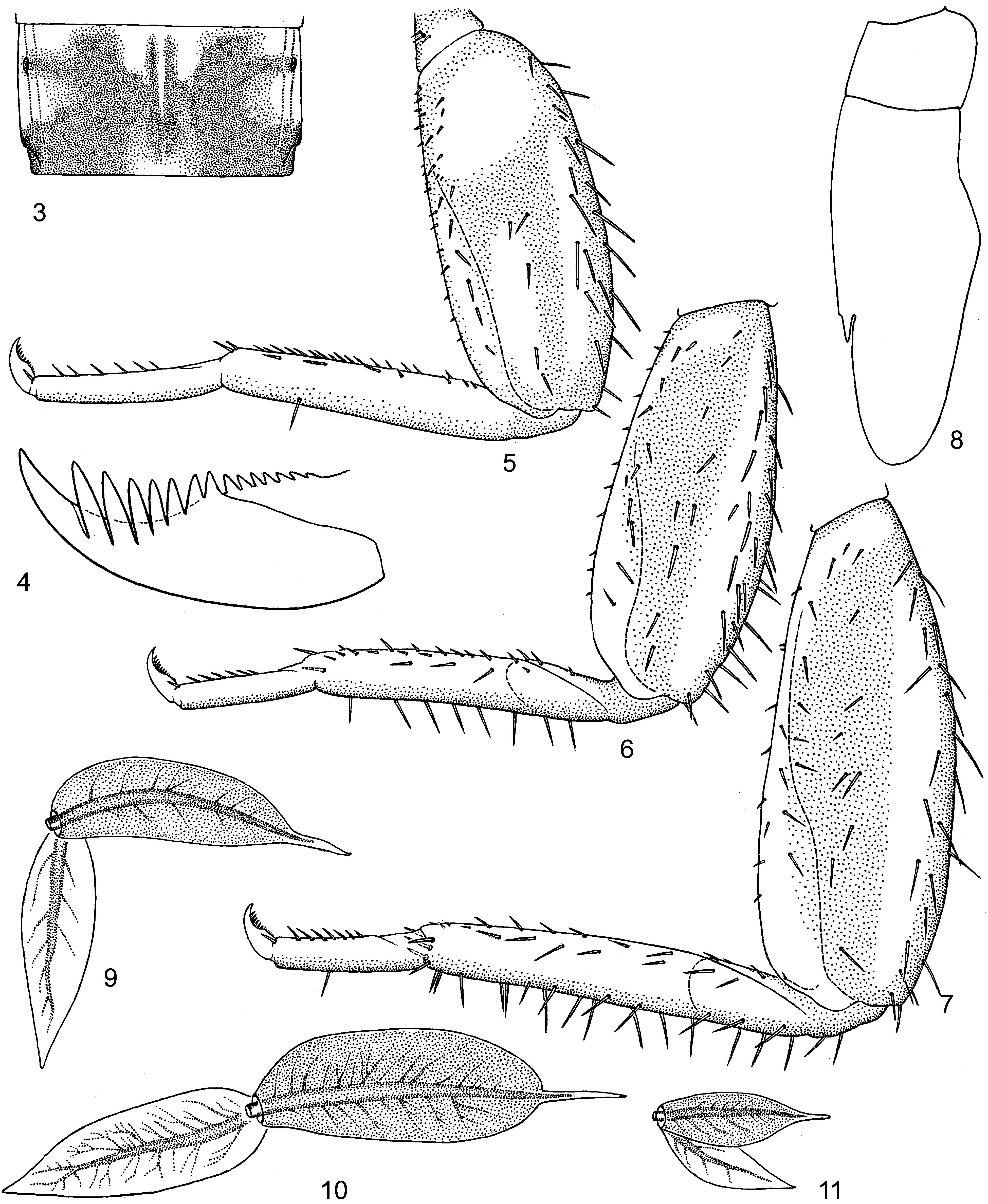
Larva. Clypeus parallel-sided or slightly widened anteriorly ( Figs 1 View FIGURES 1 – 2 , 24 View FIGURES 24 – 29 ); labrum not widened, with median incision shallow or non-expressed; number of median denticles odd—5 or 3; both dorsal transverse setal rows straight and located near anterior margin, distal row very dense and irregular, proximal row more or less regular ( Fig. 1 View FIGURES 1 – 2 ). Mandibles with outer margin smoothly curved, with hair tuft near middle ( Figs. 25, 26 View FIGURES 24 – 29 ). Maxilla slightly broad apically, with 8–12 pectinate setae in ventro-apical row ( Fig. 27 View FIGURES 24 – 29 ). Labium with glossae partly inserted in cavities of paragossae, not projected neither ventrad, nor dorsad of paraglossae ( Fig. 28 View FIGURES 24 – 29 ). Patella-tibial suture developed on middle and hind legs, being absent on fore leg ( Figs 5–7 View FIGURES 3 – 11 ). Tergalii of all pairs I–VII similar, each tergalius with both lamellae pointed, without additional processes; shape of tergalii varies from thread-like ( Pescador & Peters 1985: Fig. 54) to wide-lanceolate ( Finlay 2000: Fig. 23 View FIGURES 19 – 23 ) or oval ( Figs 10 View FIGURES 3 – 11 , 30).
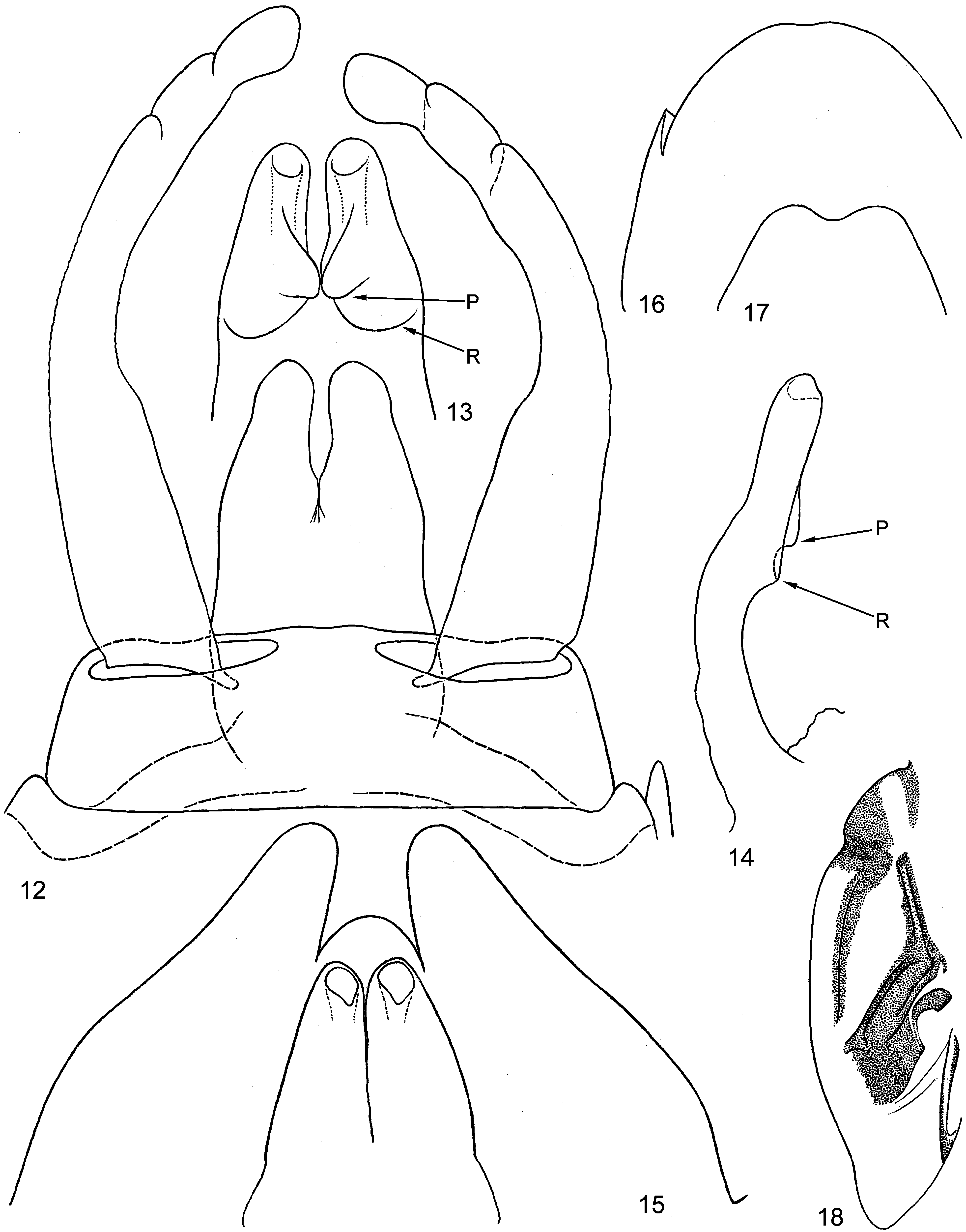
Subimago. Pigmented area anteriad of mesonotal suture forms a narrow stripe along medioparapsidal suture ( Fig. 18 View FIGURES 12 – 18 ). First tarsal segment covered with microtrichia; tarsal segments II–V covered with pointed microlepides.
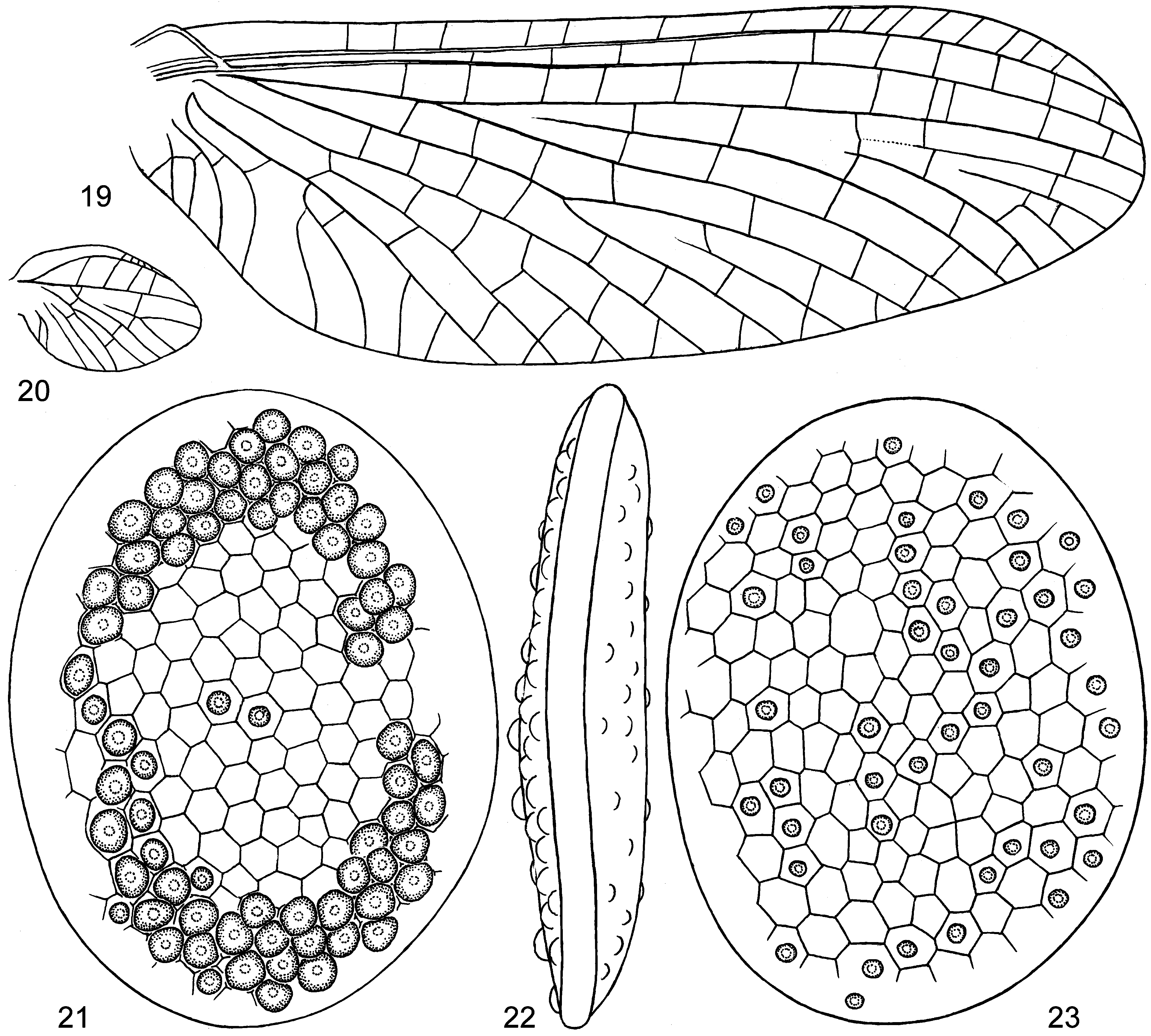
Imago. Patella-tibial suture developed on middle and hind legs, absent on fore leg. On each leg both claws pointed. Hind wing without prominent costal projection and with subcostal vein long ( Fig. 20 View FIGURES 19 – 23 ). Penis with peculiar structure ( Figs 13, 14 View FIGURES 12 – 18 ): dorsal surface of each penis lobe crossed by a transverse semicircular ridge, with mediallydirected sclerotized process close to this ridge; unpaired proximal portion about 1/2–2/3 of total penis length ( Figs 12–14 View FIGURES 12 – 18 ; Demoulin 1955: Figs 6 View FIGURES 3 – 11 b', 7b', 8c'; Pescador & Peters 1985: Fig. 7 View FIGURES 3 – 11 ; Finlay 2000: Figs 8 View FIGURES 3 – 11 , 27 View FIGURES 24 – 29 ).
Remarks. Among larval characters of Nousia , an enlarged distal denticle of the claw is mentioned ( Pescador & Peters 1985); in various species it is either strongly enlarged, or indistinctly enlarged ( Pescador & Peters 1985: Figs 50 and 48), or non-enlarged ( Fig. 4 View FIGURES 3 – 11 ).
In the key to leptophlebiid genera of South America, Nousia imagos are separated from Penaphlebia Peters & Edmunds 1972 and Rhigotopus Pescador & Peters 1982 by the absence of a direct joining of vein ICu1 with vein CuA on the fore wing ( Domínguez et al. 2006). This statement is based on the diagnosis of Nousia by Pescador & Peters (1985), who stated that "vein ICu1 free or attached at base by a cross vein to vein CuA ... ". Actually, the vein ICu1 is often directly jointed with CuA ( Fig. 19 View FIGURES 19 – 23 ; Needham & Murphy 1924: Fig. 97; Demoulin 1955: Figs 6 View FIGURES 3 – 11 a, 7a, 8a) and occasionally can have a free base ( Pescador & Peters 1985: Fig. 1 View FIGURES 1 – 2 ; Finlay 2000: Fig. 2 View FIGURES 1 – 2 ) or be attached at the base by a cross vein to vein CuA ( Demoulin 1955: Fig. 9 View FIGURES 3 – 11 ).
Subgenus Araucophlebia subgen. n.
Type species: Nousia (Araucophlebia) latifolia sp.n.
Diagnosis. Larval characters:
(1) Long spine-like setae on outer margin of tibia are present not only on hind legs, but also on middle legs ( Fig. 6 View FIGURES 3 – 11 ) (in contrast to most other Leptophlebiidae ).
(2) Tergalii have dorsal lamella oval, with sharply separated slender apical process ( Figs 10 View FIGURES 3 – 11 , 30). In other Nousia tergalii have both lamellae lanceolate and without terminal process ( Pescador & Peters 1985: Figs 51–54; Finlay 2000: Fig. 23 View FIGURES 19 – 23 ).
(3) Denticles on posterior margins of abdominal terga I–IX are reduced. In other Nousia denticles on posterior margins of abdominal terga are well developed ( Pescador & Peters 1985: Fig. 58; Finlay 2000: Fig. 22 View FIGURES 19 – 23 ).
Male imaginal character:
(4) Penis has simple outline of lateral sides; median sclerotized processes (peculiar for Nousia — see above) are massive ( Fig. 12 View FIGURES 12 – 18 ). In other Nousia lateral sides of penis have protuberances, and median sclerotized processes are small ( Pescador & Peters 1985: Figs 6–13 View FIGURES 3 – 11 View FIGURES 12 – 18 ; Finlay 2000: Fig. 26 View FIGURES 24 – 29 ).
Egg character:
(5) Eggs have unusual disk-like shape ( Figs 21–23 View FIGURES 19 – 23 ). In other Nousia eggs have a usual ellipsoid shape ( Pescador & Peters 1985: Figs 56–57; Finlay 2000: Fig. 28 View FIGURES 24 – 29 ).
Composition. One species, Nousia (Araucophlebia) latifolia sp. n.
No known copyright restrictions apply. See Agosti, D., Egloff, W., 2009. Taxonomic information exchange and copyright: the Plazi approach. BMC Research Notes 2009, 2:53 for further explanation.
|
Kingdom |
|
|
Phylum |
|
|
Class |
|
|
Order |
|
|
Family |
Nousia Navas 1918
| Kluge, Nikita J. 2014 |
Atalonella
| Needham & Murphy 1924 |
N. delicata
| Navas 1918 |