
Merodon elegans Hurkmans, 1993
|
publication ID |
https://doi.org/ 10.1163/18759866-20191414 |
|
DOI |
https://doi.org/10.5281/zenodo.8368090 |
|
persistent identifier |
https://treatment.plazi.org/id/03A787C5-FFDD-B149-83C1-B0C2F2EBFCDA |
|
treatment provided by |
Felipe |
|
scientific name |
Merodon elegans Hurkmans |
| status |
|
Identity of Merodon elegans Hurkmans View in CoL View at ENA
Merodon elegans was described by Hurkmans (1993) based on the large number of specimens collected in the Western Mediterranean. A recent study of all known Merodon types resulted with discovery of two names related to the same taxon. Vujić et al. (2011) cited this species for Turkey under one of these names, M. biarcuatus Curran, 1939 based on the holotype found in AMNH. Syntype of M. femoratus Sack, 1913 found in ZMHB, resolved question about the oldest name that should be used for this species.
syn. n. biarcuatus Curran, 1939
syn. n. elegans Hurkmans, 1993
Types. Merodon femoratus Sack, 1913: 446 . Typelocality. Corsica, Greece, Asia Minor. Described based on unspecified number of males and females. One syntype was found in ZMHB: France, Corsica “Mann” “855”, a male designated here as lectotype .
Merodon biarcuatus Curran, 1939: 6 . Typelocality. Morocco. Holotype (studied). ♀ forest of Namora , near Rabat, Morocco (AMNH), with clear apomorphic character, broad metafemur, ventrally covered with long whitish pile as in M. femoratus .
Merodon elegans Hurkmans, 1993 . Holotype (studied). ♂ Italy, Sicilia, “V. S. v. d. Goot! Erna rif. Filiciusa 1400-1500m 22-28.vii.1961 / Lampetia spinipes det. V. S. v.d. Goot 1963 I Type A” (NMNL), conspecific with M. femoratus .
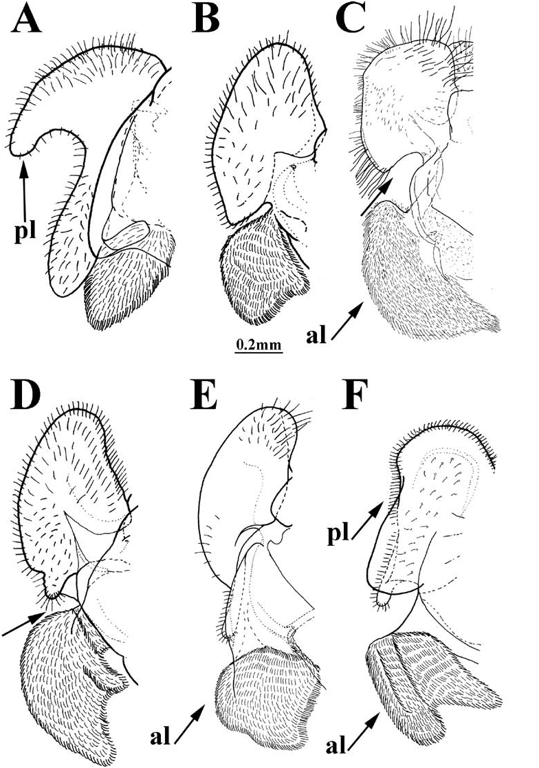
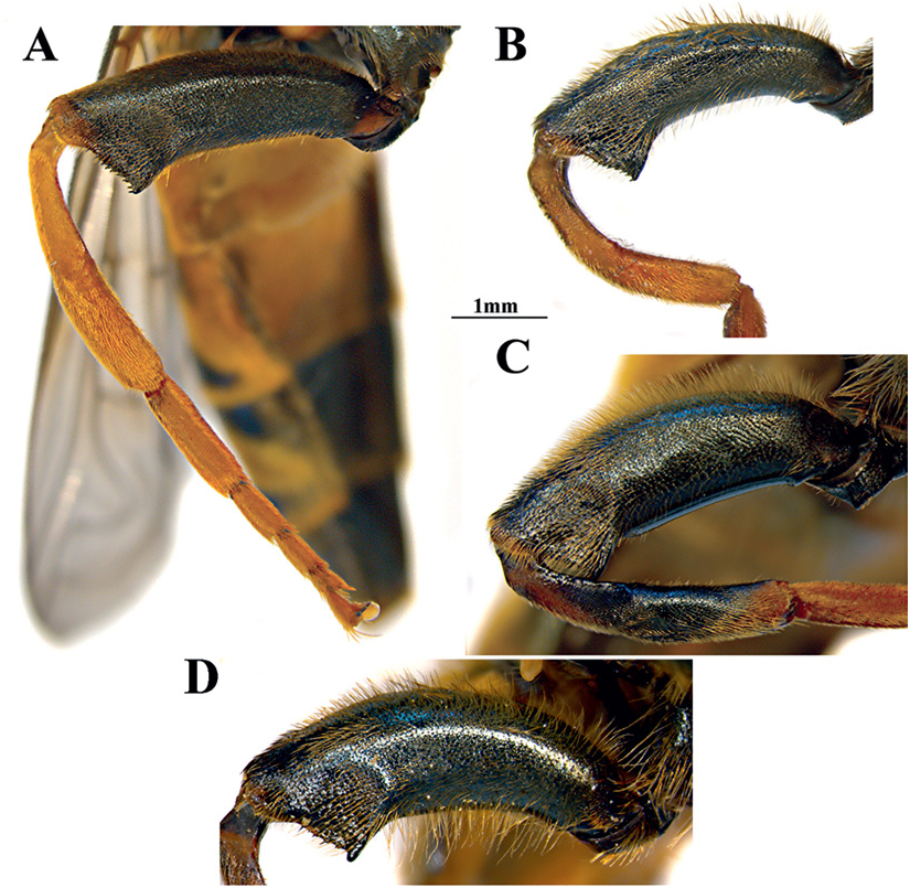
Diagnosis. Medium sized species (11–13 mm) similar to M.avidus complex,from which can be distinguished by broad metafemur (narrower in other members from M. avidus complex (fig. 37A–B)), ventrally covered with long whitish pile (fig. 37D), and by deep incision between anterior and posterior surstyle lobe in male genitalia (fig. 13C: marked with arrow) (absent in M. avidus complex fig. 32C).
Distribution. Northern Africa ( Algeria, Morocco, Tunisia), south and southwest Europe ( Croatia, France, Portugal, Spain, Italy).
IdentiFIcation keys of males of M. nigritarsis and M. avidus groups and Merodon crassifemoris
Following a key to the males of the M. nigritarsis group (Vujić et al., 2013) we present an updated identification key for all known members of the M. nigritarsis and M. avidus groups. Identification of females is very difficult and will be the subject of future studies, using a combined integrative approach.
1. Posterior part of mesocoxa without long pile ( Merodon avidus-nigritarsis lineage) (fig. 34B: cxp) ..................................................... 2
– Posterior part of mesocoxa with long pile (fig. 34A: cxp) .................................................... other Merodon lineages (not treated here)
2. Species with white microtrichose vittae on black scutum and white microtrichose fasciae on dark terga; at least tergum 2 with a pair of reddish-orange maculae laterally; abdomen elongated, narrow and tapering, always longer than scutum and scutellum together; legs without spinae or other protuberances; male genitalia: anterior surstyle lobe more or less rhomboid shape, except in the alagoezicus subgroup where it is transformed into a narrow, elongated, strongly curved projection ( M. nigritarsis and M. avidus groups + M. crassifemoris ) ....................... 3
– Species with different combination of characters ......................................................... other species groups belonging to Merodon avidus-nigritarsis lineage (not treated here)
3. Tarsi dark brown/black dorsally and orange/brown ventrally ( M. nigritarsis group) (fig. 14E–H) ..........................................................9
– Tarsi yellow dorsally and ventrally (M. avidus group) (fig. 14A–D) ............................... 4
4. Posterior surstyle lobe with hump in apical half (fFig. 32A: marked with arrow); anterior surstyle lobe short and rounded, oval (fig. 32A: al)............................ M. rutitarsis sp. n.
– Posterior surstyle lobe simple; anterior surstyle lobe longer, rhomboid shape (fig. 32C: al) .....................................................5 5. Metafemur broad, ventrally covered with long whitish pile ( Fig. 37D View FIGURE 37 ); surstyle with deep incision between anterior and posterior lobes (fig. 13C: marked with arrow) ........................................................... M. femoratus
– Metafemur less broad, without long ventral pilosity (fig. 37A–C); surstyle without deep incision (fig. 32C).................................6
6. Body pile golden; metafemur with very short pile (fig. 37A) .............................. M. megavidus
– Body pile yellow to pale/grayish; metafemur with longer pile (fig. 37B–C) ............. 7
7. Distribution: western Mediterranean; clearly defined with genetic data (see Popović et al., 2015) .................................................... M. ibericus – Distribution: Europe, except Iberian Peninsula ................................................................8
8. Tergum 2 with a pair of whitish, microtrichose spots; terga 3 and 4 with broad microtrichose fasciae (fig. 38A); tibiae usually pale (fig. 37B); body pile slightly shorter, especially on the tergum 4 (fig. 39A); tergum 3 with a pair of orange, lateral, triangular maculae, anterior part of tergum 3 is also predominantly orange-red, except medially, where it is narrowly black (in darker specimens at least, small orange areas are present antero-sublaterally) .......................... M. avidus
– Tergum 2 shiny, without microtrichia; terga 3 and 4 with narrow microtrichose fasciae (fig. 38B); tibiae always partly dark (fig. 37C); body pile longer (fig. 39B); tergum 3 black (in some specimens orangered anterolaterally, but with a black posterior margin) .............................. M. moenium
9. Metafemur narrow (about 4.5 times longer than wide); anterior surstyle lobe with strong interior accessory lobe (fig. 12A) ......................................................... M. nitidifrons
– Metafemur broad (as on fig. 20A–B); anterior surstyle lobe without or with small interior accessory lobe............................... 10
10. Anterior surstyle lobe transformed to narrow, long, curved, pointed extension (fig. 12B–F)........................................................... 11
– Anterior surstyle lobe rhomboid or triangular shape .................................................... 16
11. Anterior surstyle lobe, sickle-shaped, curved downwards, with pointed apex directed towards base of epandrium (fig. 27A: al) .................................................. M. obstipus sp. n.
– Anterior surstyle lobe curved upwards (as on fig. 12B: al) ................................................ 12
12. Apical part of metatibia with clear ventrolateral lamella (fig. 36B: la) .................................13
– Apical part of metatibia without clear ventrolateral lamella (fig. 36A) ................15
13. Posterior surstyle lobe two times as long as wide, straight (fig. 12B: pl); anterior surstyle lobe long, narrow, with rounded curve (fig. 12B: al); ................................ M. alagoezicus
– Posterior surstyle lobe S- shaped; anterior surstylelobewithangularcurve(fig.12C–D) .......................................................................... 14
14. Anterior surstyle lobe with additional basal extension (fig. 12C: marked with arrow) ........ ........................................................ M. satdagensis
– Anterior surstyle lobe without additional basal extension (fig. 12D) ......... M. schachti 15. Abdomen covered with pale pile; tergum 2 without white microtrichose maculae; posterior surstyle lobe broader basally and narrower in apical part (fig. 12E: pl) ...................... ...................................................... M. hakkariensis
– Abdomen with short black pile on posteromedial part of tergum 3 and medial parts of tergum 4; tergum 2 usually with a pair of white microtrichose spots; posterior surstyle lobe the same width along the entire length and with lamellar structure (fig. 12F: marked with arrow) ....................... .............................................................. M. lucasi
16. Face with a bulge below antennae (fig. 4D: marked with arrow); posterior surstyle lobe hook-like (fig. 13A: pl); metafemur very broad .................................................... M. crassifemoris
– Face without bulge ..................................... 17
17. Pile on metafemur very short on ventral surface; surstylus on fig. 13B ............ M. angustus
– Pile on metafemur longer on ventral surface; surstylus of different shape ........... 18
18. Metafemur and metatibia extremely curved; male genitalia: posterior and anterior surstyle lobe separated by deep incision (fig.13D: marked with arrow) ............................................ ............................................................ M. testaceus
– Metafemur and metatibia less curved; male genitalia: posterior and anterior surstyle lobe not deeply divided ................. 19
19. Metafemur broad and covered with long anteroventral and posteroventral pile, as long as half of width of metafemur (as on fig. 20A) ............................................................. 20
– Metafemur narrower and covered with shorter pile, usually on posteroventral surface much shorter or absent ..............24
20. Anterior surstyle lobe 2.5 times shorter than posterior surstyle lobe (as on fig. 23A: al) ..... ................................................................................21
– Anterior surstyle lobe less than 2 times shorter than posterior surstyle lobe (as on fig. 35C: al) ................................................... 22
21. Lateral orange maculae on tergum 2 large, cover 2/3 of the posterior margin (fig. 11C); anterior surstyle lobe is about as long as wide (fig. 23C: al); lingula shorter (fig. 24C, D: l); distribution: Apennine Peninsula ................... ............................................................. M. toscanus
– Lateral orange maculae on tergum 2 smaller, reaching the posterior margin only at outer corners of tergum (fig. 21A); anterior surstyle lobe is about 2 times wider than long (fig. 23A: al); lingula longer and pointed upward (fig. 24A, B: l); distribution: Anatolian Peninsula and Greece.......................................... M. longisetus sp. n.
22. Anterior surstyle lobe elongated, triangular; posterior surstyle lobe elongated and narrow (fig. 35C: al) ............................................................ ..................................................... M. femoratoides
– Anterior surstyle lobe shorter, not triangular; posterior surstyle lobe shorter ..........................................................................23
23. Anterior surstyle lobe square-shaped ( Fig. 13E View FIGURE 13 : al); lateral projections on hypandrium gradually tapering to the tip (fig. 30C: lp) ....................................................... M. quadraticus
– Anterior surstyle lobe high boot-shaped (fig. 29A: al); lateral projections on hypandriumnarrowonlyinapical1/4 (fig.30A: lp) ............................................ M. cohurnus sp.n.
24. Posterior surstyle lobe broad, anterior surstyle lobe very short (fig. 13F: al, pl) ................ ......................................................... M. taniniensis
– Posterior surstyle lobe narrower, anterior surstyle lobe longer ................................... 25
25. Posterior surstyle lobe shorter, 1.5 times as long as wide (fig. 35A: pl) ................................... .......................................................... M. nigritarsis
– Posterior surstyle lobe longer, 2.5 times as long as wide (fig. 35B: pl) .............................. .................................................... M. latifemoris
No known copyright restrictions apply. See Agosti, D., Egloff, W., 2009. Taxonomic information exchange and copyright: the Plazi approach. BMC Research Notes 2009, 2:53 for further explanation.