
Rhiginia lourdesae Gil-Santana & Forthman, 2021
|
publication ID |
https://doi.org/10.11646/zootaxa.4952.2.1 |
|
publication LSID |
lsid:zoobank.org:pub:FD1B0F80-4662-48C0-BBF5-B00003BE7437 |
|
DOI |
https://doi.org/10.5281/zenodo.4673953 |
|
persistent identifier |
https://treatment.plazi.org/id/03A787E1-9208-E74B-A1E6-FF0EA0696983 |
|
treatment provided by |
Plazi |
|
scientific name |
Rhiginia lourdesae Gil-Santana & Forthman |
| status |
sp. nov. |
Rhiginia lourdesae Gil-Santana & Forthman , new species
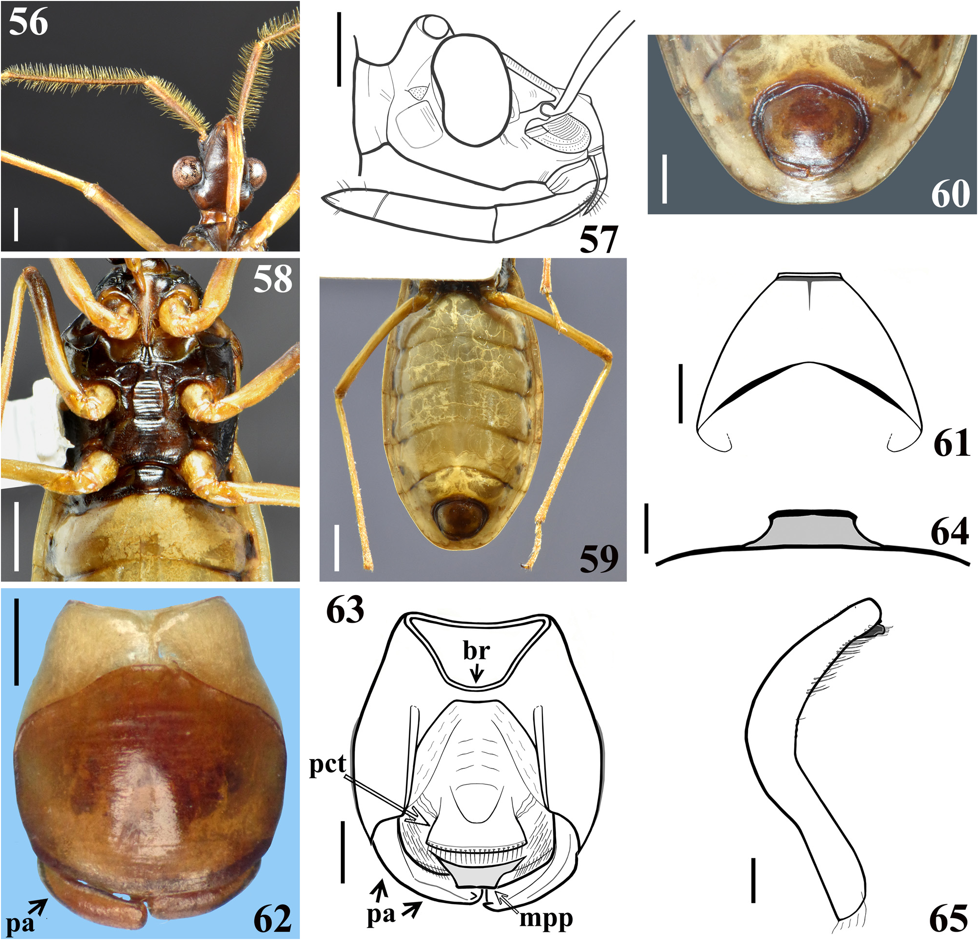
( Figs. 52–70 View FIGURES 52–55 View FIGURES 56–65 View FIGURES 66–70 )
Diagnosis. The male of this species can be distinguished from all other species of Rhiginia by the large body size, smooth and polished pronotal surface, bicolored femora, subrectangular head and flat frons in lateral view, large eyes, large ocelli separated by a distances less than the diameter of an ocellus, prominent anterolateral protuberances on the pronotum, anterior pronotal lobe approximately half the length of the posterior lobe, and abdominal sternite II with slight medial transverse striations. This species is most similar to R. cinctiventris but can be recognized by the more prominent anterolateral protuberances on the pronotum (indiscernible or slightly rounded in R. cinctiventris ), proximally diverging veins of the Cu+1A membranal cell (subparallel in R. cinctiventris ), proximal margin of the Cu+1A membranal cell about six times wider than the M+Cu membranal cell (two times in R. cinctiventris ), transversely striate abdominal sternite II (longitudinal ridges in R. cinctiventris ), and bicolored legs (uniformly dark brown-black in R. cinctiventris ). Of the species in the cruciata -group, R. cruciata and R. lourdesae sp. nov. both have bicolored legs, but the latter can be distinguished from the former by the large ocelli that occupies more than half of the ocellar tubercle in lateral view and that are separated from one another by less than the diameter of an ocellus, a subrectangular head shape, a flat frons, prominent anterolateral pronotal protuberances, an anterior pronotal lobe that is shorter than the posterior lobe, the absence of longitudinal ridges on abdominal sternite II, proximally diverging veins of the Cu+1A membranal cell, and much larger width at the proximal margin of the Cu+1A membranal cell relative to the M+Cu membranal cell.
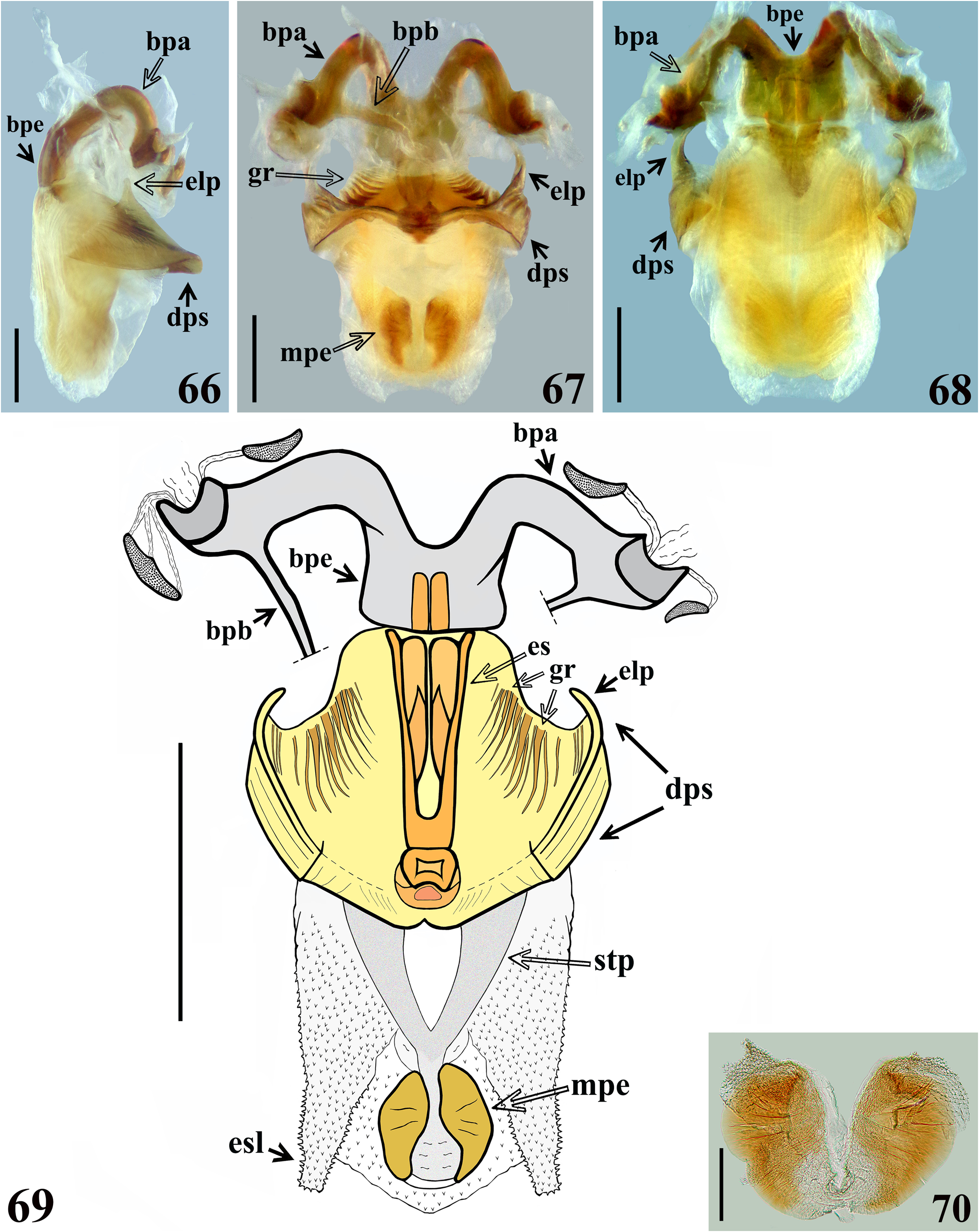
Description. Male ( holotype). MEASUREMENTS (in mm): total length, to tip of abdomen: 20.2, to tip of hemelytra: 19.6. Head (excluding neck): length: 3.1, height (maximum distance between upper and lower margin of the head): 1.7; length of anteocular portion (measured in lateral view): 1.5; length of postocular portion (measured in lateral view): 0.6; width across eyes: 2.9; minimum dorsal interocular distance (synthlipsis): 1.2; eye, dorsal view: width: 0.8, length: 1.0; distance between external margins of ocellar tubercle: 1.1; distance between ocelli: 0.25; maximum width of ocellus: 0.3; length of ocellar tubercle on midline: 0.4; maximum distance between antennal tubercles:1.5;lengths of antennal segments:scape:2.9, pedicel:4.4,basiflagellomeres I:2.0,II:1.3,distiflagellomeres: I: 0.7, II: 0.6, III: 0.5, IV: 0.6; lengths of labial segments: II (first visible): 1.5, III: 1.6, IV: 0.7. Thorax: pronotum: anterior lobe: length: 1.1, maximum width: 3.0, posterior lobe: length 2.3, maximum width: 4.9; scutellum: length: 1.9, maximum width at base: 2.4. Fore legs: femur: length: 4.3, maximum width at midportion: 0.6; length of tibia: 4.8; length of spongy fossa: 0.5; length of tarsus: 1.4. Middle legs: femur: length: 4.2, maximum width at subapical portion: 0.6; length of tibia: 5.0; length of spongy fossa: 0.3; length of tarsus: 1.6. Hind legs: femur: length: 6.7, maximum width at subapical portion: 0.6; length of tibia; 7.5; length of tarsus: 1.9.Abdomen: length: 10.5, maximum width: 7.0. COLORATION ( Figs. 52–56, 58–60 View FIGURES 52–55 View FIGURES 56–65 ): General coloration dark yellowish to pale orange with darkened to blackish markings or regions. Head ( Figs. 52–56 View FIGURES 52–55 View FIGURES 56–65 ): Dorsal surface mostly pale orange; antennifers darkened at inner and outer surfaces; ocellar tubercle with a faint reddish tinge around ocelli and somewhat darkened between them; lateral and ventral portions of head mostly dark, brownish black to brownish; ventrolateral protuberance posterior to eye pale brownish, even paler area just above and posterior to it; labrum mostly darkened; labium pale; neck pale orange dorsally and somewhat darker ventrally with a lateral brownish stripe; scape and pedicel brownish, basal portion of the former pale, flagellomeres pale orange to yellowish, the first basiflagellomere somewhat darker and tip of last distiflagellomere with a faint reddish tinge. Thorax ( Figs. 52–54 View FIGURES 52–55 , 58 View FIGURES 56–65 ): Pronotum mostly dark yellowish to pale orange with all margins darkened brownish to blackish, more extensively so at posterior margin, while the central portion of anterolateral angles are pale; transverse and mid-longitudinal sulci almost mostly dark, except the distal portion of the latter on midportion of posterior lobe, which becomes paler; the areas lateral to the midlongitudinal sulcus on anterior lobe and those anterior and posterior to transverse sulcus (the latter more extensively) darkened too; scutellum blackish; pleurae and sterna mostly brownish to blackish brown; lateral portions of supracoxal lobes, dorso-posterior portion of propleura and small portions of metasternum just anterior to hind coxae orange; stridulitrum, posterior margins of prosternal process and postacetabular area of prosternum somewhat paler. Legs: Generally yellowish to pale orange; fore coxae with a pair of faint dark longitudinal spots on anterior surface; middle and hind coxa with a single, small, longitudinal and similarly colored spot on anterolateral surface; darkened portions on approximately distal third, fourth, and apex of fore, middle, and hind femora, respectively, and more faintly at apex of tibiae. Hemelytra: Mostly grayish-dark, at basal region with a large pale yellowish area including basal angle, extending to basal portion of lateral (costal) margin and a short stripe lateral to clavus, continuous to the narrow pale claval suture; the latter meeting an irregular pale marking at base of inner discal vein (1A vein), which is also pale at approximately its basal third; another large yellowish pale irregular marking at median portion of hemelytron, extending a little upwards on corium and downwards on the membrane at the basal area of the M+Cu cell, including the base of the veins. Abdomen ( Figs. 52–53 View FIGURES 52–55 , 58–60 View FIGURES 56–65 ): Mostly dark yellowish to pale orange; inner portion of dorsal connexivum with small darkened spots at approximately median portion of the distal half of segments III–VI, spots subrounded in segments III–V and slightly larger and elongated on segment VI; sternite II blackish, except pale at lateral portions, adjacent to connexivum; sternites III–VI with mid-lateral dark irregularly shaped spots, just below and median to the respective spiracles, these spots are darker on sternites III–IV, becoming progressively paler in the following segments; on sternite VII a similar but very fainter and smaller dark spot, almost not visible; suture between segments II and III blackish, except at lateral margins, adjacent to the connexivum; sutures between segments III–VI with a pair of lateral darkened spots, in which the dark coloration extends for a short distance over the posterior segment in each case, and the more medial spot is slightly larger than the lateral spot, the latter approximately located at the level of inner half of the mid-lateral spots described above; suture between segments VI and VII narrowly and continuously darkened at level of the pairs of markings present on precedent sutures, although for a shorter distance medially; lateral borders around genital capsule and the latter darkened. VESTITURE ( Figs. 52–60 View FIGURES 52–55 View FIGURES 56–65 ): Body mostly glabrous. Head ( Figs. 52–57 View FIGURES 52–55 View FIGURES 56–65 ): Few sparse, curved, somewhat stout brownish setae on ventrolateral portions of base of first visible labial segment; last labial segment with few scattered erect, thin, pale setae on its distal half. All antennal segments, except the mid-dorsal portion of scape, which is glabrous, covered by very numerous short, oblique or adpressed thin setae, pale to yellowish; scape, pedicel and basiflagellomeres also covered by numerous long, erect setae, which are approximately twice (scape), twice or twice and half (pedicel) or as long as (basiflagellomeres) the width of the respective segment, forming a pubescence, with more numerous setae on scape and pedicel,where they are yellowish to golden colored, while on basiflagellomeres these setae are less numerous and paler; some additional few sparse longer erect straight pale setae, scattered on pedicel and basiflagellomeres, approximately thrice and twice as long as these segments, respectively; distiflagellomeres with some longer, scattered, erect, straight setae two to four times longer than the width of the segment, golden to yellowish. Thorax ( Figs. 52–54 View FIGURES 52–55 , 58 View FIGURES 56–65 ): Pronotum and scutellum glabrous, except for a tuft of pale brownish setae on inner margin of posterior border of pronotum beside lateral margin of scutellar base; pleurae and hemelytra glabrous; thoracic sterna mostly glabrous; borders of posterior prolongation of prosternum with numerous thin, longer, dark golden setae; some scattered pale thin setae on postacetabular area of prosternum laterally to the prosternal process; a patch of short, pale setae on medial wall of middle coxal cavity. Legs: Coxae glabrous; all trochanters with two rows of yellowish to golden, curved, short thin setae on approximately distal two-thirds of ventral surface, in the same portion where there are papillae, the latter very shallow, almost imperceptible; femora mostly glabrous; base of ventral surface of fore femora with a single elongated, somewhat curved, pale seta just posterior to median crest; all femora with a small mid-dorsal subapical curved brownish seta; fore and middle femora with similar apical single seta on mid-dorsal surface and a few on the former and a single one on the latter at lateral positions; fore and middle femora with a ventral fringe of minute, adpressed, thin, pale setae inserted on the median ventral crest; tibiae glabrous at approximately basal half to two-thirds of dorsal surface, with a mid-ventral fringe of short, straight, somewhat stouter, pale brownish setae; at approximately distal third, all tibiae generally covered by golden to darkened setae around the segment, which become somewhat more numerous and longer towards apex; tarsi covered with numerous yellowish and golden setae, which are longer on ventral surface. Abdomen ( Figs. 52–53 View FIGURES 52–55 , 58–60 View FIGURES 56–65 ): Glabrous. STRUCTURE ( Figs. 52–70 View FIGURES 52–55 View FIGURES 56–65 View FIGURES 66–70 ): Integument mostly shiny and smooth. Head ( Figs. 52–57 View FIGURES 52–55 View FIGURES 56–65 ): Moderately elongated, slightly shorter than pronotum (excluding neck); anteocular portion somewhat more than twice longer than postocular portion (excluding neck); ratio between the total length (including neck) and maximum width across eyes of the head: 1.06; minimum distance between eyes in dorsal view (synthlipsis) longer than the width of each eye; base of antennifers adjacent to the anterior margin of the eyes, their integument with moderately shallow transverse subparallel striations; between them, integument presenting several parallel transverse thin shallow parallel impressions, forming shallow striations, similar to those on antennifers; frons appearing flat; vertex not elevated; eyes prominent, rounded in dorsal view, suboval in lateral view; antennae inserted closer to anterior margin of eyes than to the apex of the head; transverse sulcus anterior to the ocellar tubercle not evident; ocellar tubercle prominent, undivided, ocelli rounded, distance between them slightly shorter than the diameter of each ocellus; scape surpassing apex of head by more than its distal half, somewhat curved and enlarged towards apex, shorter than pedicel and longer than anteocular portion of head; pedicel almost straight, slightly curved at midportion; flagellum slender, divided into two basiflagellomere and four distiflagellomere pseudosegments; basiflagellomeres with almost the same width as the pedicel, the first basiflagellomere longer than the second; distiflagellomeres thinner than basiflagellomeres, subequal in length; clypeus wider at basal portion, denticulate in lateral view; integument of maxillary plate rugose; postocular region with a conspicuous ventrolateral subquadrate protuberance, just behind posterior margin of eye; labium thick, length ratio between labial (visible) segments 2.1:2.2:1.0; labial segment II (first visible) somewhat curved, almost as long as the segment III, its apex a little posterior to the level of the anterior margin of eyes; labial segment III almost straight, its apex approximately at level of posterior limit of the head (excluding neck); labial segment IV shorter, tapering; ventral surface of head with scattered and irregular thin shallow transverse linear impressions, somewhat more numerous and deeper at anterior portion, near labial base and to the protuberance of postocular region; constriction between postocular region and neck distinct. Thorax ( Figs. 52–54 View FIGURES 52–55 , 58 View FIGURES 56–65 ): Pronotum smooth, shiny; collar thin; anterolateral protuberances rounded and small; anterior lobe rounded on anterior margin, shorter and narrower than posterior lobe; mid-longitudinal sulcus on fore lobe thin and narrow, slightly deeper at posterior half, ending somewhat above the median fovea at median portion of transverse sulcus, with the remaining posterior part of the mid-longitudinal sulcus represented by punctations, the two more anterior ones deeper and larger, followed by several progressively smaller and shallower punctations towards posterior margin, shortly exceeding the distal half of the hind lobe, where the sulcus becomes a very thin line reaching the posterior margin (almost imperceptible); transverse sulcus distinct, shallow, interrupted at median portion by the median fovea and a pair of short, curved shallow ridges beside the median fovea; transverse sulcus mostly smooth dorsally, somewhat wrinkled at mid-lateral portion, continuing laterally on propleura, where it is formed by somewhat larger and deeper depressions, more numerous at ventral half, forming a somewhat curved lateral furrow which ends a short distance anteriorly to the posterior margin of propleura; posterolateral furrows of pronotum distinct and formed by thin transverse striations, which end above a rugose area near the posterior margin, and extends to posterior margin and medially approximately to the level of lateral margin of scutellum; humeral angles rounded; posterior margin of pronotum somewhat prolonged posteriorly at the level of inner half of clavus, below a rugose area described above; scutellum sculptured, with a shallow, wrinkled, median depression; scutellar prongs moderately short, narrowly separated at the base and convergent towards their apices; supracoxal lobes of propleura and mesopleura somewhat prominent, those of metapleura not; integument of anterior portion of propleura with few deep irregular punctations; integument of mesopleura mostly smooth; metapleura coarsely rugose, with several linear subparallel irregular ridges, dorsal margin thickened and curved; prosternum wider on approximately anterior half in which there is a pair of short, rounded, lateral processes directed forward and narrowing posteriorly; on posterior half, the prosternum forms a cylindrical median process, which surpasses fore coxae for a short distance, with median portion occupied by the stridulitrum and lateral margins with a rugose integument; postacetabular area of prosternum with smooth integument; between postacetabular area of prosternum and a pair of shallow flat depressions of mesosternum, a shallow transverse ridge, which forms at its median portion a pair of slightly converging longitudinal ridges ending anteriorly just posterior to apex of process of prosternum; remainder of mesosternum with integument mostly smooth; middle coxae bordered by elevated margins anteriorly and medially, between them a moderately elevated subrectangular area, with integument marked by several thin and shallow irregular transverse lines extending anteriorly to meet the posterior margins of the aforementioned ridges, where it is shallower and narrower; metasternum moderately swollen at central portion, integument with several thin, transverse lines, some of them incomplete or interrupted; area just anterior to hind coxa somewhat elevated; fore coxae close, separated by a distance shorter than the width of each of them; middle and hind coxae distant from each other by a distance approximately equivalent to 2 and 1.6 times the width of each of them, respectively; all trochanters with a ventral pair of rows of very shallow papillae (almost imperceptible) along approximately the distal two-thirds of median portion; femora and tibiae generally slender; hind legs longer; fore and middle femora somewhat curved at distal third; middle and hind femora somewhat thickened subapically; hind femora straight; fore and middle femora with a ventral median shallow and narrow crest, slightly larger basally and ending approximately at the distal fifth of the segment; tibiae straight; fore tibiae conspicuously thicker at apex, in which the anterior margin is prominent, with a mesal comb; mid and hind tibiae slightly thicker at apex; spongy fossa on fore and middle tibiae small; tarsi slender, three-segmented; length ratio between tarsal segments is approximately the following: fore tarsus: 1:2.5:3.4, middle tarsus: 1:2.7:3.1 and hind tarsus: 1:4:4; hemelytra generally dull, moderately shiny at basal portions in which the coloration is yellowish pale, its apex far from posterior margin of last tergite by approximately 0.5 mm; inner discal cell of hemelytral membrane (Cu+1A cell), with veins diverging from one another proximally; the proximal margin of the Cu+1A membranal cell about six times wider than the outer discal cell (M+Cu cell). Abdomen ( Figs. 52–53 View FIGURES 52–55 , 58–61 View FIGURES 56–65 ): Connexivum with posterolateral angle between segments II and III slightly prominent; dorsal connexival integument finely transversely wrinkled, except on the lateral margin, in which the integument is smooth; sternites with smooth, shiny integument; sternite II narrower than the following segments, integument somewhat rugose, somewhat elevated at mid portion, separated from sternite III partially by canaliculae which are more prominent just lateral to the median elevation of sternite II, less developed posterior to this elevation and progressively more laterally in direction to connexivum, becoming absent at approximately the lateral third of sternite adjacent to the connexivum; sternites III–VII shiny, mostly smooth, finely, shallowly transversely wrinkled, except on median portion; intersternite furrows more prominent at lateral portion; the furrow between sternites VI and VII somewhat more marked at median portion; spiracles on sternites II–VII elliptical, diagonally oriented in relation to the abdominal margin, approximately 0.2 mm in its maximum length, closer to the connexival suture by a distance approximately similar to its maximum length, except about three times its maximum length on sternite VII; on sternite II, the spiracle is located approximately at the midportion of basal half of the segment; on sternites III–VI, the spiracles are placed slightly posterior to the medial point between the intersegmental furrows; on sternite VII, the spiracle is closer to the level of the basal portion of pygophore than to the intersegmental furrow; segment VIII ( Fig. 61 View FIGURES 56–65 ) not visible externally, sclerotized on ventral portion, which becomes wider towards curved posterior margin; dorsal portion of segment VIII membranous and narrower; spiracles on dorsal margin of ventral portion of segment VIII. Male genitalia ( Figs. 62–70 View FIGURES 56–65 View FIGURES 66–70 ): Pygophore in ventral and lateral views: exposed portion of pygophore subpentagonal ( Figs. 60, 62 View FIGURES 56–65 ) and rounded, respectively, integument glabrous, smooth and shiny; not pigmented in the ventral non-exposed portion; in dorsal view ( Fig. 63 View FIGURES 56–65 ): between anterior and posterior genital openings, a well-sclerotized dorsal (transverse) bridge ( br); membranous areas of posterior genital opening smooth; proctiger ( pct) subrectangular, posterior margin slightly curved, with an apical row of straight setae; medial process of pygophore ( mpp) sclerotized, trapezoidal, short, apical margin straight, even more sclerotized ( Fig. 64 View FIGURES 56–65 ). Parameres ( pa) mildly exposed when genital capsule is in situ or in ventral view ( Figs. 60, 62 View FIGURES 56–65 ), their apices in contact in resting position; symmetrical, elongated, very curved at approximately middle third, with apex truncated and a short subapical blunt tooth on ventral margin; mostly glabrous, with a few subapical very short straight setae on upper surface and a row of somewhat curved, moderately long setae on apical fourth of ventral margin, which are somewhat more numerous below and beside the subapical tooth ( Fig. 65 View FIGURES 56–65 ). Phallus ( Figs. 66–69 View FIGURES 66–70 ): articulatory apparatus with basal plate extension ( bpe) much shorter than basal plate, the latter with moderately short and curved basal plate arms ( bpa) connected by a narrow basal plate bridge ( bpb); dorsal phallothecal sclerite ( dps) symmetrical, enlarged to the apex, sinuous in center of anterior margin and somewhat sinuate laterally to the anterior margin; mid-lateral portions with several grooves ( gr); apico-lateral portions mostly smooth, slightly wrinkled laterally, moderately thickened, with an elongated prominent curved inwards lateral process ( elp), rounded at its tip ( Figs. 66–69 View FIGURES 66–70 ); endosomal struts ( es) formed by a pair of subparallel arms, larger at basal portion, slight converging toward apex of dps, united at base and fused at apex, above which a moderately enlarged and ovoid portion ( Figs. 67, 69 View FIGURES 66–70 ); endosomal wall longitudinally striated on basal portion, ventrally ( Fig. 68 View FIGURES 66–70 ) smooth basally, becoming progressively spiny towards apex (spines minute) ( Fig. 69 View FIGURES 66–70 ); endosoma with two processes ( Figs. 67, 69–70 View FIGURES 66–70 ): a basal wide, subtriangular process ( stp) and a median process ( mpe) at the apex; median process of endosoma ( mpe) formed by a pair of suboval, flat, somewhat sclerotized portions, narrowly united at its apex, with this point of union and surrounding parts even less sclerotized than most part of the process ( Figs. 67, 69–70 View FIGURES 66–70 ); endosomal wall with a small pair of subapical, lateral membranous lobe ( esl) ( Fig. 69 View FIGURES 66–70 ).
Distribution. Mexico.
Etymology. The new species is named in honor of the beloved mother of the junior author (HRG-S), Maria de Lourdes da Silva Gaspar Gil Santana, who gave him all of her full support when he began to study insects in his childhood and always endorsed his desire to become an entomologist.
Type material. Holotype: MEXICO, Quintana Roo, Cobá , vii. 1979, Fernandéz, J. leg., ( 1 ♂) ( MNRJ).

| MNRJ |
Museu Nacional/Universidade Federal de Rio de Janeiro |
No known copyright restrictions apply. See Agosti, D., Egloff, W., 2009. Taxonomic information exchange and copyright: the Plazi approach. BMC Research Notes 2009, 2:53 for further explanation.
|
Kingdom |
|
|
Phylum |
|
|
Class |
|
|
Order |
|
|
Family |
|
|
SubFamily |
Ectrichodiinae |
|
Genus |