
Psithyristria paracrassis, Lee, Young June, 2010
|
publication ID |
https://doi.org/10.5281/zenodo.198106 |
|
DOI |
https://doi.org/10.5281/zenodo.6202322 |
|
persistent identifier |
https://treatment.plazi.org/id/03A88784-FFE7-9366-59C6-15659D7BFDDC |
|
treatment provided by |
Plazi |
|
scientific name |
Psithyristria paracrassis |
| status |
sp. nov. |
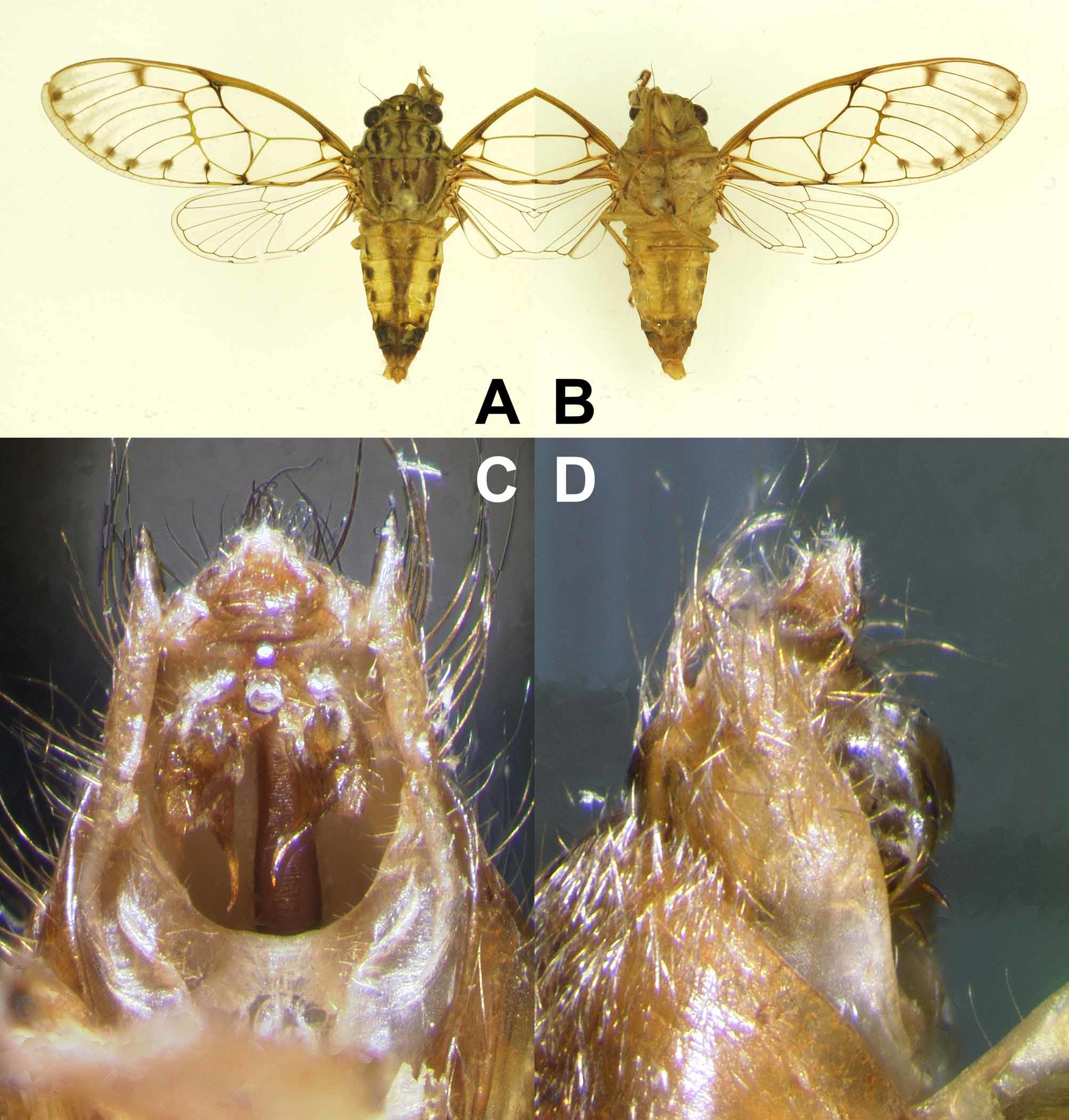
24. Psithyristria paracrassis View in CoL sp. nov.
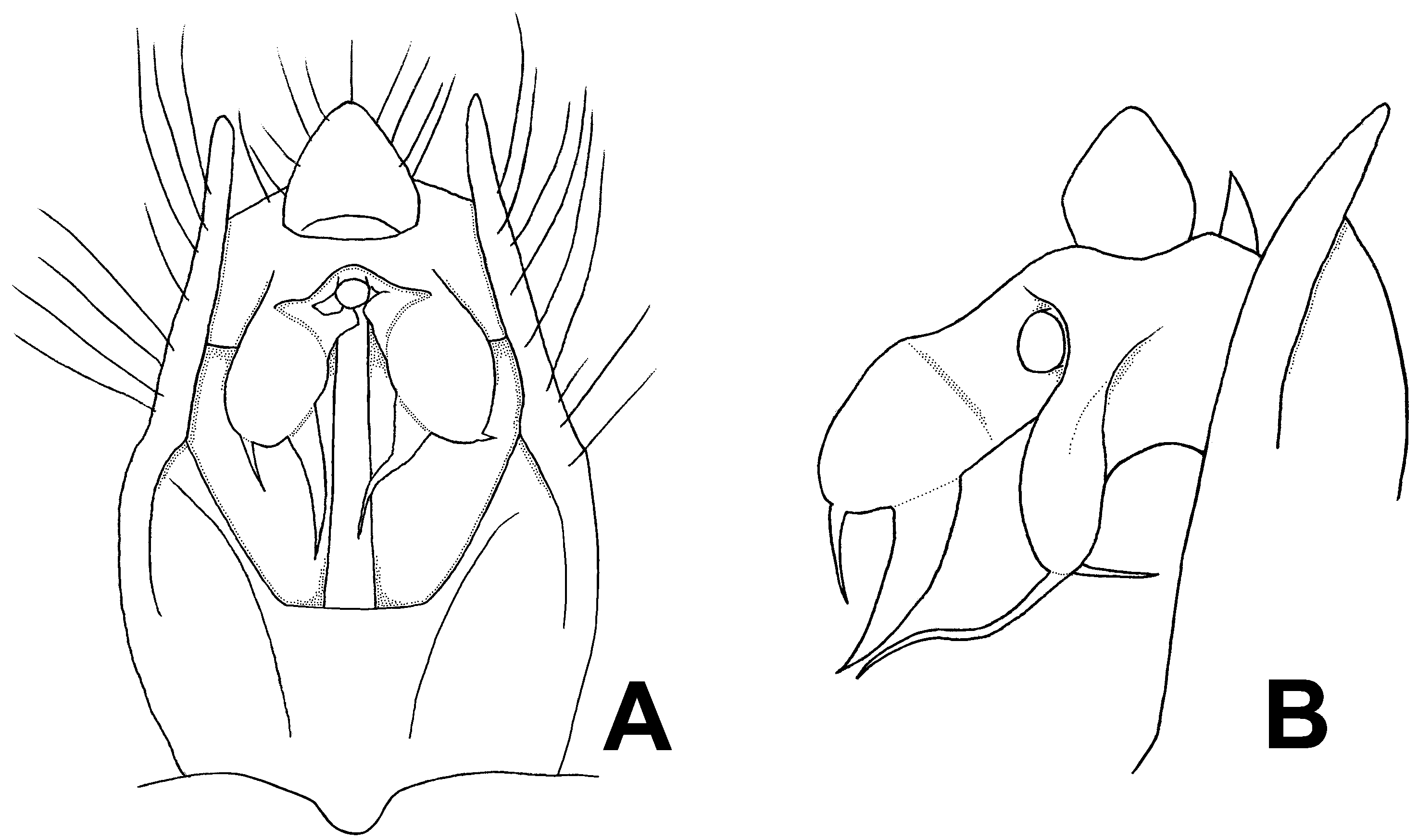
( Figs 6 View FIGURE 6 , 7 View FIGURE 7 )
Type material. Holotype: male ( Fig. 6 View FIGURE 6 ), S. Luzon, 10 km E. of Irosin, Mt. Bulusan lake, 280 m, 12°44.872’N 124°05.925’E, 12 III 2009, J.H. Lourens & K. Cerny ( IRSNB).
Etymology. The specific name refers to the morphological similarity of this species to P. crassinervis .
Measurements of types (in mm, 1 male). Length of body: 18.7; length of forewing: 21.0; width of forewing: 8.0; width of head including eyes: 5.2; width of pronotum: 6.2; wing span: 47.5.
Diagnosis. This species has the enlarged and elevated junctions of the forewing M4 and M3+4, of M3 and ulnar cell 3 and of M2 and M1+2 as in P. crassinervis and is nearly indistinguishable from crassinervis in external morphology except for its smaller body (about 18.7 mm long versus 20.2–22.1 mm long in crassinervis ) and the reduced forewing ulnar cell 2. However, this species differs from all its congeners in having an unusual shape of the uncal lobe, which has a rounded apex (without a lateral spine as in crassinervis ) and a pair of narrow, sharply pointed fin-like projections extending downward from the apex and from the paramedian margin of the uncal lobe ( Figs 6 View FIGURE 6 C, D, 7).
Description of male ( Figs 6 View FIGURE 6 , 7 View FIGURE 7 ). Head light green with following marks: median black mark enclosing ocelli, with its anterior end reaching frontoclypeal suture and posterior end reaching pronotum; pair of fuscous patches on sides of the median mark, narrowly connecting with the median mark and their lateral sides widely connecting with compound eyes; pair of fuscous patches on supra-antennal plates; pair of fuscous minute spots posterolaterally. Distance between lateral ocelli and compound eyes about twice as wide as distance between lateral ocelli. Postclypeus moderately swollen. Antennae mostly fuscous. Ventral part of head light green to greenish ochraceous. Postclypeus with fuscous fasciae along anterior six to seven transverse grooves laterally. Anteclypeus without distinct marks. Lorum without distinct marks. Gena with small fuscous spot at about middle of lateral margin. Rostrum fuscous apically; reaching posterior margin of hind coxae.
Pronotum light green. Inner area of pronotum with following black to fuscous marks: pair of central longitudinal fasciae broadened at both anterior and posterior ends; pair of fasciae between anterior parts of paramedian fissures and posterior ends of lateral fissures, disconnected in the middle; pair of thick fasciae along lateral fissures; pair of curved fasciae along lateral parts of anterior margin and lateral margins of inner area. Pronotal collar with pair of indistinct spots at lateral inner corner and pair of indistinct spots sublaterally. Anterolateral pronotal collar not dentate.
Mesonotum light green to greenish brown, covered with silvery hairs especially on lateral and posterior parts and with following marks: median longitudinal fuscous fascia broadened posteriorly to reach anterior margin of cruciform elevation; pair of inwardly curved fuscous fasciae along parapsidal sutures; pair of small roundish fuscous spots on scutal depressions; pair of broad longitudinal dark brown fasciae on lateral sides of the roundish spots all the way to anterior margin and to posterior margin of mesonotum; pair of longitudinal short fasciae between the inwardly curved fasciae and the broad longitudinal fasciae, with their anterior ends reaching anterior margin of mesonotum. Cruciform elevation greenish ochraceous without distinct marks. Thoracic sternites greenish ochraceous without marks.
Legs greenish ochraceous to ochraceous. Fore-, mid- and hind tarsi ochraceous to brown. Fore-, mid- and hind pretarsal claws brown, but apically fuscous.
Wings hyaline. Forewing M2 derived from ulnar cell 3, very close to junction of M2 and M1+2. M3 derived from ulnar cell 3, closer to junction of M4 and M3+4. M4 derived from junction of M4 and M3+4. Ulnar cell 3 about 1.7 times as large as ulnar cell 2. Medial cell about 1.3 times as large as ulnar cell 3. Medial cell slightly smaller than cubital cell. M1+2 slightly curved anteriorly. CuA1 about three times as long as CuA2. Infuscation present on bases of apical cells 2–4 and on CuA1 and CuA2. Spot appearing on each hind margin of RA2, RP and M1–4, forming a series of spots on subapical margin of forewing. Smoky distal suffusion usually present inside and along apical margins of apical cells 1–6. Basal membrane and hind wing jugum reddish ochraceous.
Operculum greenish ochraceous; semicircular. Opercula widely separated from each other.
Abdomen much longer than distance from head to cruciform elevation; covered with silvery or fuscous hairs. Tergites 2–6 greenish ochraceous with median longitudinal brown fascia, with its lateral parts usually looking darker than medial part because of fuscous hairs. Tergites 7 and 8 dark brown to fuscous. Tergites 3–6 with pair of fuscous spots laterally. Caudal margin of tergite 3 about as wide as anterior margin of mesonotum. Timbal cover brown; wider than long in median width, semicircular but inner margin nearly straight. Timbal largely exposed medially. Abdominal sternites mostly greenish ochraceous except sternites VI–VIII. Sternite VI brown caudally. Sternites VII and VIII mostly brown.
Genitalia ( Figs 6 View FIGURE 6 C, D, 7): Pygofer barrel-shaped in ventral view. Uncal lobes short, separated from each other at base, terminating in single rounded apex and with pair of narrow, slightly curved, sharply pointed finlike projections directed downward from apex and from paramedian margin of uncal lobe, in ventral view, of which the paramedian projection is long but the apical one short, about one-third as long as the paramedian one. Dorsal beak long, triangular.
Distribution. Philippines (S. Luzon).
| IRSNB |
Institut Royal des Sciences Naturelles de Belgique |
No known copyright restrictions apply. See Agosti, D., Egloff, W., 2009. Taxonomic information exchange and copyright: the Plazi approach. BMC Research Notes 2009, 2:53 for further explanation.
|
Kingdom |
|
|
Phylum |
|
|
Class |
|
|
Order |
|
|
Family |
|
|
SubFamily |
Cicadinae |
|
Tribe |
Cicadini |
|
Genus |