
Pfitzneriella rawlinsi, ♂, Grehan & C. Mielke, 2018
|
publication ID |
https://doi.org/10.11646/zootaxa.4497.1.1 |
|
publication LSID |
lsid:zoobank.org:pub:890E26F1-1B03-4F13-9299-A5E8BED8439D |
|
DOI |
https://doi.org/10.5281/zenodo.5957841 |
|
persistent identifier |
https://treatment.plazi.org/id/03A8D74F-FF94-FFF9-C9C2-FA464740F81E |
|
treatment provided by |
Plazi |
|
scientific name |
Pfitzneriella rawlinsi |
| status |
sp. nov. |
Pfitzneriella rawlinsi sp. n.
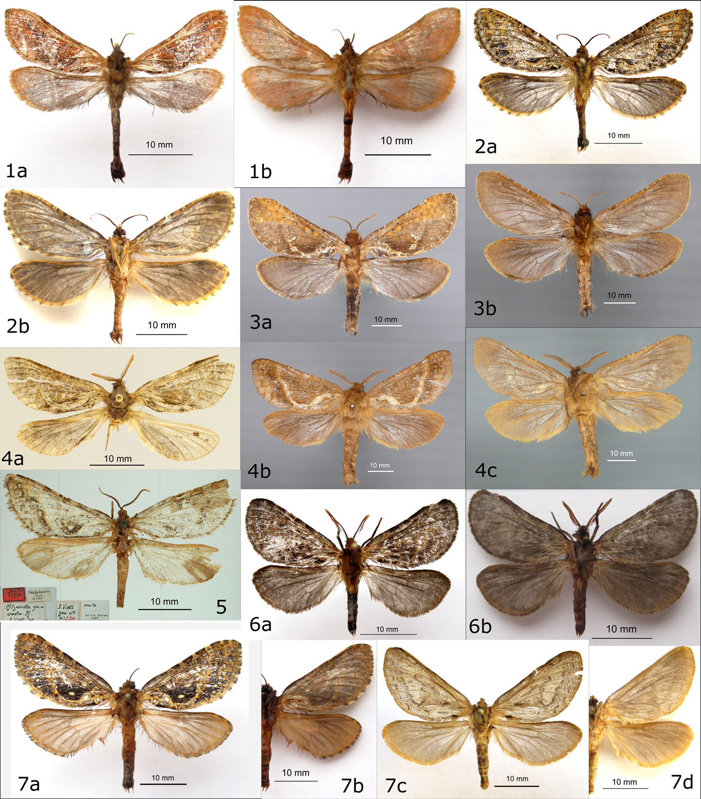
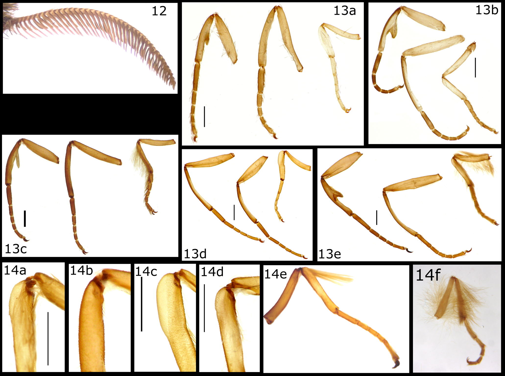
( Figs 4a, 4b, 4c View FIGURES 1–7 , 12 View FIGURES 12–14. 12 , 9c View FIGURES 8–9 , 10d View FIGURE 10 , 11d View FIGURE 11 , 13c, 14b View FIGURES 12–14. 12 , 15b, 16d View FIGURES 15–16 , 20 View FIGURES 17–25 )
Diagnosis. Pale yellowish brown FW and white cubital band most similar to P. olafi sp. n. and possibly P. remota (FW of lectotype in poor condition), and distinct from mottled yellowish brown markings on grayish brown background in P. antonkozlovi sp. n., P. antonkozlovi sp. n., and P. yuliyakovalevae sp. n. Basal white semicircular band of FW more continuous than in P. olafi sp. n. Within the cubital region of the posterior FW the white band encloses a semicircular brown patch that is bounded by CuA 2 in both P. rawlinsi sp. n. and P. remota but the patch in P. rawlinsi sp. n. lacks the interior white spot adjacent to CuA2 that is present in P. remota . Apex of P. yuliyakovalevae sp. n. also distally expanded, but forming a rectangular shape located further from the median. Presence of a basal valva spur also contrasts with other Pfitzneriella species where the spur is absent (also the condition in Dugdaleiella gen. n. and Kozloviella gen. n.). Anterior margin of saccus with deep concave depression that is absent or indistinct in other Pfitzneriella species. The unfused posterioventral apex of pseudotegumen forms a small, rounded, and laterally extended flange in contrast to pointed apex of P. antonkozlovi sp. n., P. olafi sp. n., and P. titarenkoi sp. n.
Description. Male ( Figs 4a, 4b, 4c View FIGURES 1–7 ): Wingspan 42 mm; FW length: 20 mm, width: 7 m; ratio 1.5: 1; HW length: 18 mm, width: 7 mm, ratio 2.5: 1.
Head. Frons and vertex covered with yellowish brown scales. Antenna bipectinate ( Fig. 12 View FIGURES 12–14. 12 ), brown, 43 flagellomeres, lateral arms about twice length of internal arms; scape covered with fine scales; eyes medium sized. Clypeus exposed, projecting anteriorly as a transverse ridge. Labial palpus three segmented, basal segment ovoid, first palpomere oval as long as the base, distal palpomere three times longer than the former ( Fig. 9c View FIGURES 8–9 ).
Thorax. Covered with yellowish brown scales, scutum III pale yellowish brown, free of scales other than posterior and medial regions, obscured from direct view by overlapping mesothoracic scales. Ventral thorax pale yellowish brown. Legs ( Fig. 13c View FIGURES 12–14. 12 ) coloured as the thorax, tibial and tarsal scales not obscuring individual tarsal segments; epiphysis present; metatibial gland absent ( Fig. 14b View FIGURES 12–14. 12 ); proleg missing tarsal segments. FW costal margin slightly convex; outer margin weakly convex merging with anal margin at CuA2. Venation hepialine ( sensu Dumbleton 1966), base of FW anal vein strongly curved anteriorly, distally curved posteriorly ( Fig. 10d View FIGURE 10 ). FW dorsal ground colour dark brown interspersed with pale yellowish brown and brown; posterior discal cell and anterior cubital cell reddish brown, posterior margin curved, forming semicircle within cubital cell, edged with thin line of dark brown and then pale yellowish white band between CuA2 and widening basally to semicircle; curved transverse band of yellowish white bordered with dark brown extending between CuA2 and M1 and M2 stalk ( Fig. 11d View FIGURE 11 ); dark brown spot at anal margin anterior to A, and posterior to outer CuA2 beyond cross vein with CuA1; marginal fringe scales missing. FW ventral ground colour yellowish to greyish brown; costal pocket present with band of narrow pale yellowish brown scales. HW dorsal greyish brown with pale orange-brown shading basally and about 2/3 costal region. HW ventral yellowish to greyish brown except for orange-brown scales between Sc and costal margin.
Abdomen. Coloured as the thorax. Abdominal tergites and sternites sclerotized. Tergosternal bar elongate triangle, anterior and posterior edges thickened, lateral tergal brace long, slightly ventrally oriented, dorsal tergal brace short, central region indistinct, tergal, posterior margin slightly convex, tergal knob absent ( Fig. 15b View FIGURES 15–16 ). Tergum VII and VIII rectangular, wider than long, posterior margin with V-shaped medial indentation; sternite VIII rectangular, wider than long, posterior margin broadly concave from latero-posterior corners ( Fig. 16d View FIGURES 15–16 ).
Male genitalia ( Fig. 20 View FIGURES 17–25 ). Tegumen fused to pseudotegumen, slightly distinct, region of fusion extending anteriorly (inwardly) into abdomen beyond lateral articulation with saccus. Pseudotegumen, strongly sclerotized, medially unfused dorsally and ventrally, dorsal region expanded into a posteriorly curved flange, lightly sclerotized, widest dorsally, narrowing laterally; ventral apical margin forming a laterally curved and serrated edge with shallow, sickle shaped teeth, largest tooth at the ventro-most apical point; and a small inner, medially projecting rounded flange. Saccus wider than long, posterior edge V-shaped, apodemal margin forming ventrally projected wall that is medially enlarged into pair of acutely pointed teeth, points oriented medially, separated by tear-drop shaped gap, anterior margin medially concave with rounded convex lateral corners. Tergal lobes absent. Valva setose, distally lobate, central inner surface with longitudinal ridges either side of triangular central concavity from near apex to base; anterobasal corner extended as ventrally directed tooth. Fultura superior mostly membranous, slightly sclerotized laterally; fultura inferior wider than long, sub-rectangular, anterior margin a shallow ‘V’ extending laterally to acutely angled corners, posterior margin shallow U-shape with rounded lateral corners, lateral margins slightly concave. Phallus membranous, without cornutus.
Female unknown.
Distribution. Southeast Ecuador. Known only from the type locality on east facing slope of the Andes along the highway between Gualaceo and General Leonidas Plaza Gutiérrez ( Figs 27a, 28 View FIGURES 27–29 ).
Ethology. Attracted to UV-light at collecting sheet.
Host plant. Larval habits and host plants are unknown.
Etymology. Named in honor of John Rawlins, Curator at the Carnegie Museum of Natural History, Pittsburgh, USA, for his longstanding logistical and moral support for ghost moth research.
Type material. Holotype male (with the following labels separated by forward slashes): ECUADOR, Morona[- Santiago, [ Limón] Indanza, 2800 m, 24.xii.1992, V.O. Becker col. /Coll. Becker, 10350/ Holotypus, Pfitzneriella rawlinsi ♂, Grehan & C. Mielke det. 2017/ Dissection JRG 200/ ( CMNH). Fig. 4a View FIGURES 1–7 .
Paratype: 1 ♂ ( Figs 4b, 4c View FIGURES 1–7 ), same data as HT ( CGCM).


| CMNH |
The Cleveland Museum of Natural History |
No known copyright restrictions apply. See Agosti, D., Egloff, W., 2009. Taxonomic information exchange and copyright: the Plazi approach. BMC Research Notes 2009, 2:53 for further explanation.
|
Kingdom |
|
|
Phylum |
|
|
Class |
|
|
Order |
|
|
Family |
|
|
Genus |