
Aschemonella tani Gooday & Holzmann, 2024
|
publication ID |
https://doi.org/10.11646/zootaxa.5419.2.1 |
|
publication LSID |
lsid:zoobank.org:pub:88353CBA-6C4D-40E3-8475-B1FCA2C48637 |
|
DOI |
https://doi.org/10.5281/zenodo.11262805 |
|
persistent identifier |
https://treatment.plazi.org/id/EE7E2E5C-6D23-4DF8-8A88-C5B4D6BEEBA2 |
|
taxon LSID |
lsid:zoobank.org:act:EE7E2E5C-6D23-4DF8-8A88-C5B4D6BEEBA2 |
|
treatment provided by |
Plazi |
|
scientific name |
Aschemonella tani Gooday & Holzmann |
| status |
sp. nov. |
Aschemonella tani Gooday & Holzmann sp. nov.
Figs 3 View FIGURE 3 , 4 View FIGURE 4
Diagnosis. Species of Aschemonella with attached test forming tubular branching structures that grow free from solid substrate. Branches are relatively wide compared to their length and in places display vague segmentation. Upstanding parts extend into basal system of flat, branching tubes that encrust parts of the substrate surface. Stercomare forms irregular, sometimes discontinuous masses, but more elongated, continuous masses run along branches. Granellare forms pale yellowish, branching strands, typically 21–36 µm wide.
Etymology. The new species is named for Dr Koh Siang Tan, Head of the Marine Biology and Ecology Laboratory at the Tropical Marine Science Institute, Singapore, who has led research by Singapore scientists in the Clarion-Clipperton Zone.
Type specimen and locality. The holotype ( Lee Kong Chian Natural History Museum, Singapore, reg. no. ZR C. FOR.0002, preserved in 10% formalin) was collected in box core BC036 (specimen RC1555 ); OMS license area, 12° 26' 45.5"N, 117° 49' 41.1"W; 4196 m water depth. A fragment was used for genetics (sequenced isolate: 21430). There were no other specimens GoogleMaps .
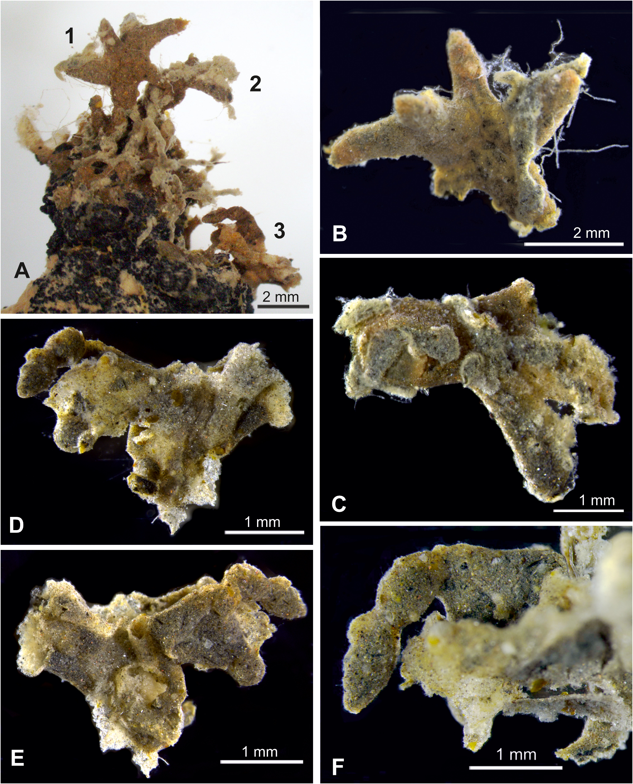
Description. Shipboard photographs. The main part of the test (labelled ‘1’ in Fig. 3A View FIGURE 3 ) stands erect at the summit of a roughly conical nodule. Its overall height is around 3.9 mm. There is a short stalk, ~ 1.45 mm long and 0.9–1.1 mm wide, that gives rise to three branches, also short and relatively wide (length 1.5–1.7 mm; width 0.60– 1.0 mm). At its top, the test bifurcates into two further branches, the longer one 1.7 mm in length and 0.63–0.77 mm wide. The structure is rusty brown in overall colour. The base of the stalk continues as an encrusting structure that spreads across part of the nodule summit. The base also gives rise to two short branches that project from the nodule surface near the summit; one is ~ 1.20 mm long and the other at least of similar length. The summit region of the nodule hosts at least two other projecting structures (labelled ‘2’ and ‘3’ in Fig. 3A View FIGURE 3 ) with a weakly segmented appearance.
Preserved fragments. Parts 1, 2 and 3 in Fig. 3A View FIGURE 3 are all recognisable among the preserved fragments. They are identical in general appearance and wall structure, suggesting that they are parts of the same organism. The wall is pale, brownish yellow, and about 40 µm thick. It is composed largely of small mineral grains (less than about 25 µm in size), mainly resembling quartz but with a scattering of blackish and reddish grains, and with sponge spicule fragments making a subordinate but important contribution. A few tests of agglutinated foraminifera are also incorporated into the wall.
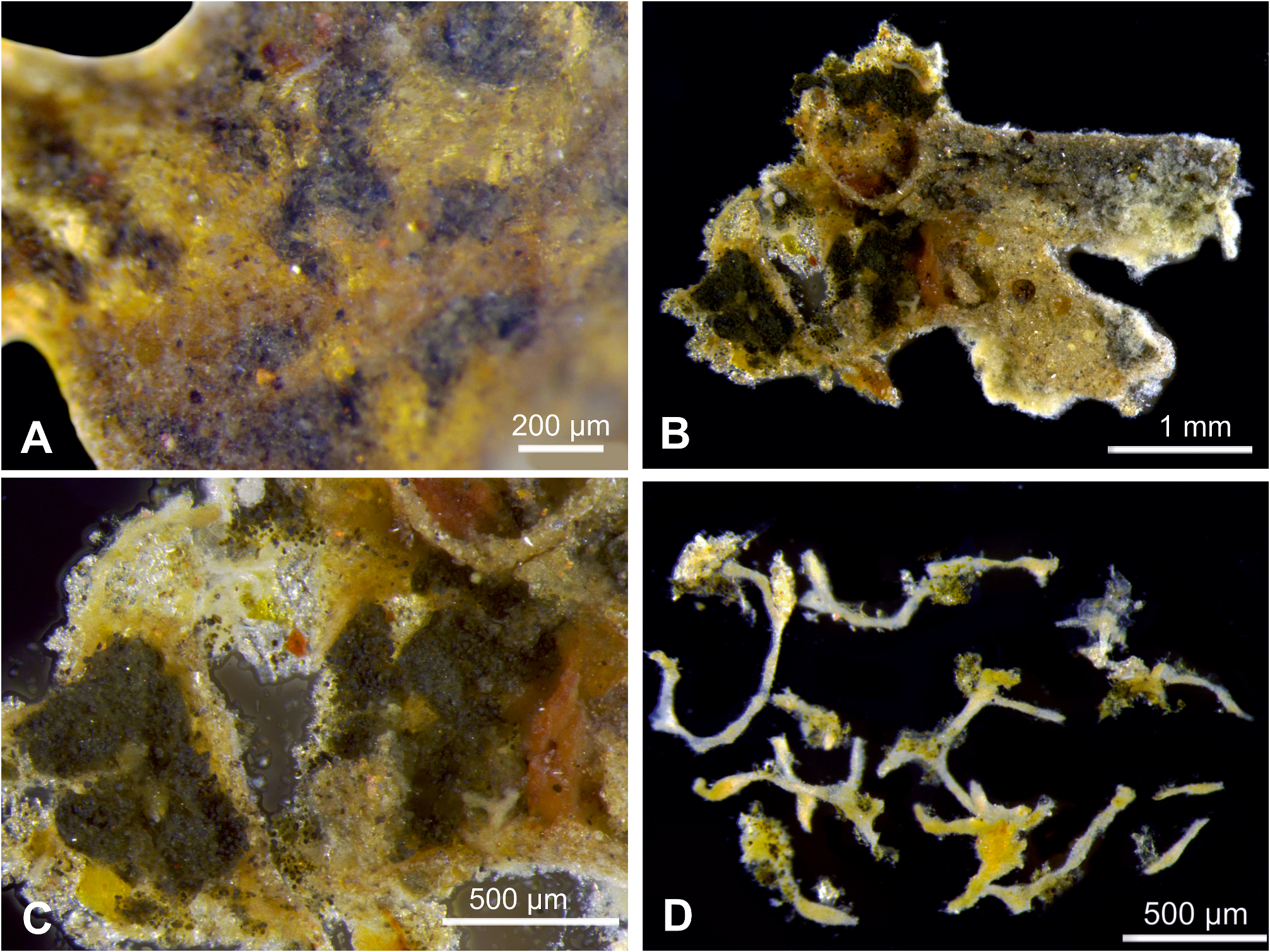
The stercomare can be seen dimly through the test wall when illuminated with transmitted light. In the central parts of the fragments ( Fig. 4A View FIGURE 4 ), it forms irregularly shaped, apparently disconnected masses up to ~800 µm in maximum extent but usually less. More elongated, continuous masses occupy the branches of the part 1 fragment ( Fig. 3B View FIGURE 3 ). Part 3 appears to have more strongly developed stercomare since the interior is filled with dark material when viewed through the test wall. The granellare forms pale yellowish, branching strands, typically 21–36 µm wide but swelling in places to 50–65 µm ( Fig. 4D View FIGURE 4 ).
The test, including the lower encrusting part, is to some extent obscured by another agglutinated structure. This is basically tubular, branches and is to some extent reticulated. The width is variable ( 0.40–0.80 mm) and there are several inflated segments. It is most likely another monothalamid species. In preserved fragments as well as in shipboard photographs ( Fig. 3A–D View FIGURE 3 ), the branches have a lighter greyish colour compared to the Aschemonella that they partly overgrow.
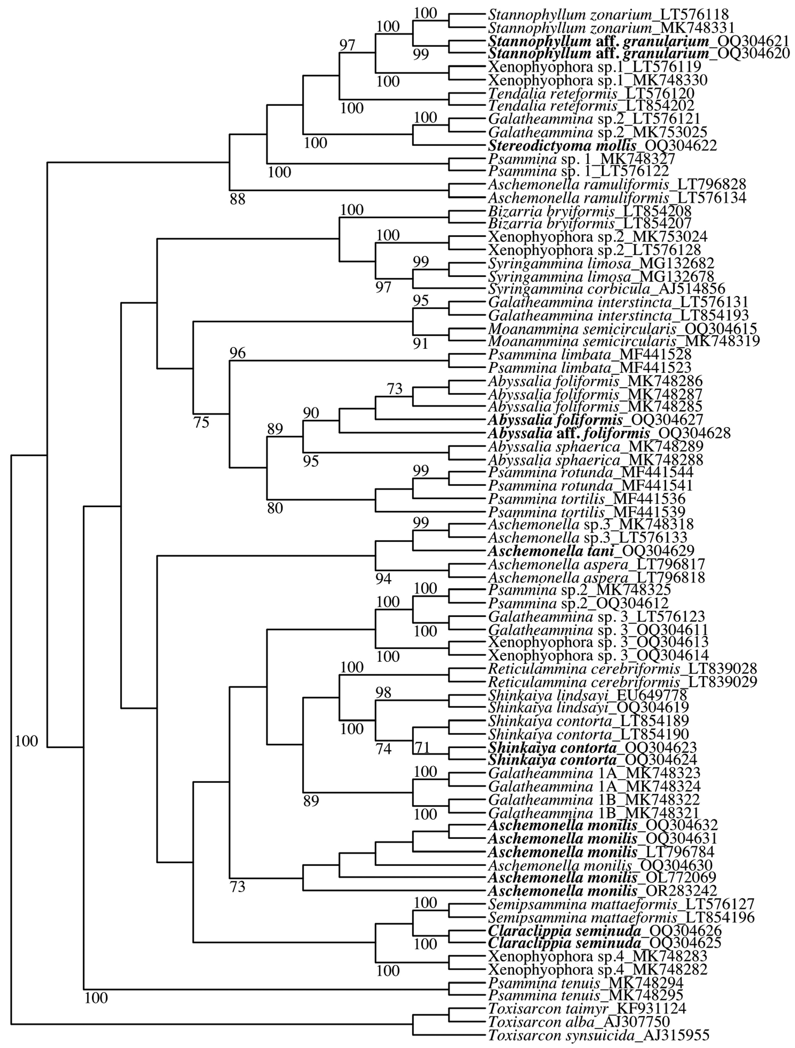
Molecular characterisation. Aschemonella tani branches at the base of Aschemonella sp. 3 with A. aspera forming a sister group to these two species ( Fig. 1 View FIGURE 1 ). The grouping is not supported by the BV. The sequenced fragment of 18S gene of A. tani contains 1028 nucleotides and the GC content is 34 %.
Remarks. The branching, basically tubular test, and the fairly large, irregularly shaped stercomare masses of Aschemonella tani , distinguish it from A. monilis , in which the test is clearly segmented and the stercomare masses resemble pellets. It is much more similar to Aschemonella aspera, another species from the CCZ that also has an approximately tubular test growing upwards from the substrate to which it is attached. However, the test is more strongly branched in the new species and has a brownish yellow wall composed of small mineral grains with a smooth outer surface, unlike that of A. aspera, in which the wall is dark grey and much more coarsely agglutinated with a rough surface composed of micronodules and mineral grains. The most similar described species is A. ramuliformis . This also forms branching tubes, but they are more elongate and regular than those of the new species (Brady, 1884; Gooday et al., 2011). There are no records of A. ramuliformis being attached to a hard substrate.
These three species ( A. aspera, A. monilis , A. ramuliformis ) are genetically distinct from A. tani ( Fig. 1 View FIGURE 1 ). Based on molecular data, the new species is most closely related to Aschemonella sp. 3 of Gooday et al. (2017a), also from the CCZ. The test of this undescribed species forms an irregular system of reticulated tubes that are sometimes vaguely segmented and either attached to a nodule surface or grow free. The lower encrusting part of our A. tani specimen, which comprised tubular structures spreading across the nodule surface, is rather similar to the attached parts of Aschemonella sp. 3 . However, the upper, free-standing part of the test does not form the same kind of reticulated structure.
| FOR |
Forssa Museum of Natural History |
No known copyright restrictions apply. See Agosti, D., Egloff, W., 2009. Taxonomic information exchange and copyright: the Plazi approach. BMC Research Notes 2009, 2:53 for further explanation.
|
Kingdom |
|
|
Phylum |
|
|
Class |
|
|
Family |
|
|
Genus |