
Aschemonella monilis Gooday & Holzmann, 2017
|
publication ID |
https://doi.org/10.11646/zootaxa.5419.2.1 |
|
publication LSID |
lsid:zoobank.org:pub:88353CBA-6C4D-40E3-8475-B1FCA2C48637 |
|
DOI |
https://doi.org/10.5281/zenodo.11247628 |
|
persistent identifier |
https://treatment.plazi.org/id/03A987A1-7B45-AF65-66C4-4212FB05DEA9 |
|
treatment provided by |
Plazi |
|
scientific name |
Aschemonella monilis Gooday & Holzmann, 2017 |
| status |
|
Aschemonella monilis Gooday & Holzmann, 2017 View in CoL
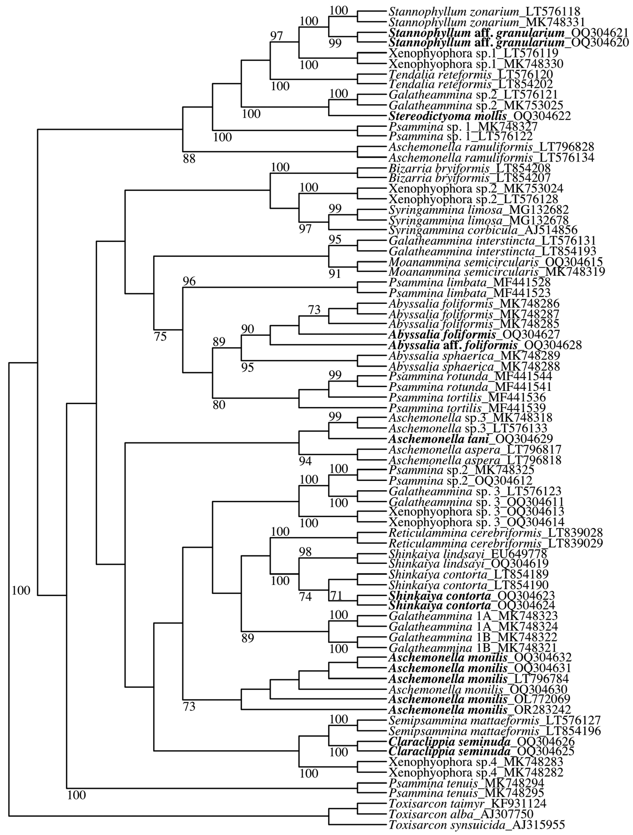
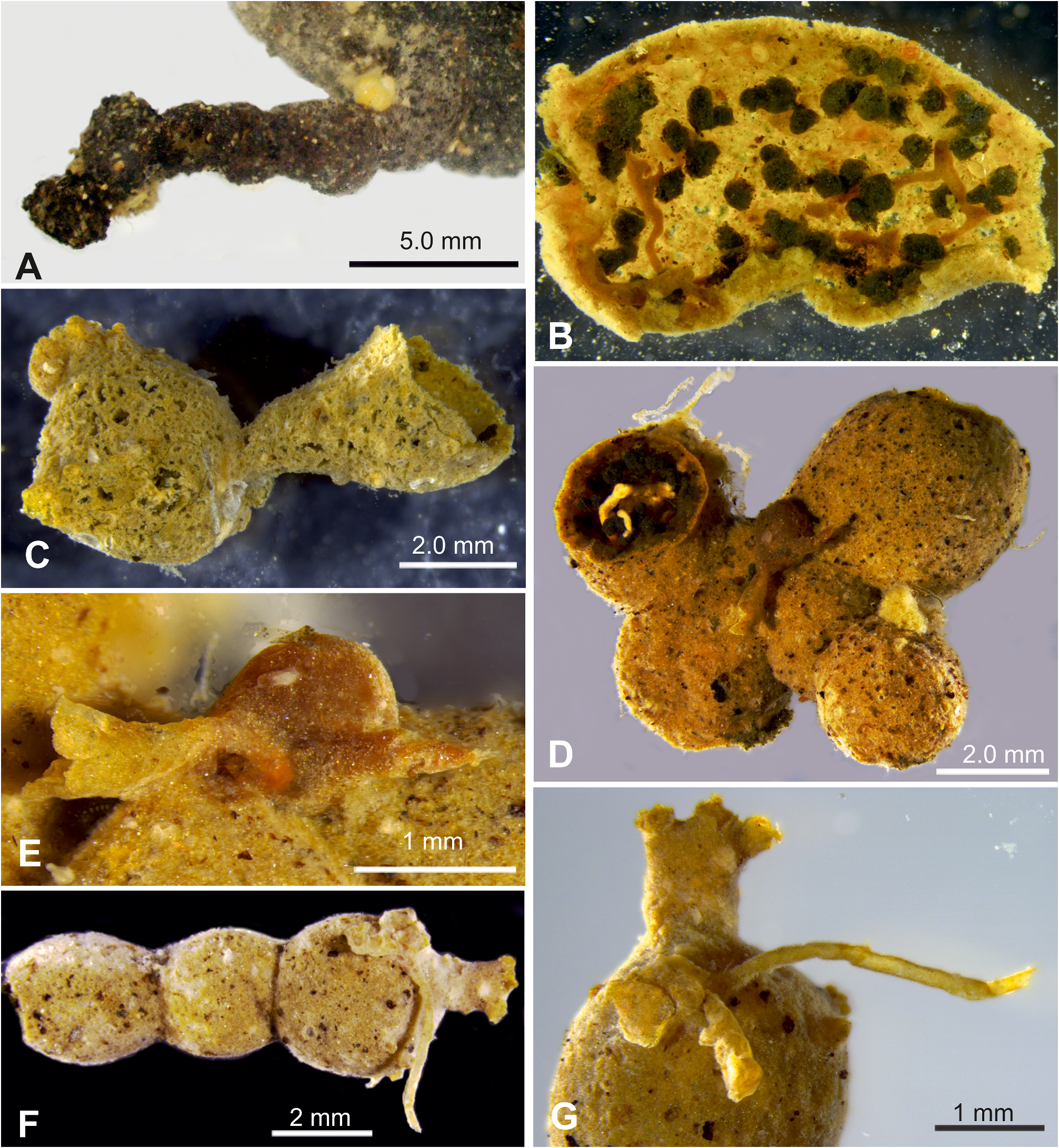
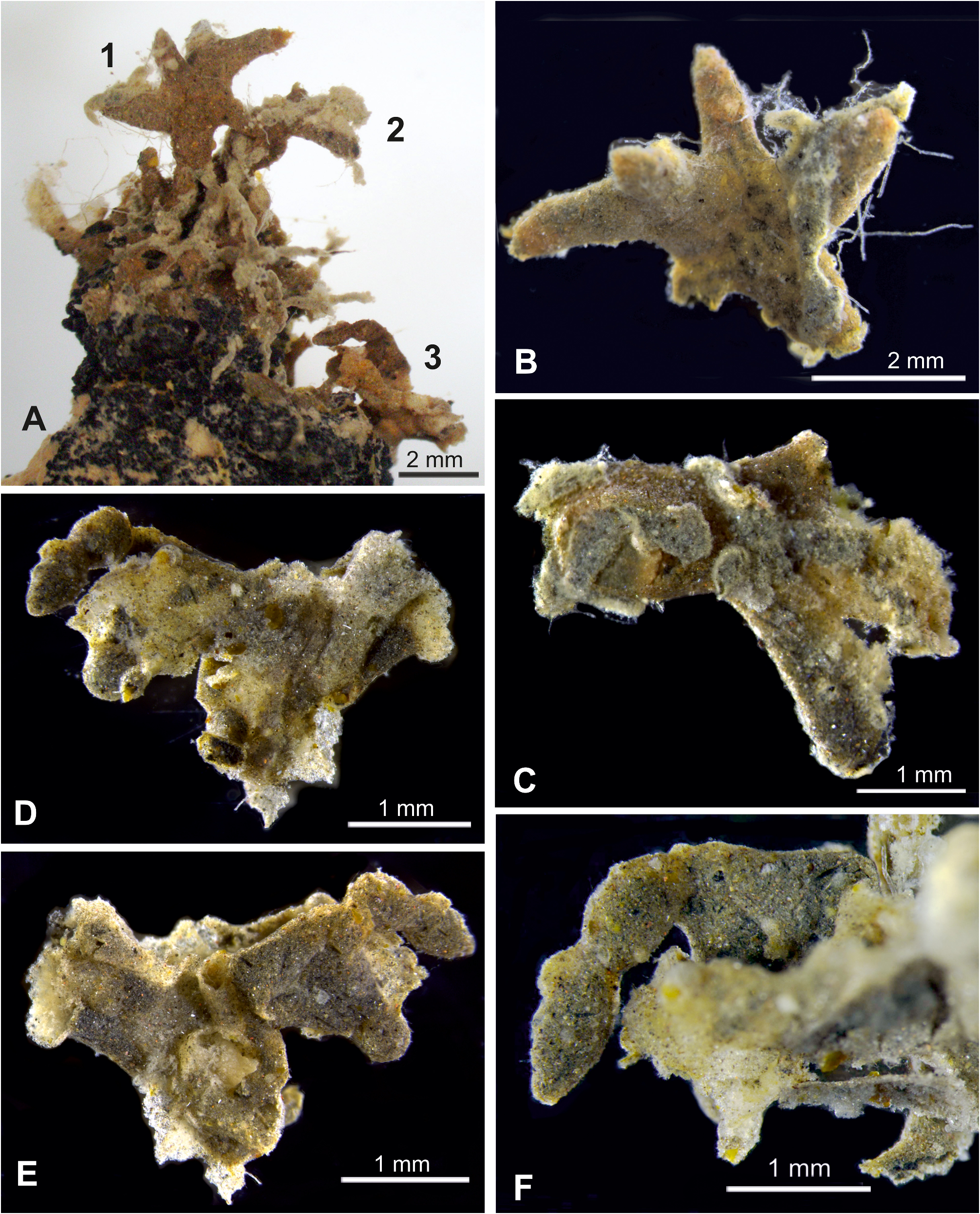
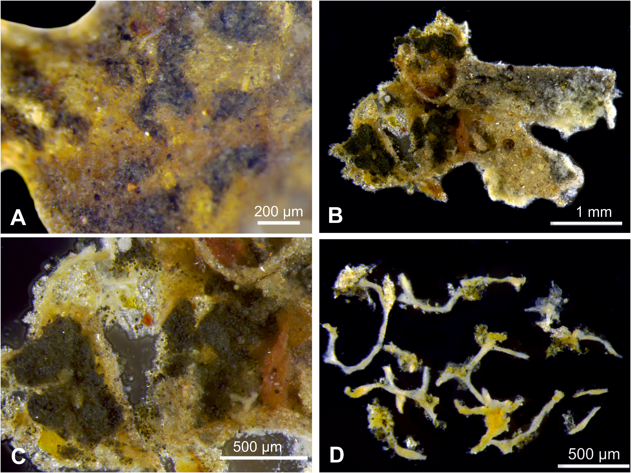
Fig. 2 View FIGURE 2 ; Supplementary Figs S1 View FIGURE 1
Aschemonella monila Gooday and Holzmann View in CoL in Gooday et al., 2017b, Figs 2A–E View FIGURE 2 , 3A–F View FIGURE 3 ; Supplementary Fig. S1 View FIGURE 1 , Figs 1–8 View FIGURE 1 View FIGURE 2 View FIGURE 3 View FIGURE 4 View FIGURE 5 View FIGURE 6 View FIGURE 7 View FIGURE 8 . Aschemonella monilis Gooday and Holzmann. Gooday et al., 2020a, p. 4 View in CoL -6, figs 2-3.
Material examined. BC001 RC0056: morphology and genetics (isolates 21438, 21439). BC010 RC049B: morphology and genetics (isolate 21431). BC025 RC1042: morphology only. BC026 RC1056: morphology and genetics (isolate 21108). BC031 RC1345: morphology only. BC040 RC1689: morphology and genetics (isolate 21435). BC040 RC1731: morphology only. BC045 RC1900.1: morphology and genetics (isolate 21444). Sequenced isolates: 21108, 21431, 21435, 21444 ( Table 2 View TABLE 2 ).
Description and remarks. Aschemonella monilis is by far the most abundant xenophyophore species in our collection. It is represented by around 34 complete and fragmentary specimens ( Table 2 View TABLE 2 ), although not all of these were examined in detail. They conform closely to the original description ( Gooday et al., 2017b). Nineteen specimens were found attached to nodules, of which three encrusted the host nodule for their entire length and the others extended upwards from the surface to a greater or lesser extent. The remaining 15 were unattached, at least when found. The majority of specimens are dark grey with either a smooth, relatively fine-grained wall or a rougher, more coarsely grained wall. Some of the latter type resemble the ‘delicate’ form distinguished by Gooday et al. (2017b) (Supplementary Fig. S1A, B View FIGURE 1 ). Other specimens are paler, dull orange to yellowish in overall colour but speckled with a variable density of dark grains ( Fig. 2C, D, F View FIGURE 2 ; Supplementary Fig. S1D View FIGURE 1 ). These lighter coloured tests have generally smoother surfaces than the darker ones. Most sequenced specimens were of the paler, smooth-walled type, but they grouped together with one having a darker, rougher wall ( Fig. 2A View FIGURE 2 ). This is consistent with the earlier genetic data (Gooday et al., 2017), and indicates that they represent the same species.
Apertural structures were observed in two specimens from BC045 ( RC1900.1 & 2). One has a smooth, blisterlike dome, measuring 1.42 × 1.00 mm, located near the junction between several chambers ( Fig. 2D, E View FIGURE 2 ). It gives rise to two tubular extensions, one 0.83 mm long and of fairly even width (~ 0.17 mm), the other 1.06 mm long and of variable width ( 0.26 to 0.45 mm). The other structure is located on the final chamber and comprises a swelling ~ 1.20 mm long that is associated with two tubes ( Fig. 2F, G View FIGURE 2 ). The longer tube is 3.5 mm in length and again of fairly even width ( 0.21 to 0.26 mm), the shorter is ~ 0.85 mm in length and 0.36 to 0.53 mm wide. The longer tube is relatively smooth, but the shorter tube has a lumpy, very uneven surface and the associated swelling has a similarly irregular shape. Several short, pustule-like tubes ~ 0.18 mm long and of similar width, are present elsewhere on the final chamber of this specimen. Similar apertural features (swellings, long tubes and clusters of short, pustule-like tubes) were described by Gooday et al. (2017b, Figs 3 View FIGURE 3 , 4 View FIGURE 4 therein).
Aschemonella monilis is widely distributed across an area spanning some 3,800 km, being common in samples from the UK-1 license area (adjacent to the OMS area), as well as present in the Russian area in the central CCZ and APEI 4, a protected area in the western CCZ ( Gooday et al., 2017a,b, 2020a). In the latter case, the three recorded specimens were morphologically atypical but genetically identical to those from the eastern CCZ. Aschemonella monilis is also the dominant faunal component in seafloor photographs from the southwestern part of APEI 6 (now APEI 3), located in the northeastern CCZ ( Gooday et al., 2017b; Simon-Lledó et al., 2019).
No known copyright restrictions apply. See Agosti, D., Egloff, W., 2009. Taxonomic information exchange and copyright: the Plazi approach. BMC Research Notes 2009, 2:53 for further explanation.
|
Kingdom |
|
|
Phylum |
|
|
Class |
|
|
Family |
Aschemonella monilis Gooday & Holzmann, 2017
| Holzmann, Maria, Barrenechea-Angeles, Inés, Lim, Swee-Cheng & Pawlowski, Jan 2024 |
Aschemonella monilis
| Gooday and Holzmann. Gooday 2020: 4 |
Aschemonella monila
| Gooday and Holzmann 1879 |