
Claraclippia seminuda Gooday & Holzmann
|
publication ID |
https://doi.org/10.11646/zootaxa.5419.2.1 |
|
publication LSID |
lsid:zoobank.org:pub:88353CBA-6C4D-40E3-8475-B1FCA2C48637 |
|
DOI |
https://doi.org/10.5281/zenodo.11247634 |
|
persistent identifier |
https://treatment.plazi.org/id/03A987A1-7B4C-AF71-66C4-4240FC31DF9D |
|
treatment provided by |
Plazi |
|
scientific name |
Claraclippia seminuda Gooday & Holzmann |
| status |
|
Claraclippia seminuda Gooday & Holzmann View in CoL gen. & sp. nov.
Figs 8 View FIGURE 8 , 9 View FIGURE 9
Diagnosis. As for genus.
Etymology. The name reflects the appearance of the type specimen, which, when freshly collected, was covered with a thin layer of fine sediment that was not retained when the specimen was preserved.
Type specimen and locality. The holotype ( Lee Kong Chian Natural History Museum, Singapore, reg. no. ZR C. FOR.0001, preserved in 10% formalin) was collected in box core BC005 (specimen RC0202 ); OMS license area, 14° 06' 38.2"N, 117° 13' 54.2"W; 4200 m water depth. A fragment was used for genetics (sequenced isolates: 21436, 21437). There were no other specimens GoogleMaps .
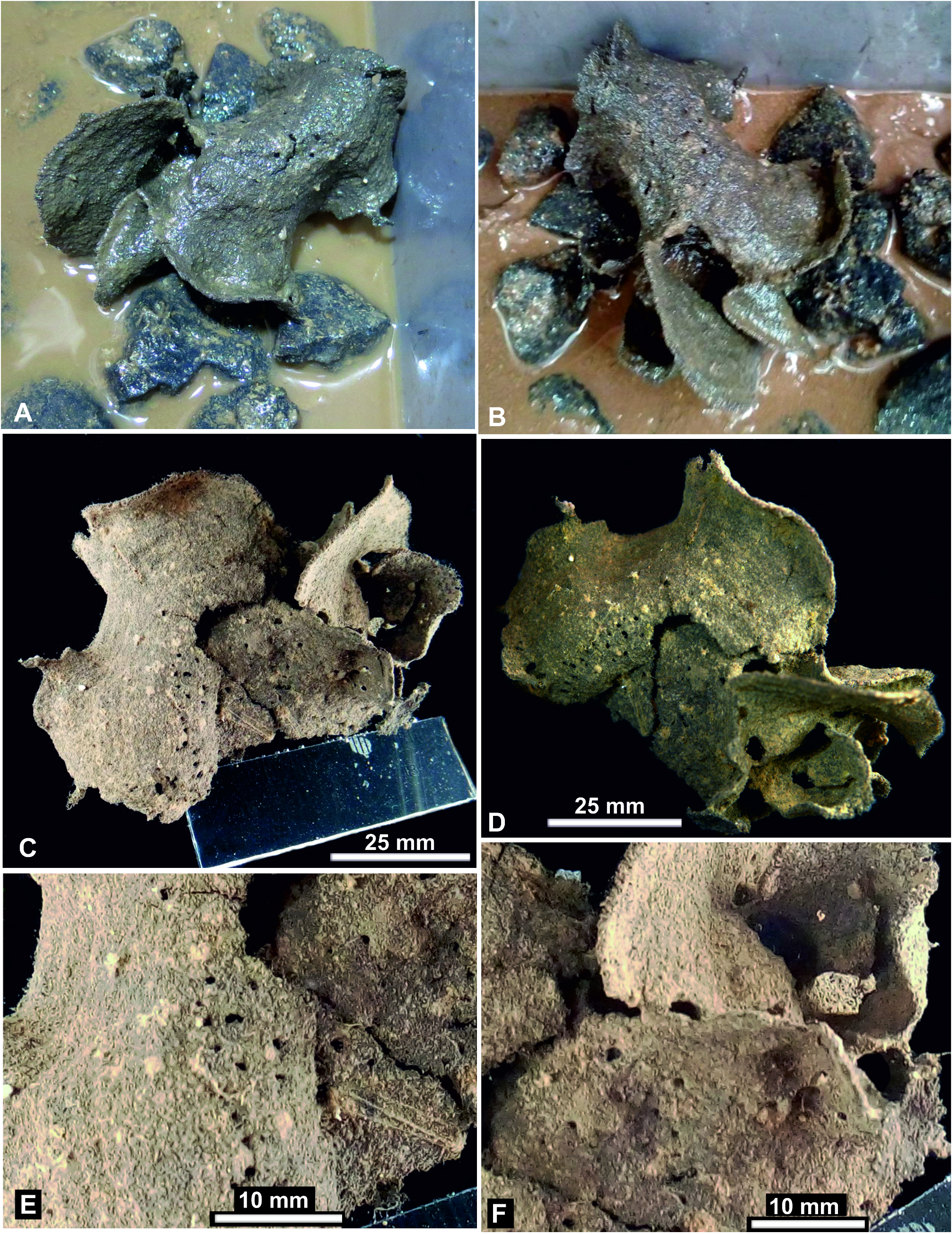
Description. Shipboard photographs. Photographs of the specimen as first seen on the box core surface shows it spread across several nodules with part of the base attached to at least one nodule ( Fig. 8A, B View FIGURE 8 ). The photograph gives the impression that the body was somewhat flexible and had collapsed from a more upright position when the overlying water was drained from the box core. The test formed a complex but basically, plate-like structure, brownish grey in colour, that included several lobes, the main part being strongly curved.
When photographed in the shipboard laboratory after removal from the box core, the specimen appeared somewhat damaged with several obvious breaks, an indication of its fragility ( Fig. 8C–F View FIGURE 8 ). It measured about 8 cm in overall maximum dimension. The largest part, which had broken into two main pieces, formed a folded, undulating plate, 6.4 cm in maximum dimension. The outer margin, which seemed largely intact, was curved with a broad concave section and two short tapering outgrowths, the larger being about 5 mm long and 3 mm wide at the base. Very vague, concentric zonation patterns were visible under low-angle lighting ( Fig. 8C View FIGURE 8 ). These had different orientations, suggesting that there were several directions of growth. The plate was also perforated by a number of small open spaces, 0.5–1.2 mm in maximum dimension, some of them arranged in a rough arc ( Fig. 8C–F View FIGURE 8 ). The other main body part visible in these photographs was more complicated. Although basically plate-like, it curved around to form what appears to be a funnel-like structure and was perforated by several relatively large open spaces, 1.4 –4.9 mm maximum dimension ( Fig. 8F View FIGURE 8 ).
The photographs ( Fig. 8 View FIGURE 8 ) show that the test surface was originally covered with a veneer of fine-grained material. In some patches this appeared to be absent, exposing the tightly packed strands of the stercomare system. Under low-angled lighting, the strands created a hair-like surface pattern, even where the fine-grained veneer was present. The margin of the structure was often fairly even, but in places, possibly where damage has occurred, it has a frayed appearance with a fringe of exposed stercomare branches.
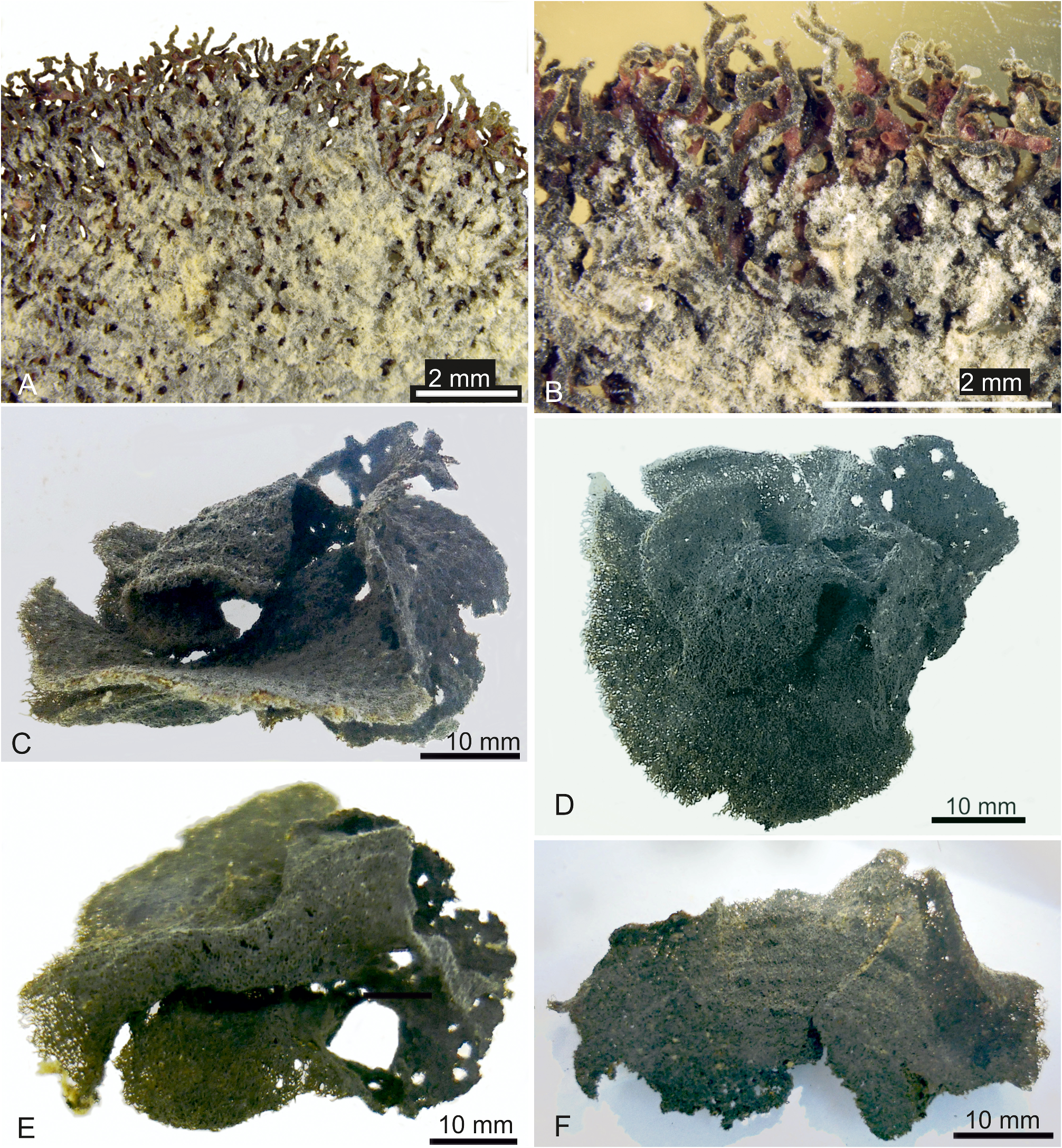
Preserved fragments. There are two main fragments that probably correspond to the two parts recognisable in shipboard photographs. Both are dark grey and somewhat flexible but very delicate. The larger fragment ( Fig. 9C–E View FIGURE 9 ) measures between 4.7 to 5.5 cm maximum dimension and 2.6 to 4.1 cm minimum dimension, depending on the viewing angle. The structure has no obvious regularity or centre of organisation. It forms a complex and irregular three-dimensional system comprising plates, in places interrupted by open spaces or merging into bars that define open spaces ( Fig. 9C, E View FIGURE 9 ). These spaces are of highly variable size, ranging from ~ 0.5 mm or less to 6.6 mm in the case of the largest one that is easily visible. The second fragment ( Fig. 9F View FIGURE 9 ) is a much simpler undulating plate, 4.8 cm long, a maximum of 2.8 cm wide, and around 1.0 to 1.2 mm thick. The plate is interrupted by a few small open spaces (up to 1.1 mm maximum dimension), most of which are concentrated in one area.
The preserved fragments are composed largely of naked stercomare, mainly in the form of closely packed strands that are most distinct around the edges where they project slightly to form a dense fringe ( Fig. 9A, B View FIGURE 9 ). Here, they are 75 to 150 µm, typically 90–120 µm, wide and branch but rarely anastomose.At least some of these sections probably represent the original margin of the test, although others appear damaged. Away from the edge, the strands lose their identity to varying extents. In places, they are still quite distinct. Elsewhere they become more tightly meshed and reticulated, with only chinks of space between them, and up to 200 µm wide. Sometimes they merge to form a more continuous sheet perforated by small open spaces.
When examined in Geneva after transport from Singapore the fragments retained some patches of the pale, fine-grained surface veneer that was seen in the shipboard photographs ( Fig. 9A, B View FIGURE 9 ). Usually, this was found filling spaces between the stercomare branches. By the time they reached Southampton, no obvious trace of the fine-grained material remained ( Fig. 9C–F View FIGURE 9 ). However, careful examination revealed a scattering of tiny mineral particles across the surface of the stercomare. Some of these grains stand out because they are white or because they glint in the light.
The granellare strands are clearly visible only around parts of the margin, where they are closely associated with the stercomare branches ( Fig. 9B View FIGURE 9 ). They are distinctly reddish and stand out in contrast to the dark grey stercomare. The organic tube that contains the cytoplasm is very thin. The strands are of irregular width, generally between ~100 and 200 µm but occasionally somewhat wider. Some peripheral strands have slightly expanded ends. Away from the margin, the reddish strands can sometimes be glimpsed in gaps within the grey stercomare system.
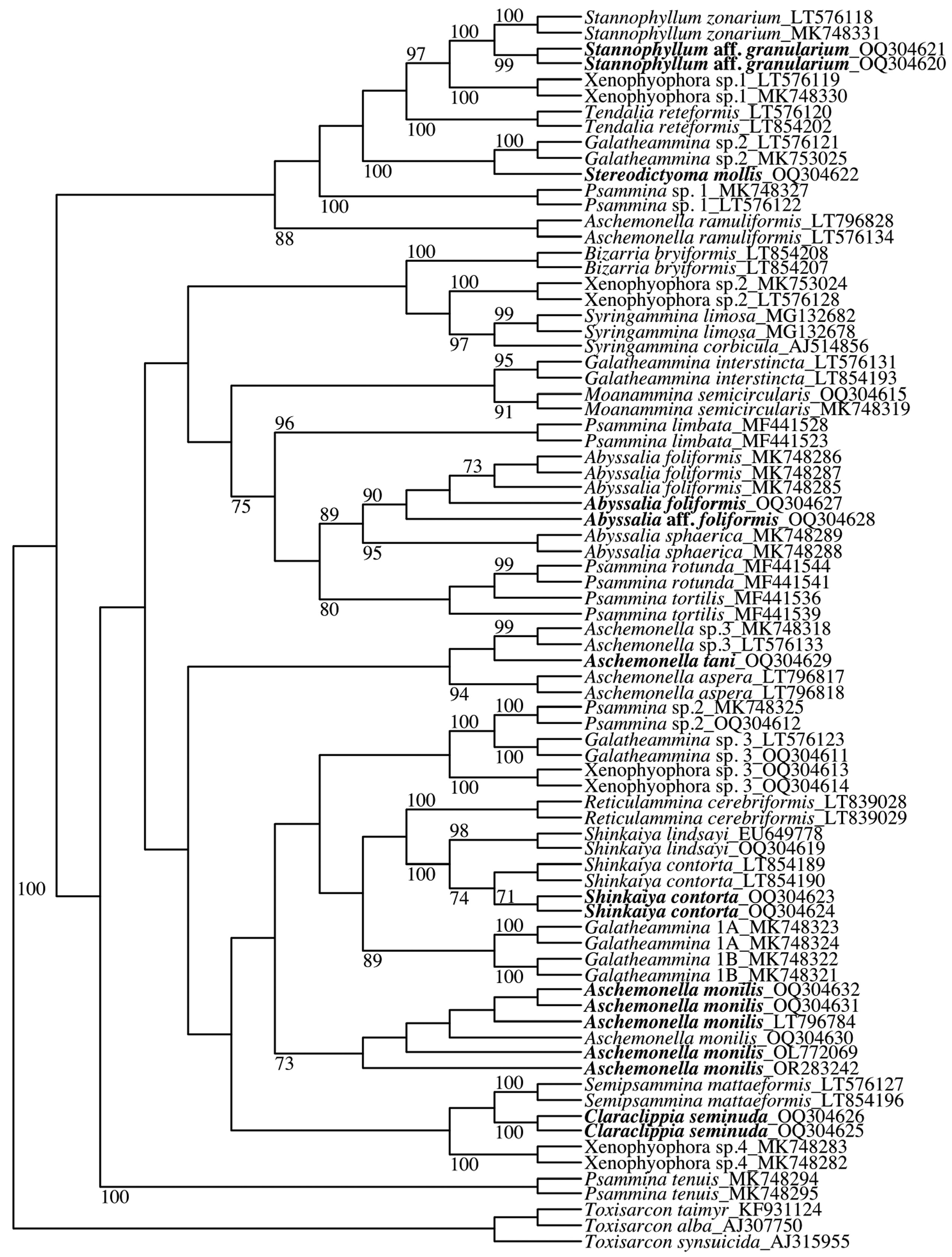
Molecular characterisation. Claraclippia seminuda (100% BV) branches as sister to S. mattaeformis (100% BV), but the grouping of the two species is not supported by the BV. The two 18S sequences of C. seminuda are identical, they contain 908 nucleotides and the GC content is 44%.
Remarks. A distinctive feature of Claraclippia seminuda is the lack of any real test. Shipboard photographs of the freshly collected specimen show a layer of fine sediment particles covering much of the surface, although this veneer was very thin and did not totally obscure the underlying stercomare. Some parts of the veneer survived transport in RNAlater to Geneva, but it had disappeared when the fragments, now preserved in formalin, were examined in Southampton a year later.
There are intriguing morphological similarities between Claraclippia seminuda and Semi psammina mattaeformis Gooday & Holzmann, 2017 , a species also described from the CCZ that lives attached as a flat structure on nodule surfaces. In particular, the stercomare of S. mattaeformis forms ‘a dense, mat-like formation comprising closely packed, convoluted masses, generally 100–200 µm in width, that appear to merge and anastomose, but sometimes are aligned to run more or less parallel……. Elsewhere, the masses are less closely packed and form a more open system of anastomosing branches (again generally 100–200 µm width)’ ( Gooday and Holzmann, 2017c). When the test is removed, these stercomare formations look remarkably similar to those of C. seminuda , although individual strands are somewhat wider and there are no obvious granellare branches. A test is present in S. mattaeformis , but it is thin, flimsy and easily detached, which tends to enhance the similarity with C. seminuda . The two species also branch as sister in the phylogenetic tree, although without bootstrap support ( Fig. 1 View FIGURE 1 ).
The construction of the body of Claraclippia seminuda largely from stercomare is a characteristic shared with Ceralasma massa . In other respects, however, the two species are quite different. The body is a rounded lump comprising wide ( 2–4 mm) stercomare branches in C. massa ( Tendal, 1972) , compared to mainly plate-like elements made up of much narrower (100–150 µm) stercomare branches in C. seminuda . The new species is also much larger (~ 8 cm), almost three times the size of the largest specimen of C. massa ( 2.8 cm; Tendal, 1972). It is more similar to Cerelasma implicata Kamenskaya, Gooday & Tendal, 2017, which has a test composed of relatively narrow, closely packed stercomare branches interwoven with granellare branches. The main difference is that C. implicata is much smaller ( 14 mm or less) and morphologically simpler, with a basal trunk attached to a nodule and an expanded, flattened, fan-shaped upper part (Kamenskaya et al., 2017). The stercomare branches are also narrower (50–60 µm) than those of C. seminuda (75–150 µm). It is possible that the small specimens described by Kamenskaya et al. (2017) are juveniles of C. seminuda , but confirmation of this hypothesis would require genetic data. Stannophyllum mollum Tendal, 1972 is another species that is largely devoid of xenophyae. However, like other members of the genus, the test is held together by fine organic fibres (linellae), forming a distinct surface layer that clearly distinguishes S. mollum from C. seminuda ( Tendal, 1972) .
| FOR |
Forssa Museum of Natural History |
No known copyright restrictions apply. See Agosti, D., Egloff, W., 2009. Taxonomic information exchange and copyright: the Plazi approach. BMC Research Notes 2009, 2:53 for further explanation.
|
Kingdom |
|
|
Phylum |
|
|
Class |
|
|
Family |
|
|
Genus |