
Abyssalia aff. foliformis, Gooday & Holzmann, 2020
|
publication ID |
https://doi.org/10.11646/zootaxa.5419.2.1 |
|
publication LSID |
lsid:zoobank.org:pub:88353CBA-6C4D-40E3-8475-B1FCA2C48637 |
|
DOI |
https://doi.org/10.5281/zenodo.11262821 |
|
persistent identifier |
https://treatment.plazi.org/id/03A987A1-7B4F-AF6D-66C4-44B1FEC0DF03 |
|
treatment provided by |
Plazi |
|
scientific name |
Abyssalia aff. foliformis |
| status |
|
Abyssalia aff. foliformis View in CoL
Figs 6 View FIGURE 6 , 7 View FIGURE 7
Material examined. BC011 RC0520 (morphology and genetics). Sequenced isolate: 21429.
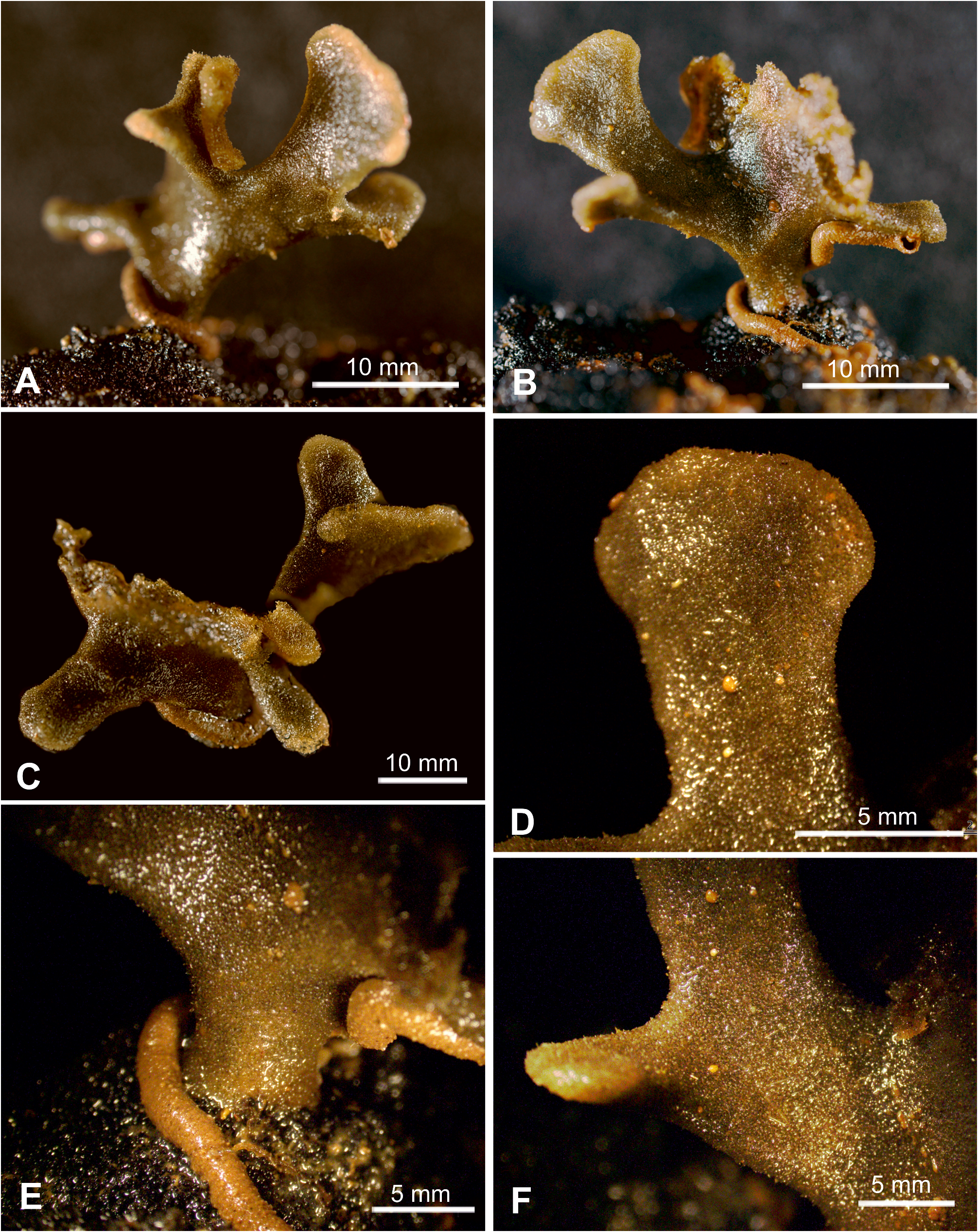
Description. Shipboard photographs. Most of our morphological information about the single specimen comes from photographs taken soon after its collection ( Fig. 6 View FIGURE 6 ). These show a complex branching, plate-like test attached to a nodule by a short, relatively wide basal stalk, about 4.3 mm wide and 4.0 mm high. The entire test is estimated to be roughly 22 mm high with a maximum horizontal span of about 58 mm. The stalk widens rapidly into a central plate-like part that gives rise to a series of elongate lobes of different sizes, radiating in different directions and in some cases appearing slightly twisted. The most prominent of these is roughly 13 mm long and widens from about 5.8 mm near the base to about 11 mm at the end. Others are shorter and do not widen to the same extent. One lobe, which can be measured accurately because there is a corresponding scale, is 11 mm long, 5.3 mm wide near the base and 8.1 mm wide near the end ( Fig. 6D View FIGURE 6 ).
The overall colour of the test in these photographs is greyish brown, with a paler rim that is most obvious around the ends of the lobes. The yellow agglutinated tube of a foraminifera, probably Saccorhiza ramosa, winds around the stem and extends along the underside of one of the lobes.
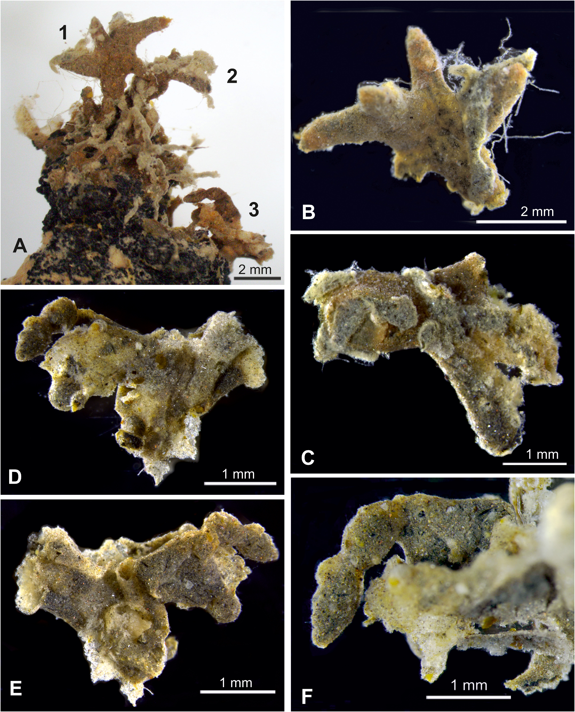
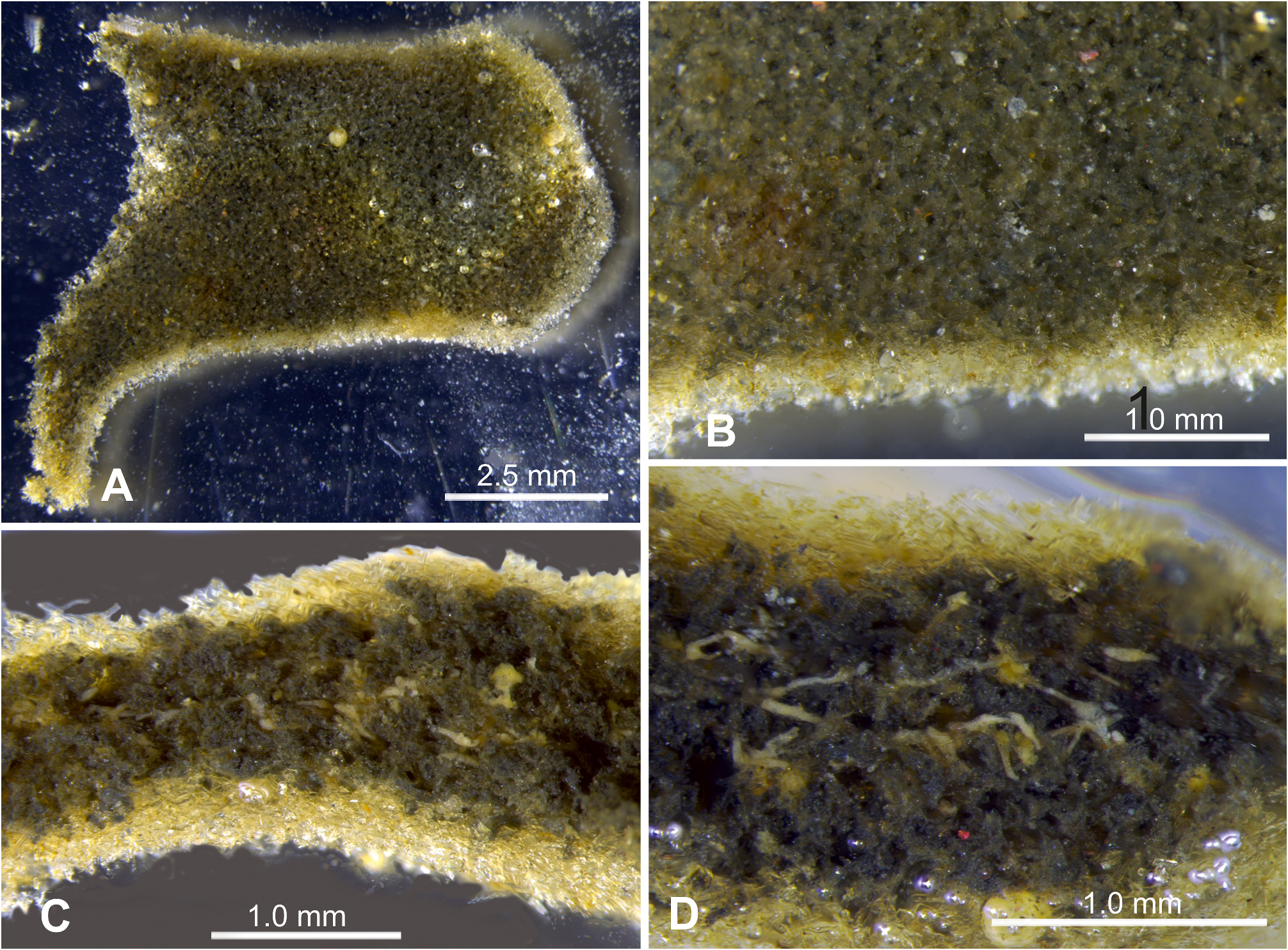
Preserved fragment. One small lobe was available for more detailed study ( Fig. 7 View FIGURE 7 ). It measures ~ 6.5 cm long, a maximum of ~ 4 cm wide, and about 1.60 to 1.75 mm thick. There is a clearly defined test wall, about 220 to 245 µm thick, composed mainly of short, sponge spicule fragments and tiny transparent mineral grains (~10–50 µm in size), as well as occasional radiolarian shells. A few agglutinated foraminiferal shells are also incorporated. The spicules form a three-dimensional mesh that creates a very distinctive, somewhat labyrinthic appearance. There are few if any internal xenophyae and the test interior is largely occupied by dense stercomare. Narrow, pale cream granellare strands are exposed on the broken end of the fragment ( Fig. 7D View FIGURE 7 ). They are generally 20–40 µm wide but sometimes wider at branching points. A few larger masses (up to ~60 µm) are also visible.
Molecular characterisation. Abyssalia aff. foliformis branches at the base of A. foliformis (90%BV) and both taxa build a well sustained (89%BV) group with A. sphaerica . The sequenced fragment of the 18S gene of Abyssalia aff. foliformis contains 1018 nucleotides and the GC content is 37%.
Remarks. This species is closely related genetically to Abyssalia foliformis , but morphologically distinct. The test is a branching structure that is considerably more complex than the leaf-like test of the type specimen of A. foliformis (Gooday et al., 2020) . The test wall of both species is composed almost entirely of sponge spicules, but the spicule framework of A. aff. foliformis is much more intricate that the relatively simple felted wall of A. foliformis .
Abyssalia aff. foliformis is very likely the same as Galatheammina sp. 7 of Gooday et al. (2017a, Supplementary Fig. S3A View FIGURE 3 ), a small, semi-circular plate, less than 1 cm in width and height, that was attached to a nodule in the UK-1 area. It was much smaller and simpler than our specimen, presumably a young individual, and lacked a basal stalk. Both share the same very distinctive wall structure comprising an intricate framework of spicule fragments and mineral gains, but since sequences were not obtained from Galatheammina sp. 7 , we cannot confirm that it represent the same species.
No known copyright restrictions apply. See Agosti, D., Egloff, W., 2009. Taxonomic information exchange and copyright: the Plazi approach. BMC Research Notes 2009, 2:53 for further explanation.
|
Kingdom |
|
|
Phylum |
|
|
Class |
|
|
Family |
|
|
Genus |