
Stereodiktyoma mollis Gooday & Holzmann
|
publication ID |
https://doi.org/10.11646/zootaxa.5419.2.1 |
|
publication LSID |
lsid:zoobank.org:pub:88353CBA-6C4D-40E3-8475-B1FCA2C48637 |
|
DOI |
https://doi.org/10.5281/zenodo.11247638 |
|
persistent identifier |
https://treatment.plazi.org/id/03A987A1-7B50-AF76-66C4-42D5FECFDB1D |
|
treatment provided by |
Plazi |
|
scientific name |
Stereodiktyoma mollis Gooday & Holzmann |
| status |
|
Stereodiktyoma mollis Gooday & Holzmann View in CoL gen. & sp. nov.
Fig. 10 View FIGURE 10 , Supplementary Fig. S3 View FIGURE 3
Diagnosis. As for genus.
Etymology. Latin mollis , meaning soft, a reference to the poorly cemented test wall.
Type material and locality. The holotype ( Lee Kong Chian Natural History Museum, Singapore, reg. no. ZR C. FOR.0003, preserved in 10% formalin) was collected in box core BC039 (specimen RC1623 ); OMS license area, 12° 22' 05.1"N, 117° 33' 01.0"W; 4157 m water depth. The specimen is in the form of numerous small fragments GoogleMaps .
The paratype ( Lee Kong Chian Natural History Museum, Singapore, reg. no. ZR C. FOR.0002, preserved in 10% formalin) was collected in box core BC040 (specimen RC1697 ); OMS license area, 12° 20' 37.4"N, 117° 28' 50.6"W; 4174 m water depth. The specimen is in the form of numerous small fragments, some of which were used for genetics (sequenced isolate 21433) GoogleMaps .
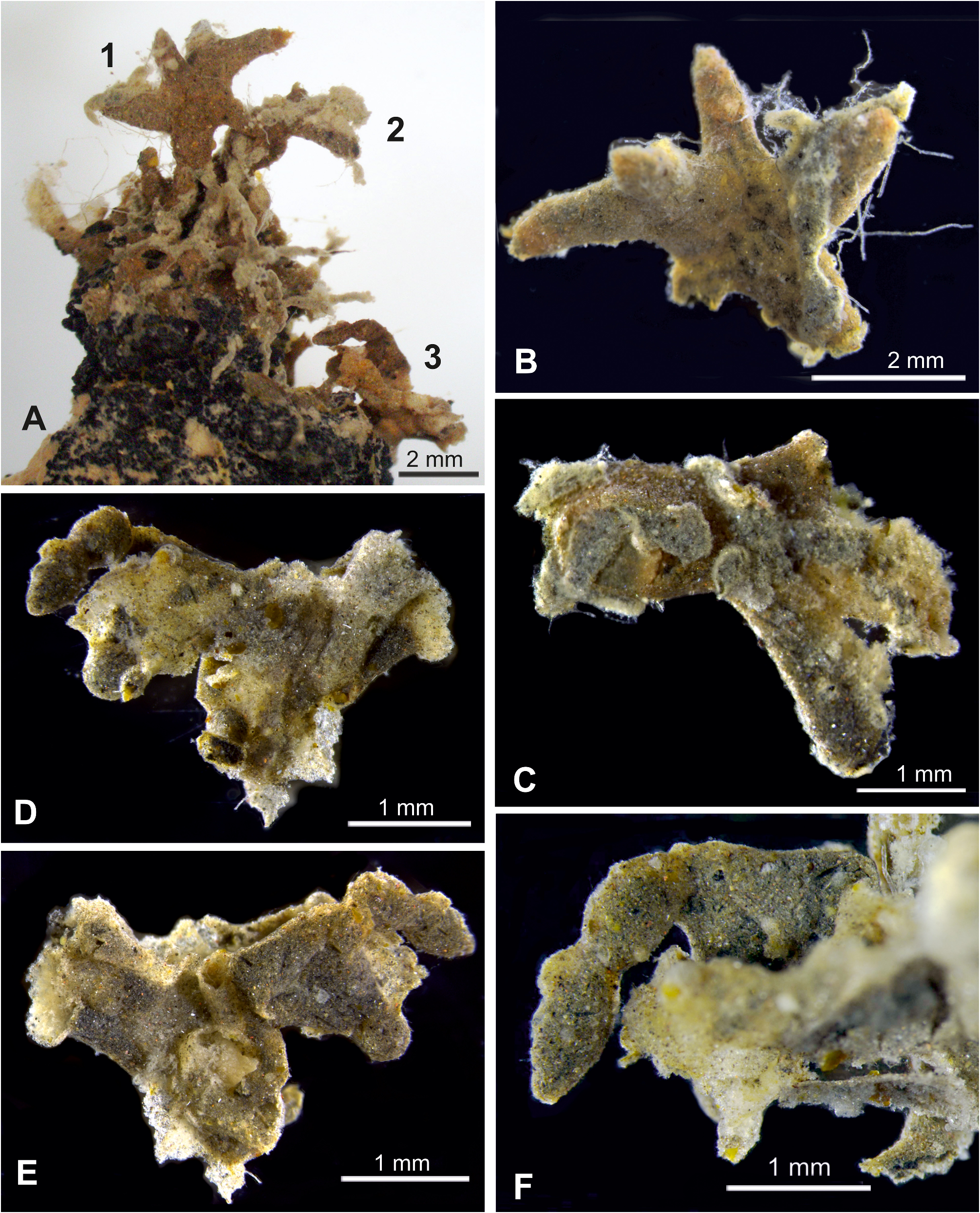
Description. Shipboard photographs. The holotype was intact when photographed ( Fig. 10A, B View FIGURE 10 ). It was pale, brownish tan in colour and attached to a nodule. The base of the test extended for about 5 mm in one direction across the nodule surface, less in other directions. The width at the base, including these encrusting parts, was about 23 mm. The maximum height was almost 11 mm, of which about 6.2 mm was elevated above the surface of the nodule. This upper part was narrower (width ~ 14 cm) than the base. The attached part of the test comprised bars that had mainly coalesced to form plates interrupted by open spaces, although retaining some identity in places. It had an uneven margin with short, projecting finger-like or lobate processes. The upper elevated section formed a three-dimensional framework that consisted mainly of bars, 0.7–1.0 mm wide, around open spaces.
The paratype was also originally attached to a nodule but broke into two fairly large fragments when removed ( Fig. 10C, D View FIGURE 10 ). One fragment measured 11.2 by 10.9 cm and had a subrectangular outline. The other measured 11.8 by 9.2 cm and had a semicircular outline; the flattened part may have been the base of the specimen. Both fragments comprised a three-dimensional framework of branches, each ~ 0.54–0.92 mm in diameter. A photograph of a nodule from the same box core (BC040) showed the remains of an encrusting xenophyophore that probably represents the same species (Supplementary Fig. S3A View FIGURE 3 ). It formed a mat-like structure covering an area measuring at least 17.5 by 15 mm. In places the surface was fairly smooth, but elsewhere it was uneven and appeared to comprise coalescing tubes, a few of which stood up for a short distance from the general surface. It was possibly the basal part of an upstanding test.
Preserved material. Both specimens were very fragile. The holotype, preserved in formalin, arrived in Southampton as small fragments, the largest a few milliimetres in size (Supplementary Fig. S3C–F View FIGURE 3 ). Fragments of the paratype, preserved in RNAlater, were initially sent to Geneva and included two larger pieces ( Fig. 10E, F View FIGURE 10 ), around 6.0 and 8.7 mm maximum dimension. Further disintegration occurred during onward transport to Southampton. Most of the surviving fragments are basically cylindrical, although sometimes coalescing to form more plate-like structures (Supplementary Fig. S3D View FIGURE 3 ). This tendency for the tubes to coalesce is also evident in the shipboard photographs of the intact holotype ( Fig. 10A, B View FIGURE 10 ).
The test wall has a very thin (no more than ~5 µm) basal layer composed of small but discernible transparent, pale yellowish grains. This is overlain by a much thicker (typically 130 to 260 µm) layer of soft, very fine-grained and easily disaggregated, sediment-like material (Supplementary Fig. S3C–D View FIGURE 3 ). The branches are tubular and there are no internal xenophyae, much of the internal space being occupied by a stercomare branch, typically 100 to 200 µm diameter (Supplementary Fig. S3F View FIGURE 3 ). Several branches often emerge from plate-like fragments or are visible along their broken edges. A narrow granellare strand is sometimes seen running parallel to the stercomare (Supplementary Fig. S3B View FIGURE 3 ). The granellare is pale yellowish, usually 30 to 50 µm diameter, and branches together with the stercomare where the tubular test elements bifurcate.
Molecular characterisation. Stereodictyoma mollis branches as sister to Galatheammina sp. 2 (100%BV). The length of sequenced fragment of 18S gene of S. mollis is 1068 nucleotides and the GC content is 30%.
Remarks. There are some morphological differences between the two specimens of Stereodiktyoma mollis . In particular, the holotype from box core included a fairly high proportion of plate-like fragments whereas fragments of the paratype were predominantly tubular. This difference is in the small preserved fragments is consistent with the appearance of the more intact specimens in shipboard photographs. However, in other respects, notably the wall structure, they are very similar and hence we consider them to represent the same species.
DNA sequences obtained from the paratype reveal a strongly supported relationship (100% BV) between Stereodiktyoma mollis and Galatheammina sp. 2 of Gooday et al. (2017c), albeit with fairly long branches in both cases. The Galatheammina species is known from a single specimen, possibly a fragment, from the UK-1 area. This forms a flat plate composed of radiolarians in a fine-grained matrix and with radiolarians also occupying the test interior, together with stercomare and granellare. The two species therefore have little in common morphologically.

| FOR |
Forssa Museum of Natural History |
No known copyright restrictions apply. See Agosti, D., Egloff, W., 2009. Taxonomic information exchange and copyright: the Plazi approach. BMC Research Notes 2009, 2:53 for further explanation.
|
Kingdom |
|
|
Phylum |
|
|
Class |
|
|
Family |
|
|
Genus |