
Shinkaiya contorta Gooday & Holzmann, 2017
|
publication ID |
https://doi.org/10.11646/zootaxa.5419.2.1 |
|
publication LSID |
lsid:zoobank.org:pub:88353CBA-6C4D-40E3-8475-B1FCA2C48637 |
|
DOI |
https://doi.org/10.5281/zenodo.11262829 |
|
persistent identifier |
https://treatment.plazi.org/id/03A987A1-7B56-AF76-66C4-42CDFBA5DFEF |
|
treatment provided by |
Plazi |
|
scientific name |
Shinkaiya contorta Gooday & Holzmann, 2017 |
| status |
|
Shinkaiya contorta Gooday & Holzmann, 2017 View in CoL
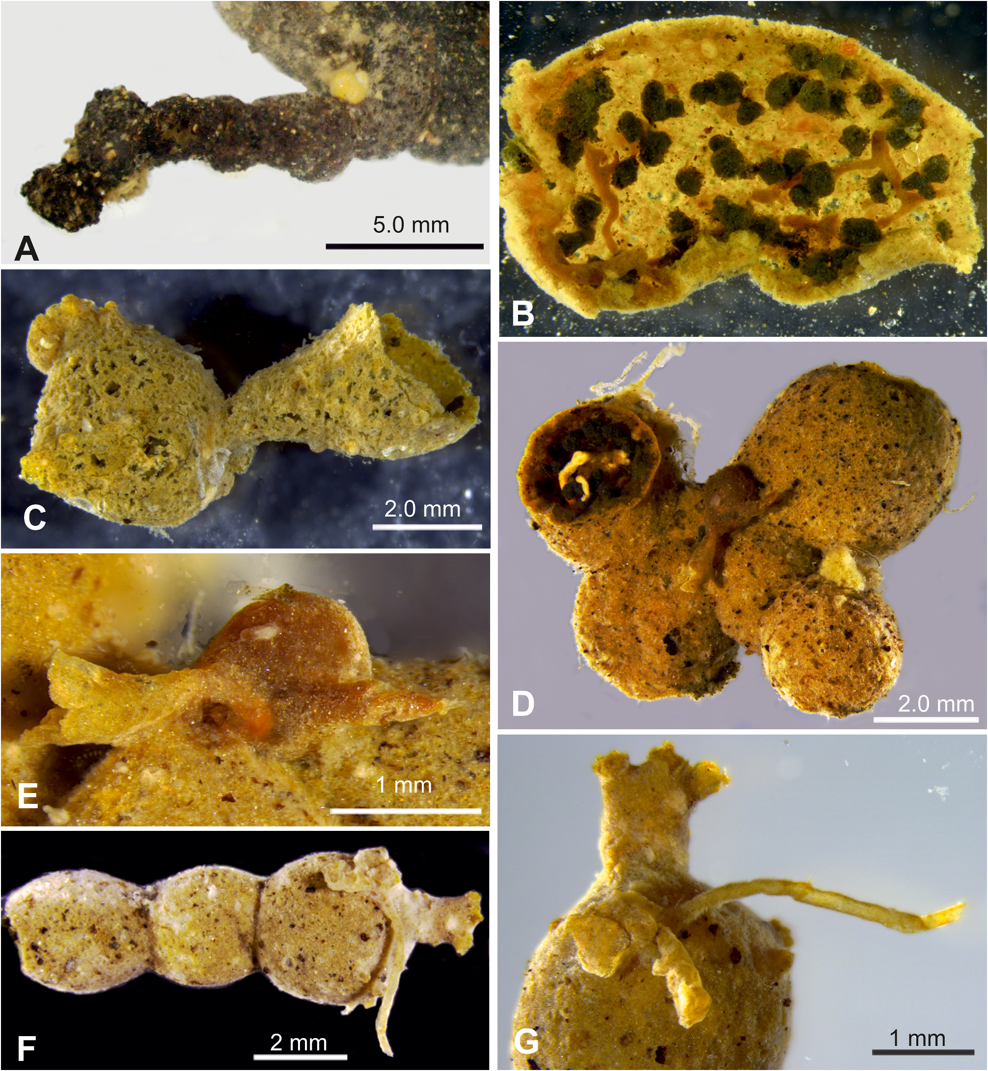
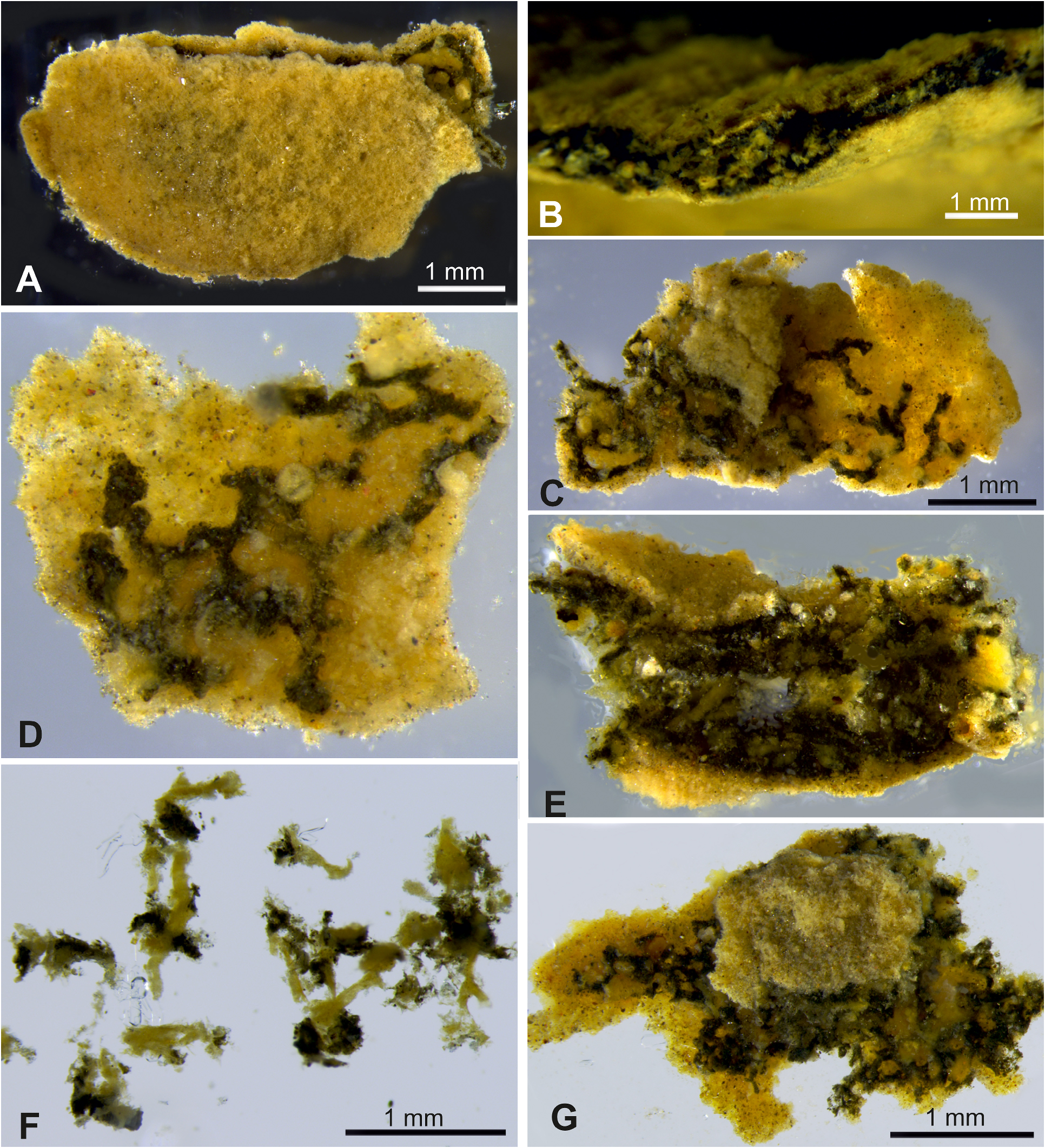
Figs 11 View FIGURE 11 , 12 View FIGURE 12
Shinkaiya contorta Gooday & Holzmann, 2017 View in CoL , in Gooday et al. 2017c, p. 727 –730, Fig. 2A–F View FIGURE 2 .
Material examined. BC004 RC0160 (morphology and genetics). Sequenced isolates: 21448, 21449.
Description. The shipboard photographs show a single plate-like fragment with an intact semicircular margin and concentric ‘growth lines’ clearly developed over parts of the surface ( Fig. 11A View FIGURE 11 ). It was originally attached to a nodule and the lower margin was broken when it was removed from the substrate. The plate was strongly undulating so that it did not lie in one plane. It was still largely intact when seen in Geneva, where it measured 44 mm in maximum dimension. By the time it reached Southampton, the fragment had broken into several smaller pieces, some almost flat but others curved, and the largest with a maximum dimension of about 15 mm ( Fig. 11B View FIGURE 11 ). They are greyish, with a smooth surface, in places overlain by patches of lighter material resembling fine-grained sediment. ‘Growth lines’ are sometimes visible. The wall is 60–95 µm thick, in a few places up to 115 µm ( Fig. 11D, E View FIGURE 11 ; 12E View FIGURE 12 ), quite soft, delicate, and very fine-grained with a scattering of darker flecks.
The test interior is occupied mainly by masses of stercomare ( Fig. 12C–F View FIGURE 12 ), some of which are attached to the underside of the wall. On detached wall fragments the stercomare forms distinctive strands, typically 50–105 µm in width, that branch and usually anastomose to varying degrees, sometimes forming dense networks ( Fig. 12C–E View FIGURE 12 ). Granellare strands are whitish, typically 45–75 µm in width and weave between the stercomare. A granellare tube is not clearly visible under stereomicroscope.
Remarks. The wavy, plate-like morphology of the preserved fragment is consistent with that of the unique holotype of Shinkaiya contorta from the UK-1 area of the CCZ ( Gooday et al., 2017c). This was an intact specimen, with a maximum dimension ( 46 mm), similar to that of the new specimen, but with a more complex structure that comprised a number of curved, plate-like elements, often with well-developed growth lines. The soft, finely agglutinated test wall, and the reticulated stercomare branches, are similar but some other test features are different. Particularly notable is that the plate itself, and particularly the wall of the plate, are much thinner (about 0.5–1.0 mm and 60–95 µm, respectively) than those of the type specimen (1.3–2.0 mm and 270–500 µm, respectively). Nevertheless, sequences obtained from the new fragment confirm its identification.

No known copyright restrictions apply. See Agosti, D., Egloff, W., 2009. Taxonomic information exchange and copyright: the Plazi approach. BMC Research Notes 2009, 2:53 for further explanation.
|
Kingdom |
|
|
Phylum |
|
|
Class |
|
|
Family |
|
|
Genus |
Shinkaiya contorta Gooday & Holzmann, 2017
| Holzmann, Maria, Barrenechea-Angeles, Inés, Lim, Swee-Cheng & Pawlowski, Jan 2024 |
Shinkaiya contorta
| Gooday, A. J. & Holzmann, M. & Caulle, C. & Goineau, A. & Pearce, R. B. & Voltski, I. & Weber, A. A. - T. & Pawlowski, J. 2017: 727 |