
Epistylis camprubii, Canals & Salvadó, 2016
|
publication ID |
https://doi.org/ 10.4467/16890027AP.16.002.4043 |
|
DOI |
https://doi.org/10.5281/zenodo.8377724 |
|
persistent identifier |
https://treatment.plazi.org/id/03A987BA-FFF0-775A-B620-FD9CBE89026A |
|
treatment provided by |
Felipe |
|
scientific name |
Epistylis camprubii |
| status |
sp. nov. |
Epistylis camprubii n. sp.
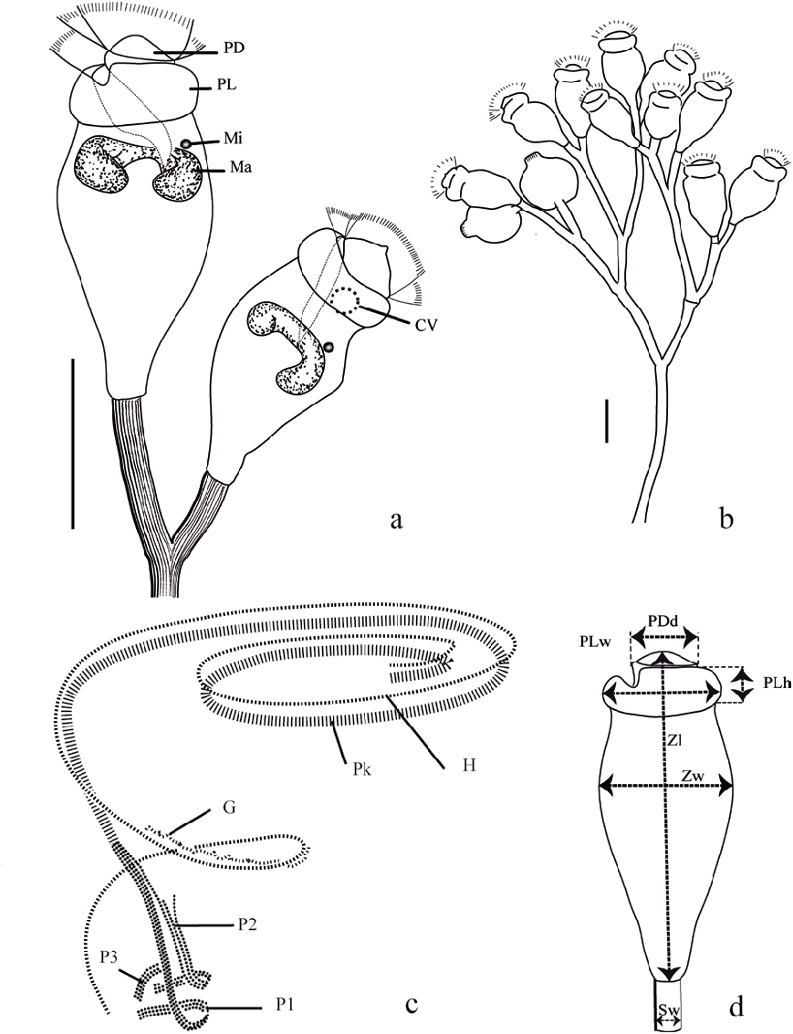
1. Diagnosis. Freshwater bacterivorous Epistylis , measuring on average 58.7 µm in length and 32.0 µm in width. The zooids are vase-shaped and constricted below the thick peristomial lip. Persitomial disc commonly rounded or pointed, very rarely umbilicated. C-shaped macronucleus located in the adoral half of the zooid and transversely oriented. Contractile vacuole placed in the adoral third of the body, on dorsal wall of infundibulum. Row 3 of P2 slightly divergent to the other rows at their abstomal ends, extending, together with row 2, approximately 2/3 of the length of row 1. Transverse silverlines numbering 106 to 136 from peristome to aboral trochal band and 33 to 48 from aboral trochal band to scopula.
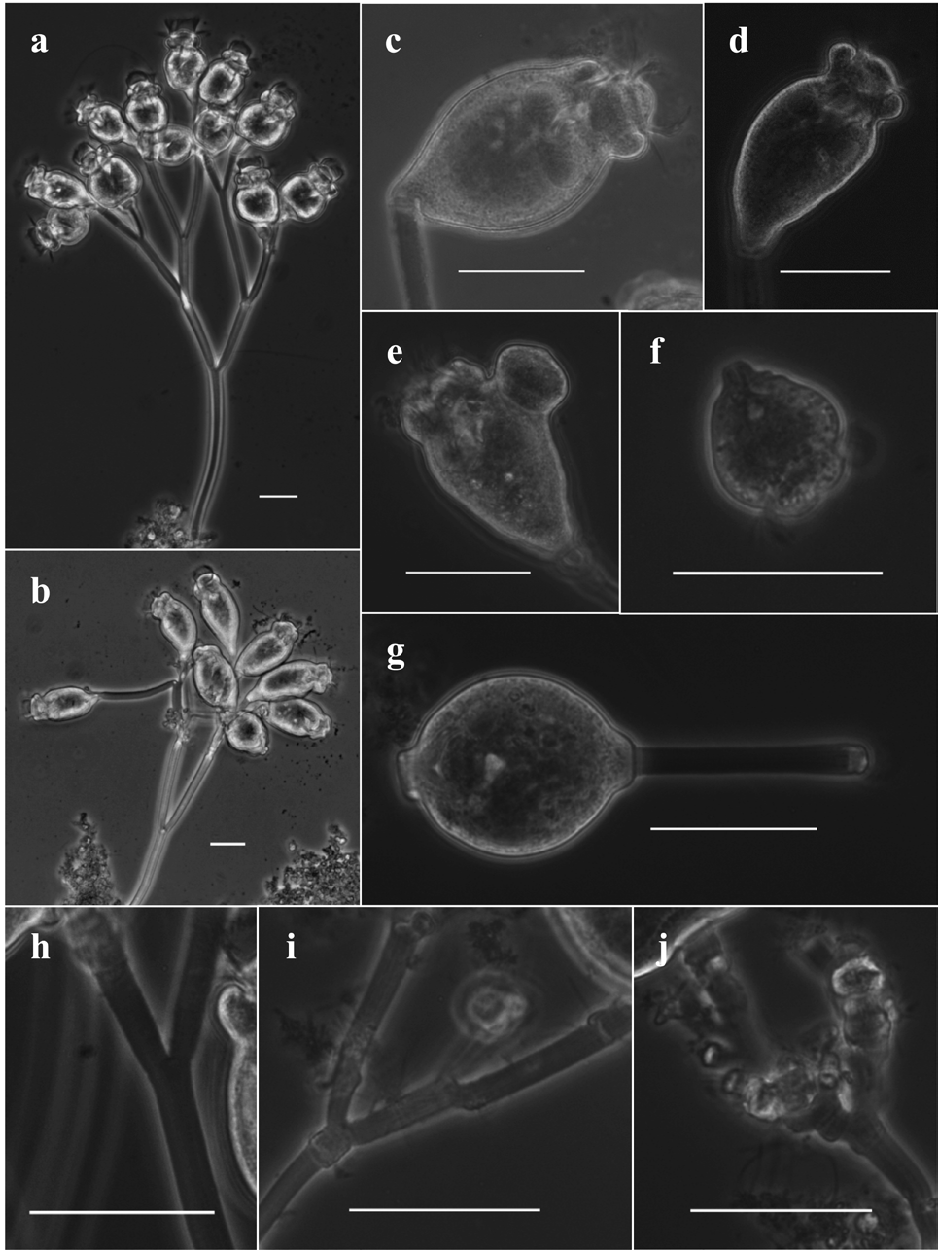
2. Description. Epistylis camprubii n. sp. is a vase-shaped peritrich, constricted below the peristomial lip. The zooid length ranges from 35.3 to 98.1 µm (average of 58.7 ± 10.1 µm) and the width from 18.0 to 65.2 µm (average of 32.0 ± 5.4 µm). The peristomial disc is commonly rounded or pointed, and very rarely umbilicated. It is possible, but not common, to observe zooids of the same colony showing different peristomial disc shapes. The diameter of the peristomial disc ranges from 11.2 to 21.3 µm (average of 15.5 ± 1.9 µm). E. camprubii shows a thick peristomial lip with a height ranging from 5.0 to 10.6 µm (average of 7.8 ± 1.2 µm) and a width from 16.2 to 31.7 µm (average of 24.2 ± 2.9 µm). The width of the stalk varies from 3.1 to 8.4 µm (average of 5.3 ± 0.9 µm). The stalk is longitudinally striated and compact, occasionally exhibits uneven thickness, and rarely shows transverse segments. Stalk thickening is often associated with separation nodes, but they can also be seen in the internodal part of the stalk ( Fig. 2h–j View Fig ). Thicker stalks or branches, as well as more frequent stalk thickening, are related to shorter stalks. E. camprubii presents a colourless cytoplasm and a transversely oriented C-shaped macronucleus located in the adoral half of the zooid. The only contractile vacuole lies in the adoral third of the body, on dorsal wall of infundibulum.
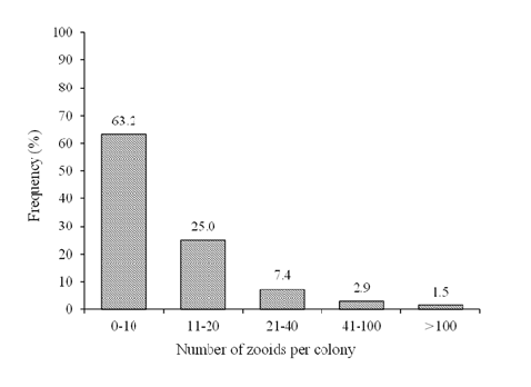
In the wastewater treatment systems where E. camprubii was found, 88.2% of the colonies showed a number of zooids between 2 and 20 ( Fig. 4 View Fig ), with a maximum value of 124. The ramification is initially dichotomic, but becomes irregular after the second level of ramification. Sometimes the ramification is totally irregular.
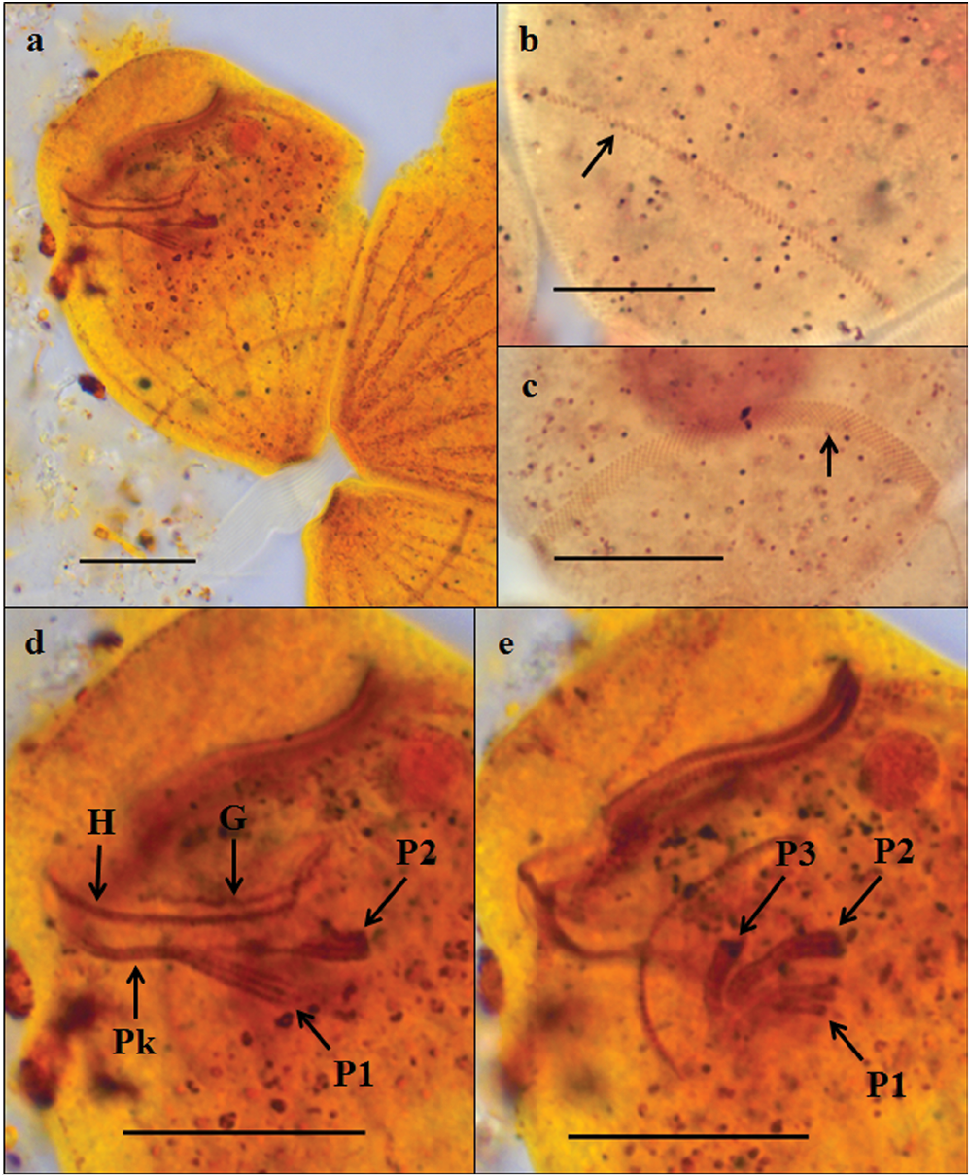
E. camprubii presents the typical oral infraciliature of the genus Epistylis . Specifically, it has very similar oral infraciliature to Epistylis chrysemydis (detailed by Foissner et al. 1992). The haplokinety and the polykinety make between one and one and a half circuits before entering the infundibulum, where they make a turn again. In the infundibulum, the haplokinety is initially accompanied by a germinal kinety. The germinal kinety is no longer observed when the haplokinety turns and follows the longitudinal axis of the zooid. The polykinety becomes three infundibular polykineties (named P1, P2, P3), each consisting of 3 rows of kinetosomes. The abstomal end of P1 comes directly from the original polykinety and its three rows are equal in length. The abstomal end of row 1 of P2 extends the entire distance to P1 and merges with it near the oral opening. Rows 2 and 3 of P2 are much shorter than row 1, extending only ~ 2/3 of the length of row 1 at their abstomal ends. Row 3 of P2 is slightly divergent from the other two rows at its abstomal end. The adstomal ends of all three rows of P2 end a short distance before the adstomal ends of P1 and P3. P3 is the shortest polykinety and presents the three rows equal in length. P3 appears after the last turn of P1 and P2 and its adstomal end is very close to the adstomal end of P1 ( Figs 1c View Fig and 3d–e View Fig ).
Epistylis camprubii presents 106 to 136 transverse silverlines from peristome to aboral trochal band and 33 to 48 from aboral trochal band to the scopula.
3. Etymology. The name camprubii refers to the surname of the Catalan biologist Jordi Camprubí Capella, in personal recognition of a life dedicated to teaching and sharing his love for biology, especially for ciliate protozoa.
4. Ecology. Epistylis camprubii is a bacterivorous peritrich. It was first observed in the aerobic reactor of an A/O SBNR-MBBR process and was initially identified as Epistylis cf. rotans ( Canals et al. 2013) . It was observed mainly inhabiting the biofilm adhered to carriers (the plastic support for the biofilm attachment), but was also found colonising the liquid phase, adhered to flocs. It was observed again attached to the biofilm and colonising the liquid phase of a PN-MBBR and PN-granular processes, both previous to an Anammox reactor.
A/O SBNR and PN processes are designed to treat high ammonium-loaded wastewater (up to over 103 N-NH 4 + mg·L –1). Specifically, the aerobic reactor of an A/O SBNR process is designed to oxidise the ammonium in the influent and accumulate the oxidised nitrogen form as nitrite, while the PN process is focused on achieving an adequate effluent for the posterior Anammox reactor, that is, a nitrite: ammonium ratio close to 1.2. The range of ammonium and nitrite values in which Epistylis camprubii was observed are shown in Table 2 View Table 2 .
Some organisms were identified cohabiting with E. camprubii : the ciliates Colpoda spp. , Cyclidium glaucoma , Opercularia coarctata , Telotrochidium matiense , Vorticellides microstoma -complex and two unidentified Hypotrichia; Tetramitus rostratus , Polytoma sp. , Anthophysa sp. and Trimastix sp. among other unidentified flagellates; nematoda; and unidentified gymnamoeba smaller than 20 µm. Most of these species, specifically the ciliates, are usually observed in conventional activated sludge wastewater treatment plants. It would not be surprising if Epistylis camprubii had been misidentified in this kind of treatment systems.
5. Type Sequence. GenBank accession number KP713786 View Materials .
6. Type material. An holotype slide with 36 specimens (registration number CRBA-28006) and a paratype slide with 52 specimens (registration number CRBA-28007) after ammoniacal silver carbonate method were deposited in the Centre de Recursos de Biodiversitat Animal ( CRBA), Facultat de Biologia , Universitat de Barcelona, Barcelona ( Spain). Another paratype slide with 35 specimens was deposited in the Natural History Museum of London, UK (registration number NHMUK 2015.4.20.1) .
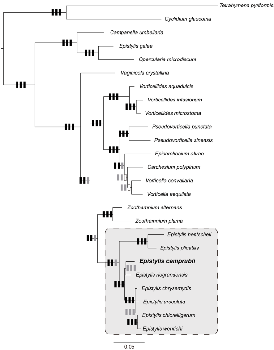
7. Phylogenetic position. The molecular analysis of the 18s rRNA gene sequences supported the inclusion of E. camprubii within the Peritrichia, regardless of the inference method implemented. All analyses agreed in supporting the monophyly of E. camprubii with the other Epistylis sequences used in the present study, with the single exception of E. galea ( Fig. 5 View Fig ).
8. Comparative diagnosis. A large number of Epistylis species have been described. Among them, just a few showed morphological similarities to E. camprubii and/or are typical species of wastewater treatment plants.
E. camprubii was formerly reported as Epistylis cf. rotans ( Canals et al. 2013) , since E. rotans is a vase-shaped peritrich that is often observed in wastewater treatment plants, and presents a longitudinally striated and transversally segmented stalk, as E. camprubii does. But further observations revealed that transverse segments are not always present in E. camprubii , in contrast to E. rotans . Moreover, E. rotans is longer (70 to 100 µm) and was described as an oligosaprobic species by Curds (1969), while E. camprubii is shorter ( Table 1 View Table 1 ), inhabits more polluted environments, and shows a high tolerance to ammonium and nitrite compounds ( Table 2 View Table 2 ). It must be noted that Foissner et al. (1992) considers E. rotans a synonym of E. procumbens , a funnel-shaped peritrich with a zooid length from 60 to 140 µm, often bent and with transverse segments before the stalk division, whose characteristics clearly do not match those of E. camprubii .
A comparison of E. camprubii and other Epistylis species of wastewater treatment plants showed that E. camprubii is shorter than E. chrysemydis (120 to 220 µm), E. coronata (70 to 120 µm), E. entzii (125 to 190 µm), E. hentscheli (110 to 170 µm), E. balatonica (90 to 100 µm) and E. plicatilis (90 to 160 µm). In addition, most of these species have a wider stalk, ranging from 7 to 20 µm width ( Foissner et al. 1992), while the stalk of E. camprubii ranges from 3.1 to a maximum of 8.4 µm when stalk thickening occurs ( Table 1 View Table 1 ). Other clearly different traits are that E. chrysemydis and E. balatonica have two lips in the peristome, in contrast to the one lip of E. camprubii . E. coronata always shows an umbilicated peristomial disc and E. plicatilis and E. hentscheli are clearly funnel-shaped.
Outside the field of wastewater treatment systems, three Epistylis species presented similar characteristics to E. camprubii : E. epistyliformis , E. thienemanni and E. variabilis ( Stiller 1971) . E. epistyliformis , the most similar species, is a vase-shaped peritrich with a clearly pointed and rarely umbilicated peristomial disc, a zooid length from 43 to 62 µm, and clearly constricted below the peristomial lip. Nevertheless, the C-shaped macronucleus of E. epistyliformis is flattened, located in half of the body, and follows the longitudinal axis of the zooid, in contrast to the transverse position of the macronucleus of E. camprubii . In addition, Stiller’s guide (1971) does not specify in the text or in the drawings whether the stalk of E. epistyliformis is longitudinally striated or not. E. variabilis shows variability in the stalk and branches that strongly resembles that of E. camprubii . Colonies of E. variabilis inhabiting calm waters present long and smooth branches, while the branches of colonies from vigorous water flows are shorter, articulated and thicker or show uneven thickness. Nevertheless, there is no information on the specific width of the stalk in E. variabilis , and the main stalk of the colony is always short, a feature that does not always occur in E. camprubii . Moreover, E. variabilis zooids are funnel-shaped, in contrast to the vase-shaped zooids of E. camprubii . Finally, E. thienemanni ( Stiller 1971, first reported as Rhabdostyla thienemanni by Nenninger 1948) is an epibiont of leeches, and is larger (67 to 120 µm), but with a shorter stalk than E. camprubii . It has the contractile vacuole located in the peristomial disc. A summary of the comparison between Epistylis camprubii and the other Epistylis species can be seen in Tables 3a and 3b View Table 3 .
9. Comments. The great variability observed in the appearance of the stalk of Epistylis camprubii could be related to water hydrodynamics, as Stiller (1971) suggested regarding the stalk variability of E. variabilis . The authors also consider that the low number of zooids observed per colony (63% of colonies showed between 2 and 10 zooids, Fig. 4 View Fig ) may also be influenced or limited by the flow, agitation speed or other hydrodynamic parameters of water, in addition to the availability of food (bacteria) in the reactor. Further research is needed in order to shed light on these unanswered questions.
| NHMUK |
Natural History Museum, London |
No known copyright restrictions apply. See Agosti, D., Egloff, W., 2009. Taxonomic information exchange and copyright: the Plazi approach. BMC Research Notes 2009, 2:53 for further explanation.
|
Kingdom |
|
|
Phylum |
|
|
Class |
|
|
Order |
|
|
Family |
|
|
Genus |