
Phyxioschema huberi, Schwendinger, Peter J., 2009
|
publication ID |
https://doi.org/10.5281/zenodo.188258 |
|
DOI |
https://doi.org/10.5281/zenodo.6214228 |
|
persistent identifier |
https://treatment.plazi.org/id/03A9B15A-FFAA-7E65-FF06-B0B8FA9BFB3D |
|
treatment provided by |
Plazi |
|
scientific name |
Phyxioschema huberi |
| status |
sp. nov. |
Phyxioschema huberi sp. n.
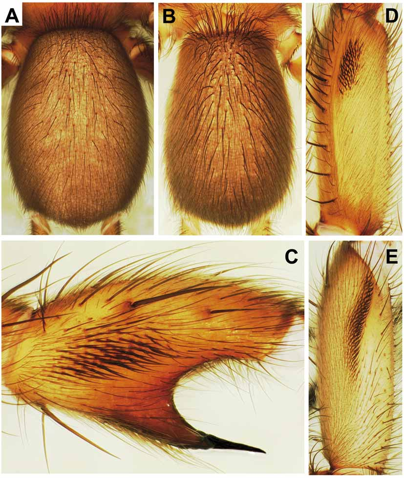
Figures 8 – 11 View FIGURE 8 View FIGURE 9 A – E View FIGURE 10 A – U View FIGURE 11 A – D , 21A, C View FIGURE 21 A – D
Material: THAILAND (southern region): Prachuap Khiri Khan Province, Kui Buri District, Khao Sam Roi Yot National Park, Phraya Nakhon Cave ( 12°11’56”N, 100°00’42”E), 100 m, male holotype (hatched X.2002, matured 4.XI.2004), 8 male paratypes (matured 26.I.2003, 25.VI.2003, 16.X.2003, VIII.2004, 15.X.2004, 12.II.2005, 13.II.2005, 28.I.2006) and 10 female paratypes, collected 10.VII.2002 (sample TH-02/05). From the same locality: 1 male paratype (matured 28.XI.2002) and 2 female paratypes, collected 22.XI.2001 (sample THMA-01/02). All specimens collected by P.J. Schwendinger; 1 male and 1 female paratypes deposited in SMF, all other specimens (including the holotype) in MHNG.
Etymology: This species is dedicated to Siegfried Huber, who brought its existence to my attention.
Diagnosis: This is the largest known Phyxioschema species. It is distinguished from the much smaller P. erawan sp. n. by the lack of metatarsal preening combs, by a lighter colouration and by relatively longer, more slender legs. Males with more distinctly asymmetrical palpal bulb; all tarsi pseudosegmented; proventral spines on tibia I situated more proximally; patella I retroventrally and distoventrally with 4 – 8 short, sigmoid spines; tibia II relatively longer and less incrassate; proventral keel on metatarsus II with sharp edge. Females with narrower spermathecae, each carrying 3 – 4 receptacles, all with long, completely sclerotised stalks and small heads carrying pores.
Description: MALE ( holotype). Colour in alcohol: Body generally light reddish brown, ventral side of all palpal articles and of leg coxae and femora lighter; eye mound black. Opisthosoma ( Fig. 9B View FIGURE 9 A – E ) grey-brown, except for orange-brown bases of long stiff dorsal bristles, indistinct light transverse stripes forming incomplete chevrons in posterior part of dorsal side, light brown genital region, yellow-orange booklung plates and light brown spinnerets with pattern of darker patches and light spots ventrally.
Habitus as in Fig. 8 View FIGURE 8 (showing paratype). Body 14.7 long. Carapace 5.5 long, 5.0 wide, oval, almost flat, covered with light adpressed hairs interspersed by darker, more erect bristles (some wavy), these longest on carapace margin, in front, on and behind eye mound. Eyes on low mound; eye group 0.70 long, anterior eye row clearly procurved, 1.19 wide, posterior eye row slightly recurved, 1.21 wide ( Fig. 10B View FIGURE 10 A – U ). Eye diameters and interdistances: AME 0.30, ALE 0.45, PME 0.27, PLE 0.36; AME–AME 0.06, AME–ALE 0.06, PME–PME 0.43, PME–PLE 0.03. MOQ 0.52 long, 0.64 wide anteriorly, 0.83 posteriorly. Fovea pit-like, with 3 slightly enlarged foveal setae anterior to it.
Chelicerae weak, grooves with 9/10 prolateral teeth and 18 median proximal denticles. Maxillae ( Fig. 10C View FIGURE 10 A – U ) 1.4 long, 0.8 wide, with pallid prolateral zone; anterior lobe indistinct, with quite wide but fairly indistinct serrula on ridge. Labium ( Fig. 10C View FIGURE 10 A – U ) 0.3 long, 0.9 wide, anterior edge distinctly setose, followed by pallid zone; posterior part pigmented, with few fine setae. Sternum ( Fig. 10C View FIGURE 10 A – U ) 3.1 long, 2.4 wide, cordate, with deeply excavated post-labial depression formed by fused anterior sigilla and labio-sternal groove, and with 3 pairs of indistinct marginal sigilla. Membrane at posterior sternal exfoliations (near posterior pair of sigilla and between coxae IV) with small amount of dark pigment.
Palps ( Fig. 10D–E View FIGURE 10 A – U ) with long stiff bristles on femur to tibia. 7+7 trichobothria in 2 rows on tibia, 11 in zig-zag row on tarsus. Palpal bulb with oval base and tapering, largely straight embolus with slightly curved tip.
Legs 2134. All tarsi pseudosegmented and armed with spines. Preening combs absent. I: tibia cylindrical, with strong spines on prolateral, ventral and retrolateral sides ( Fig. 10G View FIGURE 10 A – U ); patella with row of 3/4 thick sigmoid spines retroventrally ( Fig. 10F View FIGURE 10 A – U ) and 1 such spine (next to a thinner, less sigmoid one) situated at a right angle to the others on distoventral margin ( Fig. 10G View FIGURE 10 A – U ), without triangular projection on retrolateral margin; femur with short, fairly wide band of hooked spinules retrodorsally ( Fig. 9D View FIGURE 9 A – E ). II: metatarsus ventroproximally with 2 widely separated keels ( Fig. 10J View FIGURE 10 A – U ), proventral one sclerotised, sharp and rounded ( Fig. 10I –J, L View FIGURE 10 A – U ), retroventral one widely rounded and strongly projecting from margin sideward ( Fig. 10J–L View FIGURE 10 A – U ); tibia moderately incrassate ( Fig. 10H View FIGURE 10 A – U ), band of elongated spinules on prolateral side fairly wide, straight, slightly inclined from longitudinal axis of tibia, reaching approximately height of distal side of ventral spur ( Fig. 9C View FIGURE 9 A – E ); ventral spur of tibia with distinctly bilobed apex carrying 2 megaspines of about equal length but on different levels: prolateral one situated more distally and inclined towards retrolateral one ( Fig. 10H View FIGURE 10 A – U ); femur with long (much longer than on femur I) band of hooked spinules proventrally ( Fig. 9E View FIGURE 9 A – E ). Spination: I: patella p1/3, r6/9, v3; tibia p11, r2/5, v7; metatarsus p1, v7; tarsus p1, r2, v 1. II: patella p2; tibia p1, v2 + v2 megaspines; metatarsus p2, v10; tarsus p1/2, r2/3, v3 / 4. III: patella p3/4, r1; tibia d2, p3, r2, v6 /7; metatarsus d6/7, p3, r3, v8; tarsus p1, r 2. IV: patella p3, r1; tibia d2, p2, r3, v6; metatarsus d7, p2/3, r3, v5; tarsus p1/2, r2/3. Trichobothria: 2 rows of 8–9 each on tibiae, 12–13 in single row on metatarsi, 11–13 in single row on tarsi. Paired claws with 11–12 teeth in sigmoid row, unpaired claw with 4–5 sessile teeth.
Opisthosoma 7.0 long, 4.5 wide; dorsal side quite densely covered with fine light hairs, stronger dark hairs and long, strong dark bristles with darkened sockets (most distinct in anterior part of opisthosoma, Fig. 9B View FIGURE 9 A – E ); ventral side only with short dark and medium-sized dark hairs. Posterior median spinnerets 0.9 long; posterior laterals 9.4 long (proximal article 2.7, median article 2.8, pseudosegmented distal article 3.9).
FEMALE ("allotype"). As the male, except for: prosoma (especially chelicerae, maxillae and tips of palpal tarsi) darker, opisthosoma lighter; pale prolateral zone of maxillae more distinct.
Body 17.7 long. Carapace 6.5 long, 5.6 wide. Eye group ( Fig. 10A View FIGURE 10 A – U ) 0.62 long, anterior eye row 1.24 wide, posterior eye row 1.30 wide. Eye diameters and interdistances: AME 0.27, ALE 0.41, PME 0.22, PLE 0.34; AME–AME 0.09, AME–ALE 0.09, PME–PME 0.51, PME–PLE 0.02. MOQ 0.46 long, 0.57 wide anteriorly, 0.85 posteriorly.
Chelicerae stronger than in male, grooves with 10/11 prolateral teeth and 27/28 median proximal denticles. Maxillae 2.0 long, 1.1 wide, serrula slightly wider and more prominent than in male. Labium 0.5 long, 1.2 wide. Sternum 3.1 long, 2.6 wide.
Palps with 8+9 trichobothria on tibia and 17 on tarsus. Tarsal claw with 12/13 teeth.
Legs 2134. All tarsi integral. Spination: I: patella p2; tibia p2, v7; metatarsus v8; tarsus p1/2, r 2. II: patella p2/3; tibia p2, v7; metatarsus p2, v8; tarsus p2, r 3. III: patella p2/3, r1; tibia d2, p3, r3, v7; metatarsus d4, p3, r2, v8; tarsus p2, r 2. IV: patella p2, r1; tibia d2/3, p2, r2/3, v5 /6; metatarsus d4/8, p3, r 2/3, v7/10; tarsus p2, r2. Trichobothria: 2 rows of 8(mostly)–10 each on tibiae, 17 in a single row on metatarsi, 13–17 in a single row on tarsi; external surface of trichobothrial pits (bothria) corrugiform ( Fig. 21C View FIGURE 21 A – D ). Tarsal organ low, with shallow concentric ridges ( Fig. 21A View FIGURE 21 A – D ). Paired claws with 9–12 teeth in sigmoid row, unpaired claw with 4–6 sessile teeth.
Opisthosoma ( Fig. 9A View FIGURE 9 A – E ) 9.3 long, 6.7 wide. Posterior median spinnerets 1.1 long; posterior lateral spinnerets 10.2 long (proximal article 3.0, median article 2.7, distal article 4.5).
Vulva ( Fig. 11A View FIGURE 11 A – D ) with 2 very narrow spermathecae, each carrying 3 receptacles with long, completely sclerotised stalks and small heads with pores. Lateral receptacle with bent stalk; median receptacle with distinctly convoluted stalk; secondary receptacle shorter than others, situated basally on the spermathecal trunk, its stalk slightly convoluted.
Palp and leg measurements: See Table 1.
Variation: Measurements of males (n=10) (females with egg sacs in parentheses; n=5): body length 13.9–16.2 (17.7–19.0), carapace length 5.5–6.3 (6.2–6.6), width 4.9–5.8 (5.3–5.6). The number of foveal setae varies from 1–3 (three only in the holotype), most of them are fairly indistinct. Patella I carries 3–5 short, sigmoid spines retroventrally and 1(mostly)–2 distoventrally; two males have an extra such spine situated more ventroproximally. The shape of the coupling spur on tibia II and the number of megaspines on it is exceptionally variable: five males (including the holotype) have two megaspines on both tibiae II ( Fig. 10H, M–O View FIGURE 10 A – U ), three males have three ( Fig. 10P–Q, S View FIGURE 10 A – U ), one male has three on the right and four on the left side ( Fig. 10R View FIGURE 10 A – U ), and one male has four on the left ( Fig. 10T View FIGURE 10 A – U ) and five on the right side ( Fig. 10U View FIGURE 10 A – U ). Males with more megaspines on tibia II also have more sigmoid spines on patella I; the male with five megaspines has eight sigmoid spines, both extremes occur on the right side. The tip of the coupling spur is more ( Fig. 10H, M–R View FIGURE 10 A – U ) or less ( Fig. 10S–U View FIGURE 10 A – U ) distinctly bilobed. For variation in the shape of the spermathecae (n=4), see Fig. 11 View FIGURE 11 A – D . The number of receptacles varies from 3(mostly)–4; the secondary receptacles are quite variable in length and shape, their stalks are slightly to strongly convoluted.
Relationships: Large size, light colouration, absence of metatarsal preening combs and life on limestone rock indicate a close relationship between P. huberi sp. n., P. s a y a m e n s e sp. n., P. eripnastes sp. n. and P. spelaeum sp. n. All four species occur in the southern half of Thailand, which has a more humid and less seasonal climate than the northern half. Judged by the strong similarities in female genitalia, the closest known relative of P. huberi sp. n. is P. s a y a m e n s e sp. n.
Distribution and habitat: All spiders examined were collected from a large chamber in a limestone cave in the Khao Sam Roi Yot N.P. ( Fig. 1 View FIGURE 1 , locality 15). The roof of this chamber has collapsed, allowing light, rain and leaf litter to enter, and trees and shrubs to grow on the floor beneath the hole in the roof. The spider webs were built into crevices and holes in the walls of the cave and under rocks and loose stones on the loamy ground. All webs were in the light parts of the cave, but not in those exposed to direct sunlight.
Phenology ( Table 3): Males matured in captivity in June, August, October, November, January and February. The first three males (captured in July 2002) matured in January, June and October 2003. This indicates that mating occurs all year round, but captive rearing for over six months to over four years could have blurred the presence of a seasonal mating period.
Mating and reproduction: One copulation was observed in captivity; it lasted for 38 minutes and was performed in the typical manner. During interlocking the male held his body straight. Females produced egg sacs (one containing 66 eggs) in early September to mid-October, about two months after being captured; a female that mated in captivity in November laid its eggs more than two and a half months later. Two males were raised from eggs; the first reached maturity after two years, the second after more than three years.


No known copyright restrictions apply. See Agosti, D., Egloff, W., 2009. Taxonomic information exchange and copyright: the Plazi approach. BMC Research Notes 2009, 2:53 for further explanation.
|
Kingdom |
|
|
Phylum |
|
|
Class |
|
|
Order |
|
|
Family |
|
|
Genus |