
Proceratophrys, Miranda-Ribeiro, 1920
|
publication ID |
https://doi.org/10.1016/j.jcz.2022.09.004 |
|
persistent identifier |
https://treatment.plazi.org/id/03AA7508-FFAA-1D33-8B57-DFC82EE505F1 |
|
treatment provided by |
Felipe |
|
scientific name |
Proceratophrys |
| status |
|
3.3. Protuberances in the genus Proceratophrys View in CoL
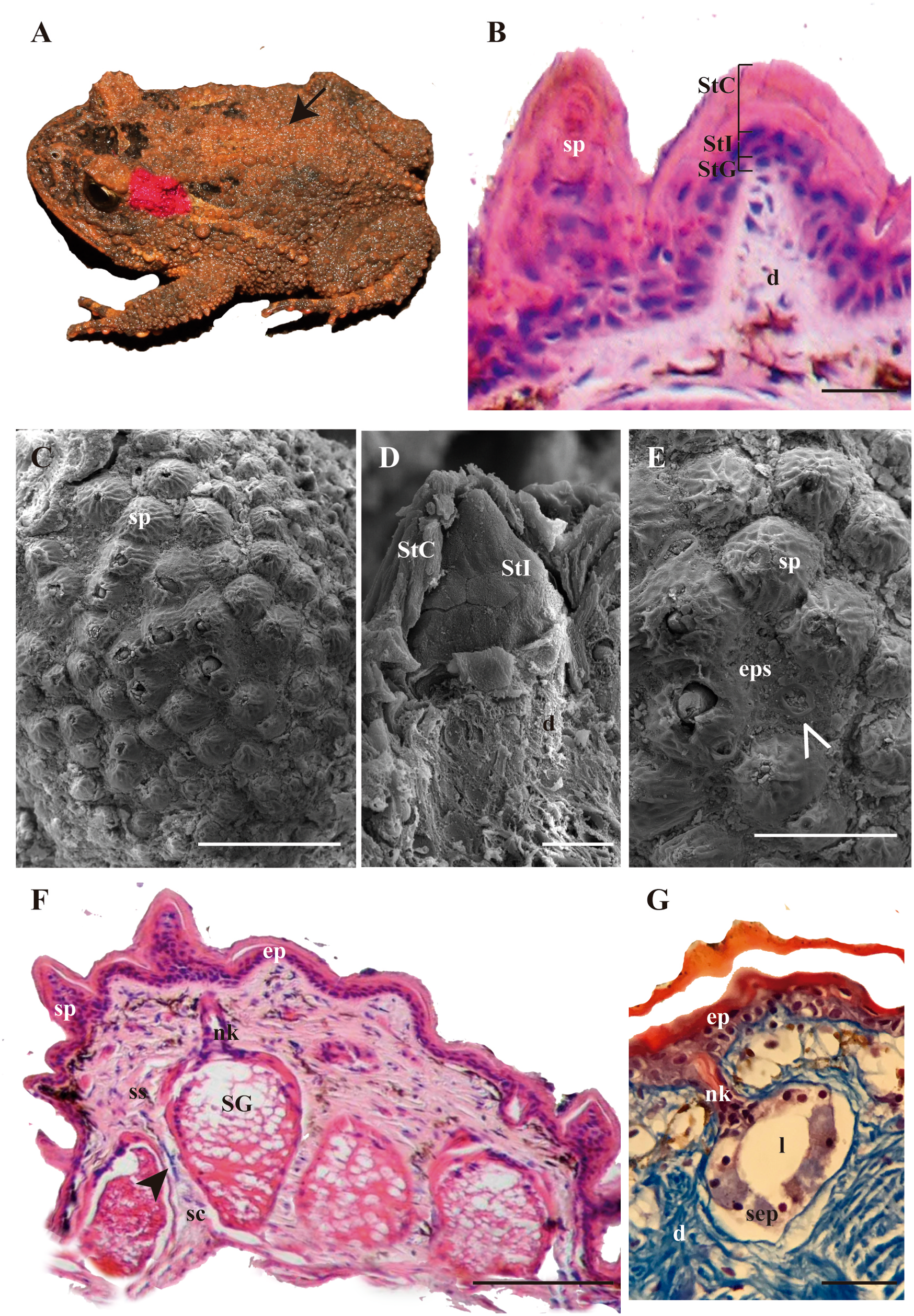
The skin of the postorbital-supratympanic and dorsal regions of the two examined species of the genus Proceratophrys ( P. avelinoi and P. brauni ) is similar, both externally and histologically. Macroscopically, the two species show small protuberances homogeneously distributed throughout these regions ( Fig. 9A View Fig ). Microscopically, these protuberances have an epidermis consisting of 3–4 cell layers: the basal layer with cylindrical cells ( stratum germinativum), a medial 1–2 layer of cubic cells ( stratum intermedium), and a very thick layer of flat, highly keratinized cells ( stratum corneum) ( Fig. 9B View Fig ). The epidermal surface is completely covered with conical spines ( Figs. 1C View Fig and 9C View Fig ). The latter formed by an increase in the thickness of the stratum intermedium, with superficial highly keratinized cells of the stratum corneum ( Fig. 9D View Fig ). This stratum reaches a size of 49.9–57.5 μm height and 57.2–60.3 μm width ( Table 1). Between the spines, it is possible to observe valleys where the glandular pores are located ( Fig. 9E View Fig ).
As for the dermis, the stratum compactum and stratum spongiosum are well defined and bound by the thin, discontinuous EK layer ( Fig. 9F View Fig ; Table 1). The alveolar glands have a circular pore ( Table 1), a short duct-
(caption on next page)
neck and a secretory epithelium with cylindrical cells surrounding a broad lumen ( Fig. 9G View Fig ). The cells possess secretory granules rich in neutral glycoconjugates and proteins ( Table 2). The syncytial glands are partially clustered with connective tissue between the secretory portions ( Fig. 9F View Fig ). They have circular pores ( Fig. 9E View Fig ; Table 4) that communicate with the secretory portions through a duct-neck lined by a bistratified epithelium ( Fig. 10A View Fig ). The secretory portion consists of a broad acidophilic secretory syncytium with ovoid nuclei ( Fig. 9F View Fig ). The secretion of these glands is characterised by an acidophilic network, and basophilic and acidophilic granules ( Figs. 9F View Fig and 10B View Fig ) with neutral glycoconjugates ( Fig. 10C View Fig ), lipids and proteins ( Tables 2 and 3).


No known copyright restrictions apply. See Agosti, D., Egloff, W., 2009. Taxonomic information exchange and copyright: the Plazi approach. BMC Research Notes 2009, 2:53 for further explanation.