
Parastichopus regalis ( Cuvier, 1817 )
|
publication ID |
https://doi.org/10.11646/zootaxa.5032.4.5 |
|
publication LSID |
lsid:zoobank.org:pub:E84B3D64-EDE7-4143-B65F-8BAED6AD4744 |
|
DOI |
https://doi.org/10.5281/zenodo.5498931 |
|
persistent identifier |
https://treatment.plazi.org/id/03AB87B4-1167-B31E-FF44-FAA6A954F802 |
|
treatment provided by |
Plazi |
|
scientific name |
Parastichopus regalis ( Cuvier, 1817 ) |
| status |
|
Parastichopus regalis ( Cuvier, 1817) View in CoL spotted morph
Figures 4–5 View FIGURE 4 View FIGURE 5 , Table 2
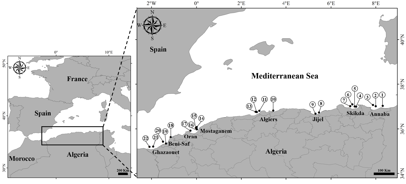
Material examined. LPVCMRMS2019.305; LPVCMRMS2019.306, Mostaganem, Algeria, 36° 05.077’ N, 00° 00.490’ E, 225 m, 03 June 2019 GoogleMaps , 2 specs. LPVCMRMS2019.307, Mostaganem, Algeria, 36° 02.611’ N, 00° 01.823’ O, 117 m, 03 June 2019, 1 spec. GoogleMaps LPVCMRMS2019.311; LPVCMRMS2019.312, Bouzedjar , Algeria, 35° 38.068’ N, 01° 08.766’ O, 96 m, 04 June 2019 GoogleMaps , 2 specs. LPVCMRMS2019.310, Arzew , Algeria, 35° 55.444’ N, 00° 00.490’ E, 77 m, 04 June 2019, 1 spec. GoogleMaps
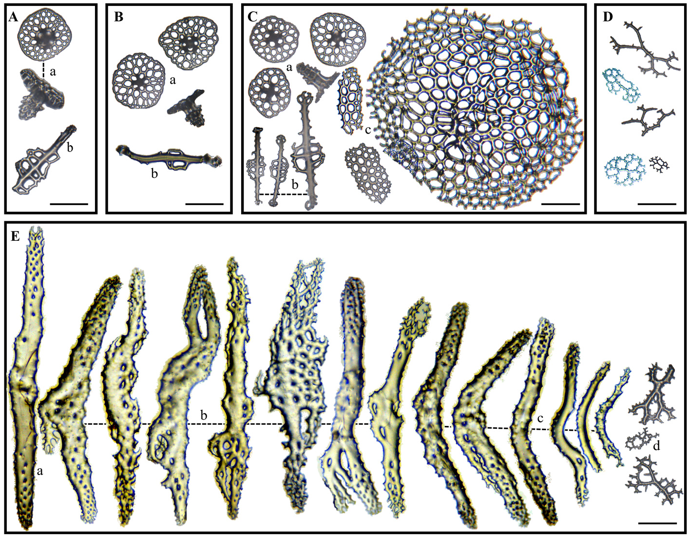
Description. Specimens ( Figures 4A and 4B View FIGURE 4 ) preserved length 14–24.90 cm, contracted width 4–7.50 cm; total weight 25.07–143.50 g. Each with 5–7 brownish to dark red blotches on the bivium when alive ( Figure 4A View FIGURE 4 1 View FIGURE 1 ) disappearing after storage in ethanol. Morphological characters similar to non-spotted specimens described above. Dorsal body wall 1.23–6.19 mm thick; ventral body wall 1.22–6.77 mm thick. Polian vesicle single, average length 17.57 mm; stone canal also single, average length about 10.71 mm. Calcareous ring about 13 mm in diameter. Longitudinal muscles flat, attached to body wall, about 6.75 mm in thickness ( Table 2).
Ossicles. Anal region ( Figure 5A View FIGURE 5 ), dorsal ( Figure 5B View FIGURE 5 ) and ventral surfaces ( Figure 5C View FIGURE 5 ) present the same type of ossicles as non-spotted specimens. Table discs ( Figures 5 View FIGURE 5 Aa; 5Ba; 5Ca) 97.926 µm in average diameter and 7099.090 µm ² in total area. Spire 4-pillared, about 67.521 µm high, 64.691 µm wide, with 3–4 cross-bars. Papillae and pedicels include perforated rods ( Figures 5 View FIGURE 5 Ab; 5Bb; 5Cb) as in the non-spotted morph, 265.026 µm in average length. Complete pedicel end plates ( Figure 5 View FIGURE 5 Cc) circular, average diameter 439.218 µm; oval end plates 197.766 µm in mean length and 84.328 µm in mean width. Cloaca ( Figure 5D View FIGURE 5 ) presents dichotomously branched rods. Tentacle rods ( Figure 5E View FIGURE 5 ) elongated ( Figure 5 View FIGURE 5 Ea), or arched ( Figure 5 View FIGURE 5 Ec), branched ( Figure 5 View FIGURE 5 Eb) and dichotomously branched rods ( Figure 5 View FIGURE 5 Ed).
Remarks. Both spotted and non-spotted Parastichopus regalis individuals degrade very quickly if they are not immediately preserved in ethanol, after collection. The dermis rapidly disintegrates and the texture of the body becomes gelatinous, spreading out on the support on which the specimen is placed ( Figure 4C View FIGURE 4 ). This reaction is probably due to the stress during fishing operation and/or exposure to light or change in pressure and/or temperature, since fished from waters more than 36 m deep. The ability to lose a piece of dermis from the body wall when in danger has been observed in many holothuroids ( Kropp 1982).
The use of formalin, although good for fixation, does not allow for better preservation because the body does not keep its original shape and the ossicles tend to corrode rapidly. Hence, alcohol is recommended although the bright red colour of the ventral surface as well as the dark spots on the dorsal surface (when present) do disappear. The thickness of the body wall can vary from one individual to another since the general texture of the body wall is very soft, thus not rigid enough to maintain the original form.

No known copyright restrictions apply. See Agosti, D., Egloff, W., 2009. Taxonomic information exchange and copyright: the Plazi approach. BMC Research Notes 2009, 2:53 for further explanation.
|
Kingdom |
|
|
Phylum |
|
|
Class |
|
|
Order |
|
|
Family |
|
|
Genus |