
Archicorynus Anderson and Marvaldi, 2013
|
publication ID |
https://doi.org/10.1649/0010-065X-67.2.61 |
|
publication LSID |
lsid:zoobank.org:pub:F9908407-F3AF-43F4-B9D2-EA892143231D |
|
persistent identifier |
https://treatment.plazi.org/id/86E9D3BC-890E-4963-99F9-5B9FFE200AB3 |
|
taxon LSID |
lsid:zoobank.org:act:86E9D3BC-890E-4963-99F9-5B9FFE200AB3 |
|
treatment provided by |
Carolina (2021-08-29 18:10:25, last updated by Plazi 2023-11-05 16:20:00) |
|
scientific name |
Archicorynus Anderson and Marvaldi |
| status |
gen. nov. |
Genus Archicorynus Anderson and Marvaldi , new genus
Type species. Archicorynus kuscheli Anderson and Marvaldi.
Etymology. The genus name is a combination of the Greek nouns ‘arche’ (first of its kind, original, primitive) and ‘koryne’ (a club or mace). The gender is masculine.
Diagnosis. Archicorynini characterized by sulcus bordering eye extended around eye, elytra with interstriae 3 (at declivity) and 5 (strongly throughout length) variously carinate and interstria 7 slightly swollen at base and slightly elevated in apical half (but not carinate) ( Figs. 1–2 View Figs ), pro- and mesotibiae with robust, strongly recurved apical tooth on inner margin ( Fig. 3 View Figs ), femora (especially pro- and meso-) strongly dorsally arched but lacking dorsal crenulations or denticles, tergite VIII of female with apical margin cleft medially, with pair of ventrally directed, curved, sharp hooks ( Figs. 5 View Figs , 10 View Figs ), and apodemes of penis (temones) short ( Figs. 6 View Figs , 11 View Figs ). The genus is also recognizable by its restricted geographical distribution (Cerro Musún, Nicaragua), characteristically elongate shape with a dirty, unkempt appearance ( Figs. 1–2 View Figs ), antennae composed of 11 segments, the basal and second more robust than 3–8 and 9–11 forming a compact club, its basal segment (9) more heavily sclerotized and darker in color. Furthermore, the body is covered with fine, elongate, appressed hairs, which in many places are erect, longer and denser and trap dirt and debris to give the beetle a scruffy, dirty, frass-covered look ( Figs. 1–2 View Figs , 8 View Figs ). These hairs are denser and clumped in a pair of paramedian inverted “Y”-shaped areas on the pronotal disc, the anterior stem of the “Y” originating as a swollen projection on the anterior margin of the pronotum behind the eye and the posterior arms diverging to about the basal one-third or one-fourth of the pronotal length. Similar fine, erect hairs are present above the eyes, around the margin of the pronotum, in a number of fine lines down the length of each tibia, along the carinate elytral interstriae, and, most distinctly, the elytra are fringed by a number of rows of these same dense, apically curved hairs that are extended laterally in a narrow plane, thus giving the epipleural region the appearance of being widely explanate. In addition, this explanate fringe is distinctly impressed ventrally on each side (giving it a sinuate or wavy appearance when viewed laterally; Fig. 2 View Figs ) opposite the metacoxa such that the apical portion of the metacoxa can swing past the epipleural fringe when at its dorsal apogee without making contact. The elytral apices appear prolonged and acuminate but again are clumps of these same dense, erect hairs ( Figs. 1 View Figs , 8 View Figs ). The ventrally curved hooks on the apical margin of tergite VIII of the females have not been observed in other weevils, but in Aralius Kuschel (Aglycyderini) the tergite VIII is multidentate at the apical margin ( Kuschel 2003: Fig. 96).
Description. Body length 3.1–4.6 mm; width 1.2–1.4 mm. Cuticle ( Figs. 1–2 View Figs ) densely, finely pilose with dense, erect hairs fringing margins of elytra and pronotum, above eyes, variously along elytral interstriae 3 and 5 where carinate, in linear rows along length of tibiae, in two anterior projections on pronotal margin, and in paramedian inverted-“Y” shapes on pronotal disc (these hairs trapping dirt and debris and individually difficult to discern). Head ( Figs. 1–2 View Figs , 8 View Figs ) with eyes small but distinct, prominent. Gular area with sutures largely covered by prosternal margin, short, separate basally, rounded and convergent (though not confluent) apically a short distance from basal margin of head. Rostrum ( Fig. 2 View Figs ) cylindrical, slightly shorter than length of pronotum in female, more so in male. Maxillary palps distinct, large, 3-segmented; prementum small, labial palps small but evident. Antennal insertions lateroventral near base of rostrum, about 1/3 length of rostrum in front of anterior margin of eyes. Antennae ( Fig. 2 View Figs ) 11-segmented, moderately long, fine, reaching just beyond anterior margin of pronotum behind eyes; segments 1 and 2 more robust, subequal in width, 3–8 much more slender (especially 3–5); 1 subequal in length to 2 and 3, segments progressively slightly shorter; club compact, 3-segmented, terminal segments tightly joined, basal one (9) more heavily sclerotized, dark, faintly shining. Pronotum widest just before base, lateral margins carinate and crenulate (but some of this due to multiple rows of apically recurved, dense fine hairs extended laterally from margin). Pronotal disc ( Fig. 1 View Figs ) with paramedian inverted-“Y”-shaped areas, anterior stem originating as swollen projection on anterior margin of pronotum behind eye and posterior arms divergent, reaching to about basal 1/3 or 1/4 of pronotal length; posterior margin low, broadly rounded. Elytra ( Fig. 1 View Figs ) 9-striate but striae indistinct, the punctures deep, large, arranged in somewhat con- fused rows; intervals flat except at base of interval 7 (slightly swollen), throughout length of 5 and at declivity of 3 (raised and carinate, accentuated by presence of rows of fine erect hairs). Ventral surface of elytra in proximal half near outer margin below wing-folding pad with submarginal ridge forming a pocket-like excavation ( Fig. 9 View Figs ). Epipleura ( Figs. 3 View Figs , 8 View Figs ) present, strongly accentuated by multiple rows of dense, apically curved hairs extended laterally in narrow fringing plane, thus giving elytral margins appearance of being widely explanate, fringe distinctly impressed dorsally (giving it a sinuate or wavy appearance when viewed laterally) opposite metacoxa, underside of this area (inside) with the pocket-like cavity. Scutellar shield moderately large, distinct. Hindwings ( Fig. 13 View Figs ) present, well-developed; anal posterior vein (AP3+4) present; veins MP4 and MP3 present, CuA2 absent; anal lobe delimited by (notchlike) anal embayment; setae of hind margin present except on anal lobe; apical field long, about 50 % of total wing length; vein r4 (between radial cell and RP) distinct; vein AA4 with distal portion after anal cell; seta at apical part of MP1+2 present; mediocubital cell rounded at apex; anal fold distinct. Legs ( Fig. 4 View Figs ) somewhat robust, tibiae straight, femora (especially pro- and meso-) dorsally strongly arched, with dorsal margin lacking crenulations or denticles. Tibiae densely pilose, with rows of linearly arranged, fine, erect hairs, width greatest towards base, tapered apically; pro- and mesotibiae with robust, strongly recurved apical tooth on inner margin. Tarsi 5-segmented, segments 1–3 progressively longer; 1 small, submoniliform, 2 slightly bilobed, 3 strongly bilobed, 4 extremely small, indistinct, at base of 3; 5 moderately elongate, moderately robust, claws robust, free, divergent; segments 1–3 with dense ventral pilosity in center (1) or on lobes (2 and 3). Thoracic venter ( Figs. 3 View Figs , 8 View Figs ) with dense, appressed, fine pilosity. Procoxae distinctly separated by projections of anterior and posterior portions of prosternum meeting between coxae. Mesocoxae widely separated by about coxal width. Metacoxae separated by about 1/3 coxal width. Metaventrite linearly impressed along midline. Abdomen ( Figs. 3 View Figs , 8 View Figs ) with 5 large, freely articulating ventrites; ventrite 1 slightly longer than 2, 2 slightly longer than 3, 3–5 subequal in length; all ventrites finely, densely pilose and punctate. Pygidium (tergite VII) shining, with rows of setae along posterior margin, apex evenly rounded. Tergite VIII ( Figs. 5 View Figs , 10 View Figs ) of female with apical margin distinctly cleft, bearing pair of sharp, ventrally curved hook-like extensions, apex of each hook well-sclerotized (dark), acuminate and divergent; sternite VIII “Y”-shaped, with apodeme very long; ovipositor with distal gonocoxites well sclerotized, styli absent; spermatheca distinct, falciform, gland larger than body, tapering to spermathecal duct inserted ventrally on bursa near or at base of common oviduct. Aedeagus ( Figs. 6–7 View Figs , 11–12 View Figs ) wellsclerotized, penis very long and slender, slightly curved ventrally; struts (temones) very short, about 1/6 length of body, pedon and tectum distinct but narrowly separated; tegmen in form of elongate loop, arms joined at base, dorsal plate broad, relatively short, apex truncate, extended towards and almost reaching apex of penis; internal sac with no apparent sclerotization.
Kuschel, G. 2003. Nemonychidae, Belidae, Brentidae (Insecta: Coleoptera: Curculionoidea). Fauna of New Zealand 45: 1 - 100.


Figs. 5–7. Archicorynus kuscheli, genitalia. 5) Female; 6) Aedeagus and tegmen, dorsal view; 7) Aedeagus and tegmen, lateral view.
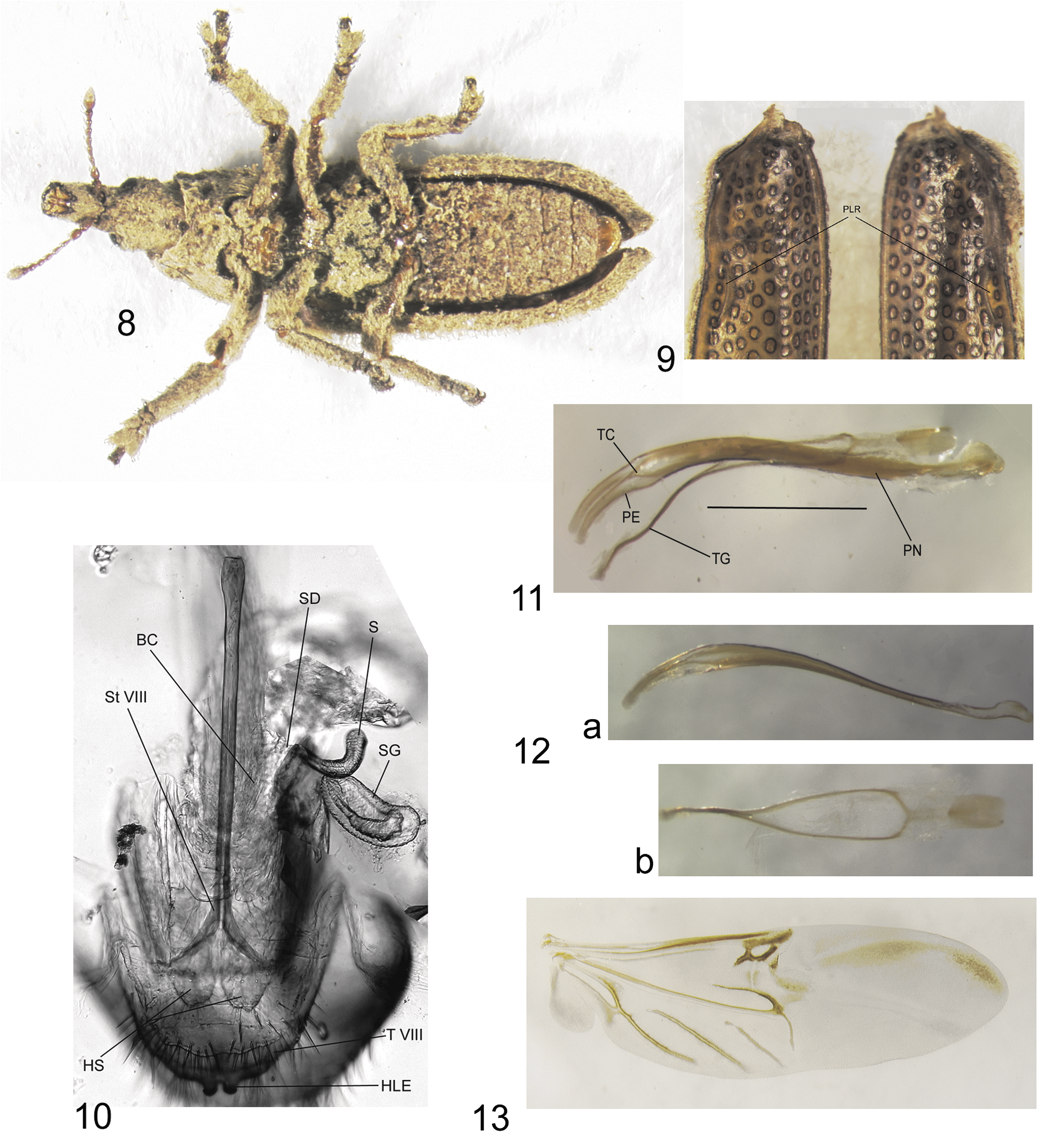
Figs. 8–13. Archicorynus kuscheli. 8) Male, ventral view; 9) Ventral surface of elytra, male (PLR = pocket-like cavity); 10) Female genitalia (BC = bursa copulatrix; HLE = hook-like extensions of T VIII; HS = gonocoxites; S = spermatheca; SD = spermathecal duct; SG = spermathecal gland; S VIII = sternite VIII; T VIII = tergite VIII); 11) Aedeagus, lateral view (PN = penis; PE = pedon; TC = tectum; TG = tegmen); 12) Penis (a) and tegmen (b), separated; 13) Hindwing, male.
No known copyright restrictions apply. See Agosti, D., Egloff, W., 2009. Taxonomic information exchange and copyright: the Plazi approach. BMC Research Notes 2009, 2:53 for further explanation.
|
Kingdom |
|
|
Phylum |
|
|
Class |
|
|
Order |
|
|
Family |