
Cecinothofagus ibarrai Nieves-Aldrey & Liljeblad
|
publication ID |
https://doi.org/10.5281/zenodo.189597 |
|
DOI |
https://doi.org/10.5281/zenodo.5681178 |
|
persistent identifier |
https://treatment.plazi.org/id/03ADE415-FFD0-FFBE-FF58-9506FCBBBD9C |
|
treatment provided by |
Plazi |
|
scientific name |
Cecinothofagus ibarrai Nieves-Aldrey & Liljeblad |
| status |
sp. nov. |
Cecinothofagus ibarrai Nieves-Aldrey & Liljeblad sp. nov.
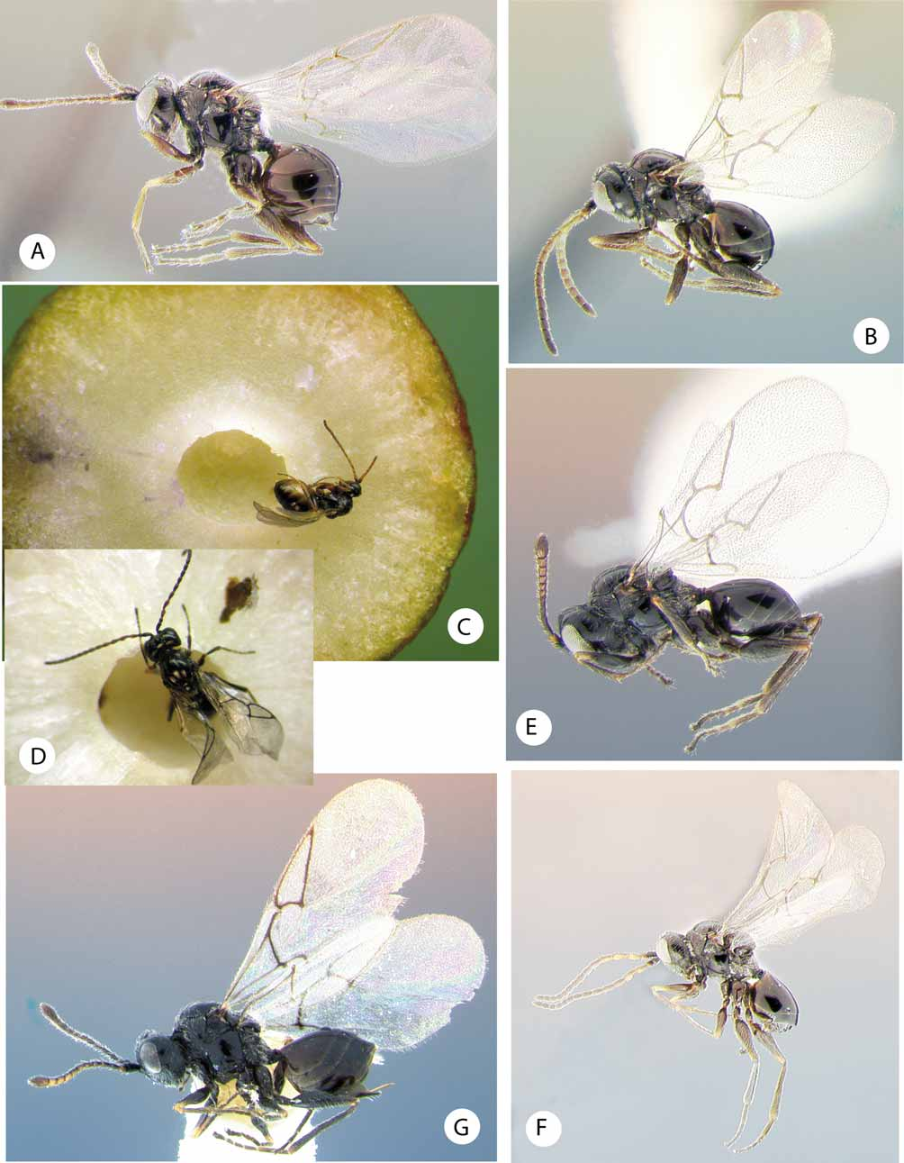
( Figs. 4 View FIGURE 4 I–K & 11E–F)
Type material. Holotype Ƥ (in Museo Chileno de Historia Natural, Santiago de Chile, card mounted) CHILE, Puerto Varas-Ensenada, 41º 12´52.55´´S 72º41´30.37´´O, 102 m; ex gall Aditrochus coihuensis on Nothofagus dombeyi (Mirb.) Blume “coigüe” ( Nothofagaceae ), gall collected 2/xii/2006. insect emerged xii/2006, J.L. Nieves-Aldrey leg. Paratypes: 13, 1Ƥ same data as holotype; 1Ƥ same data, except gall collected at Ensenada-Ralún. Additional material: one male preserved in ethanol, same data as holotype. One paratype 3 in MCHN, the remaining paratypes in Museo Nacional Ciencias Naturales, Madrid ( Spain). Non-type material: 1Ƥ, Chile, PN Nahuelbuta, 961 m., 12.xii.2001, canopy fogging Nothofagus dombeyi . Arias et al. leg. ( UCRC); 13 Chile, Malleco, 1500 m 110 km W Curacautin, 12.xii.1984. Nothofagus -Araucaria forest . S & J Peck leg. ( CNC). 13 ARGENTINA, Esquel-Chobut ex gall on Nothofagus dombeyi , 03.xii.00. S. Rizzuto leg. ( MNCN). This specimen agrees with the diagnostic antennal characters of P. ibarrai but resembles P. gallaecoihue in coloration and notauli characteristics.
Etymology. Named after Hector Ibarra, Chilean colleague, who helps us study galls on Nothofagus in Chile.
Diagnosis. Closely allied to P. gallaecoihue . Differs by body almost completely shining black ( Fig 11 View FIGURE 11 E); legs and antenna blackish or dark brown. The main morphological difference refers to the antennal conformation. While in P. ibarrae F2 of the male antenna is conspicuously expanded distally ( Fig. 4 View FIGURE 4 I), it is only slightly modified in P. gallaecoihue . Females are readily differentiated by the relative lengths of, antennomeres A2–A4.
Description. Body length 2.55 mm (range 2.5–2.6; N = 2) for females; 2.8 mm (range 2.7–2.9; N = 2) for males. Coloration of female, body shining black; antenna dark brown; legs black except apex of femora, tibia and tarsi brown. Forewing hyaline, veins dark brown. Male similar in coloration to female, but metasoma and flagellum not as dark.
Female. Head, in dorsal view ( Fig. 4 View FIGURE 4 J) 1.8 times wider than long. Gena not expanded behind compound eye; in dorsal view almost as long as compound eye. POL 1.8 times longer than OOL, posterior ocellus separated from inner orbit of eye by about 1.8 times its diameter. In anterior view, head more or less subquadrate or slightly trapezoid, 1.1 times wider than high, lateral margin of gena not forming a continuous arch with outer margin of compound eye. Face with sparse, long setation, denser in lower face, almost lacking in median area of frons; facial strigae radiating from clypeus medially absent; laterally well marked, reaching close to ventral margin of compound eye; strong vertical median carina present, running from ventral margin of clypeus almost reaching ventral margin of toruli. Upper face (frons) and vertex almost entirely smooth and glabrous. Ocellar plate slightly raised; malar space about 0.6 times height of compound eye. Clypeus indistinct, ventral margin straight, not incised. Subocular impression indistinct, visible as a shallow furrow. 6– 8 regular, vertical carinae present ventrolaterally in a depression on the gena. Anterior tentorial pits conspicuous; epistomal sulcus and clypeo-pleurostomal lines indistinct. Antennal socket (torulus) situated at about mid height of compound eye; distance between antennal rim and compound eye 0.5 times width of antennal socket including rim. Occiput with coriaceous sculpture, dorsally pubescent, without dorsal occipital carina, sharp, well marked genal occipital carina present. Posterior tentorial pits narrow, slit-like. Hypostomal sulci meeting at hypostoma. Distance between occipital and oral foramina 0.7 times height of occipital foramen.
Mouthparts. Mandibles strong, exposed; right mandible with three teeth; left with two teeth. Cardo of maxilla visible, maxillary stipes about 2.3 times longer than wide. Maxillary palp five-segmented. Labial palp three-segmented. Lateral margins of oral fossa with a band of 3–4 rows of white setae.
Antenna 0.5 times length of body, 12-segmented; flagellum slightly broadened towards apex; last flagellomere distinctly wider than penultimate; slightly truncate at apex, ending in a semicircular point. Antennomeres with sparse setation, about as long as width of flagellum basally. Placodeal sensilla indistinct, visible only on F7–F10. Relative length of antennomeres: 30:13:18:22:18:19:15:14:14:16:15:42; pedicel 1.2 times longer than wide; F1 0.8 times length of F2. Ultimate flagellomere 2.4 times longer than wide, 1.3 times wider han penultimate and 2.8 times longer than F9, ending in a semicircular, truncate apex.
Mesosoma. Pronotum in anterior view, almost glabrous medially, strongly pubescent laterally. Ratio of length of pronotum medially/length laterally = 0.4. Pronotal plate distinct; dorsal part distinctly set off, anterolateral margins marked and moderately projecting laterad ( Fig. 4 View FIGURE 4 J). Admedian pronotal depressions oval/transverse, open laterally, separated by as much as median length of pronotum. Lateral surface of pronotum smooth; sparsely pubescent by long, white setae. A few short, horizontal rugae running from lateral margin of pronotal plate.
Mesonotum. Mesoscutum ( Fig. 4 View FIGURE 4 J) 1.2 times wider than long; shining, without visible median sculpture, at most some superficial, delicately coriaceous sculpture present laterally. Pubescence in the form of long setae present only along margins of notauli. Median mesoscutal impression absent. Notauli complete, sinuate, not strongly converging posteriorly, not reaching the transscutal fissure, wider in posterior half. Separation of notauli at transscutal fissure relatively wide, 0.5 times separation at anterior margin of mesoscutum. Anteroadmedian signa weakly visible. Mesoscutum and scutellum separated by a narrow transscutal fissure. Scutellar foveae indistinct, visible only as a shallow, smooth and glabrous depression. Scutellum, in dorsal view more or less pentagonal in shape; in lateral view strongly convex. Dorsal surface of scutellum coriaceous with some rugae, more marked on lateral and posterior areas and almost absent medially. Posterodorsal and posterior margins of axillula distinct. Mesopleuron beneath mesopleural triangle smooth and glabrous excep for horizontal furrow or mesopleural impression in lower part; mesopleural impression relatively wide but incomplete, not reaching ventral margin of mesopleural triangle. Mesopleural triangle distinctly impressed and densely pubescent; dorsal margin diffuse anteriorly, not meeting area near prepectus, instead meeting posterolateral margin of pronotum well below prepectus.
Metanotum. Metascutellum distinctly constricted medially. Area posterior to median constriction of metascutellum not divided by a median vertical bar. Median metascutellum narrower than metanotal trough. Metanotal trough smooth, pubescent.
Metapectal-propodeal complex. Metapleural sulcus reaching posterior margin of mesopectus at about mid height of metapectal-propodeal complex. Lateral propodeal carinae narrow, parallel. Width of median propodeal area 0.8 times length. Lateral and median propodeal areas smooth, pubescent. Nucha dorsally with some irregular rugae.
Legs. Profemur with process of 4–5 rows of sharp, closely spaced, deep costulae visible as swelling in basal third of profemur. Tarsal claw with basal, small lobe, not distinctly developed into secondary tooth.
Forewing ( Fig. 4 View FIGURE 4 K). As long as body. Radial cell closed along anterior margin, about 3 times longer than wide; radius (Rs) straight, reaching anterior margin of wing. Areolet indistinct; vein Rs+M weakly visible, directed towards lower half of median vein; M weakly indicated anteriorly. Fringe of long setae along apical margin of wing.
Metasoma. Metasoma shorter than head plus mesosoma; in lateral view 1.7 times longer than high; laterally compressed. Abdominal petiole dorsally smooth, ventrally with deep longitudinal grooves. T1 flapshaped; laterally 2 times higher than long, with some longitudinal rugae. T2 covering about half of metasoma; anteromedian area of T2 with group of a few long setae. Projecting part of hypopygial spine 3.4 times longer than high; apical pubescence of hypopigial spine projecting beyond apex, subapical setae longer than apical ones, together forming a small tuft.
Male. Similar to female except as follows. Male antenna ( Fig. 4 View FIGURE 4 I) with 15 segments. Flagellum not distinctly expanded towards apex. F1 slightly broadening from base to apex; F2 curved basally, strongly e x p a n d e d i n a p i c a l t h i r d; F 3 n o t m o d i f i e d. R e l a t i v e l e n g t h o f a n t e n n o m e r e s: 19:9:19:24:20:18:16:15:13:15:13:14:12:13:20. Placodeal sensillae present on all flagellomeres. Metasoma smaller than that of female; 1.5 times longer than high; T2 0.4 times length of metasoma. Anteromedian area of T2 with group of 3–4 setae.
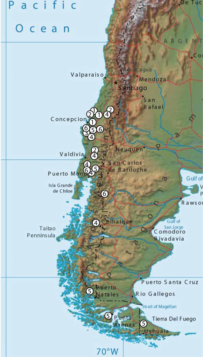
Distribution. Recorded from Nothofagus dombeyi forests in Argentina and Chile ( Fig. 15 View FIGURE 15 ). Since two cynipid species share the same host gall and plant, the potential distribution is similar to that of the related species C. gallaecoihue .
Biology. As with C. gallaecoihue , this species is an inquiline or parasitoid in bud galls induced by Aditrochus coihuensis (Chalcidoidea, Pteromalidae ) on Nothofagus dombeyi (Mirb.) Blume (Nothofagaceae) . N. dombeyi , commonly named “coihue”, is an evergreen tree native of Southern Argentina and Chile. The host gall induced by Aditrochus coihuensis is a large spherical bud gall ( Fig. 12 View FIGURE 12 C), sometimes with an apical point ( Fig. 12 View FIGURE 12 A). The surface is covered by small, blister-like, brown protuberances (lenticeles). The gall is locally common. It is variable in size, measuring between 4–20 mm with an average for the larger galls of 10– 12 mm. A sectioned gall ( Fig. 12 View FIGURE 12 B) shows the central larval chamber, the sclerenchyma wall of the larval cell and the outer layers of parenchyma and sclerenchyma. This is similar to the structurally complex oak galls induced by some species in the genera Cynips and Andricus .


No known copyright restrictions apply. See Agosti, D., Egloff, W., 2009. Taxonomic information exchange and copyright: the Plazi approach. BMC Research Notes 2009, 2:53 for further explanation.
|
Kingdom |
|
|
Phylum |
|
|
Class |
|
|
Order |
|
|
Family |
|
|
Genus |