
Scolelepis magnicornuta, Williams, Jason D., 2007
|
publication ID |
https://doi.org/10.5281/zenodo.176375 |
|
DOI |
https://doi.org/10.5281/zenodo.6242425 |
|
persistent identifier |
https://treatment.plazi.org/id/03AF87BA-6E24-4D7D-FF7A-FA9121CEF9F9 |
|
treatment provided by |
Plazi |
|
scientific name |
Scolelepis magnicornuta |
| status |
sp. nov. |
Scolelepis magnicornuta View in CoL sp. n.
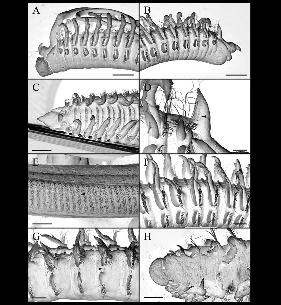
( Figs. 14–15)
Material examined. Holotype. Philippines, Diniwid Beach, Boracay, 11°60´N, 121°54´E, sandy beach, 13 Apr 1999 ( USNM 1096808).— Paratypes, same data as holotype (three complete specimens, two anterior ends, two posterior ends in alcohol, USNM 1096809; two anterior ends on SEM stubs, one middle section on SEM stub, one pair of palps on SEM stub, USNM 1096810); same location as holotype, 14 Apr 1999 (one anterior end in alcohol, ZRC 2006.0223).
Etymology. The species epithet magnicornuta (derived from the Latin adjective magni for great, large and Latin adjective cornutus for horned) refers to the large, conical occipital tentacle found in this species.
Diagnosis. A species of Scolelepis exhibiting notosetae on setiger 1, notopodial hooded hooks and large occipital tentacle. Palps long, with small, smooth basal sheath, palps with two weakly separated transverse rows of cilia. Prostomium conical, with two pairs of eyes. Caruncle ending bluntly at middle of setiger 1, nuchal cilia in semi-circular pattern around base of palps, cilia extending onto sides of large, conical occipital tentacle. Postsetal notopodial lamellae nearly completely fused with branchiae from setiger 2 to middle setigers, lamellae elongate, rounded, distal end pointed and free from branchiae, lamellae and branchiae separated posteriorly. Bidentate notopodial hooded hooks from setiger 38–55, up to seven in posterior setigers. Bidentate neuropodial hooded hooks from setiger 25–30, up to 13 in middle to posterior setigers. Pygidium small, rounded, with dorsal anal open.
Description. Holotype of 82 setigers, largest complete specimen, 42.1-mm long, 1.9-mm wide by setiger 20; setigers 24–72 with eggs. Body widest anteriorly, gradually tapering to posterior end; body nearly rectangular in cross section. Color in alcohol opaque white, no pigmentation present.
Prostomium conical, extending anteriorly to sharp point, continuing posteriorly as short caruncle, ending bluntly at middle of setiger 1 along a low, indistinct margin; reduced peristomial wings surrounding base of palps ( Figs. 14 A, B, 15A–C). Two pairs of eyes obscured, deeply embedded, in nearly straight row between bases of palps; large, conical occipital tentacle present ( Fig. 15 View FIGURE 15 B–D). Palps extending to setiger 15–20, with two weakly separated transverse rows of cilia along ventral surface, long rows of cilia approximately 41-µm long, short rows approximately 26-µm long (short rows on medial side), with long rows and short rows in approximately 1:1 ratio, rows of mucus secreting cells (represented by tubular necks) proximal to transverse ciliary rows, rows extending to distal ends of palps, median ciliated groove lacking ( Figs. 15 View FIGURE 15 E, 18C). Palps with small, smooth basal sheath. Nuchal cilia in semi-circular pattern around base of palps, cilia extending onto lateral sides of base of occipital tentacle ( Fig. 15 View FIGURE 15 C–D).
A B C D E J K F G H I
F-I
Setiger 1 well developed with rounded notopodial and neuropodial postsetal lamellae, notosetae and neurosetae present; patch of cilia present dorsal to the notopodial lamellae. Postsetal notopodial lamellae nearly completely fused with branchiae from setiger 2 ( Fig. 15 View FIGURE 15 A–B), lamellae elongate, rounded, distal end pointed and free from branchiae, fused with branchiae along about 90% of length in anterior setigers, degree of fusion decreasing in posterior setigers ( Figs. 14 C–E, 15F–G). Postsetal neuropodial lamellae of setiger 1 with triangular lobe ( Figs. 14 B, 15A–B), lamellae of setigers 2–18 with rounded lobe ( Figs. 14 C, 15A–B, F), developing notch dividing lamellae by setigers 20-25, with broad dorsal lobe and small ventral lobe, dorsal lobe with pointed dorsal end and rounded ventral end ( Figs. 14 D, 15G), in posterior setigers dorsal lobe becomes broader and ventral lobe is reduced ( Fig. 14 E). Lateral organs between notopodial and neuropodial postsetal lamellae present from setiger 1 to posterior setigers ( Fig. 15 View FIGURE 15 G).
Notosetae of setiger 1 and subsequent setigers in two vertical rows of limbate capillaries ( Fig. 14 I); dorsal notosetae of these rows longer than those below, 1–2 notopodial hooded hooks from setiger 38–55, with up to seven notopodial hooded hooks in posterior setigers; notopodial hooded hooks bidentate with acute main fang and single accessory tooth ( Fig. 14 H). Neurosetae of setiger 1 and subsequent setigers in two vertical rows of limbate capillaries ( Fig. 14 G), 1–6 neuropodial hooded hooks from setiger 25–30, with up to 13 neuropodial hooded hooks in middle to posterior setigers; hooks bidentate with acute main fang and single accessory tooth ( Fig. 14 F).
Branchiae from setiger 2 to end of body, fused with postsetal notopodial lamellae but with tips free in anterior setigers ( Fig. 15 View FIGURE 15 A–B, F–G), separated from notopodial lamellae in posterior setigers, with band of cilia along inner edge and joined to corresponding branchiae on opposite side by two bands of cilia across dorsum, anterior band broader than posterior band ( Fig. 15 View FIGURE 15 C).
Pygidium small, rounded, with dorsal anal opening ( Figs. 14 J, K, 15H).
Remarks. Scolelepis magnicornuta sp. n., belongs to a group of nine species including S. antipoda ( Augener, 1926) ; S. foliosa (Auduoin & Milne-Edwards, 1833) ; S. lingulata Imajima, 1992 ; S. magnus Ozolin'sh, 1990; S. occidentalis Hartman, 1961 ; S. oligobranchia Khlebovitsch, 1959 ; S. quadridentata Maciolek, 1987 ; and S. sagittaria Imajima, 1992 that possess an occipital tentacle, notosetae on setiger 1, and notopodial hooded hooks. Scolelepis magnicornuta sp. n. is unique among these species in having a large occipital tentacle with cilia extending along the sides. Among the species indicated above, S. magnicornuta sp. n. most closely resembles S. antipoda , S. lingulata , S. occidentalis , and S. sagittaria in having the tips of the notopodial lamellae free from the branchiae, whereas other species in this group exhibit complete fusion of the notopodial lamellae and branchiae in the anterior setigers. Scolelepis magnicornuta sp. n. is distinguished from S. antipoda based on the body size and initiation of neuropodial hooded hooks. S. antipoda is a large species with up to 194 setigers and with neuropodial hooded hooks from setiger 47–50 whereas S. magnicornuta sp. n. has up to 82 setigers and neuropodial hooded hooks from setiger 25–30. Scolelepis lingulata exhibits obliquely protruding neuropodial interramal lobes in posterior setigers for which it gets its name; such structures are absent in S. magnicornuta sp. n.. Scolelepis occidentalis has unidentate or bidentate notopodial hooded hooks, and a short occipital tentacle, whereas S. magnicornuta sp. n. has only bidentate notopodial hooded hooks and a large occipital tentacle. Finally, S. magnicornuta sp. n. is distinguished from S. sagittaria based on the form and the setiger on which the neuropodial hooded hooks first appear; the hooks are tridentate and begin on setiger 52–58 in S. sagittaria , whereas in S. magnicornuta sp. n. the neuropodial hooded hooks are bidentate and begin on setiger 25–30. As found in S. alisonae sp. n., the palps of S. magnicornuta sp. n. exhibit two weakly separated long and short transverse rows of cilia in approximately 1:1 ratio (see Discussion).
Two specimens of S. magnicornuta sp. n. collected in April had developing eggs within the body from setigers 24–72.
Distribution. Sandy beach in Boracay of the Aklan province in the Philippines; shallow subtidal (< 5 m).
No known copyright restrictions apply. See Agosti, D., Egloff, W., 2009. Taxonomic information exchange and copyright: the Plazi approach. BMC Research Notes 2009, 2:53 for further explanation.
|
Kingdom |
|
|
Phylum |
|
|
Class |
|
|
Order |
|
|
Family |
|
|
Genus |