
Scolelepis villosivaina, Williams, Jason D., 2007
|
publication ID |
https://doi.org/10.5281/zenodo.176375 |
|
DOI |
https://doi.org/10.5281/zenodo.6242431 |
|
persistent identifier |
https://treatment.plazi.org/id/03AF87BA-6E27-4D70-FF7A-F91A21CEF841 |
|
treatment provided by |
Plazi |
|
scientific name |
Scolelepis villosivaina |
| status |
sp. nov. |
Scolelepis villosivaina View in CoL sp. n.
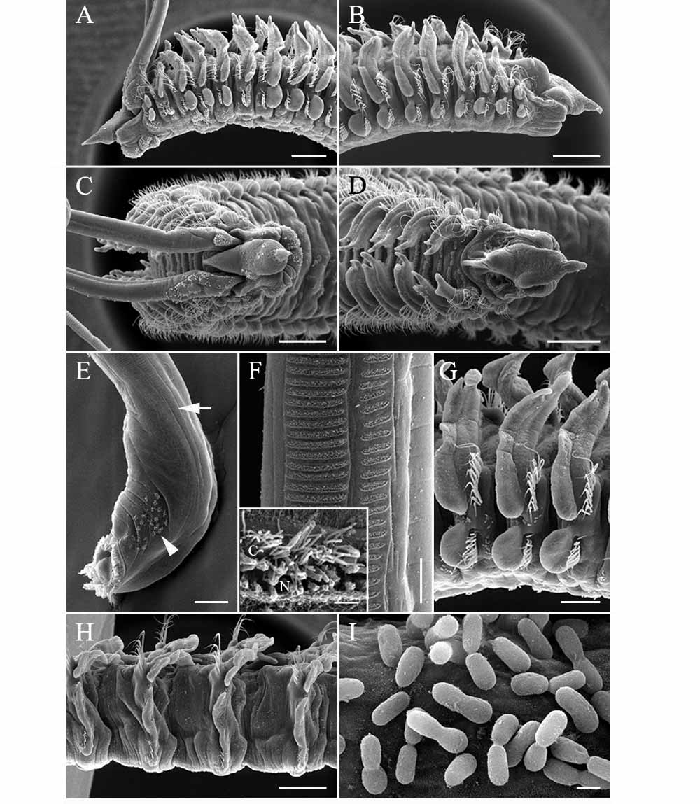
( Figs. 16–17)
Material examined. Holotype. Diniwid Beach, Boracay, 11°60´N, 121°54´E, sandy beach, 13 Apr 1999 ( USNM 1096813).— Paratypes, same data as for holotype (two anterior ends in alcohol, USNM 1096814; two anterior ends in alcohol, ZRC 2006.0225; two anterior ends and one pair of palps on SEM stubs, USNM 1096815).
Etymology. The species epithet villosivaina (derived from Latin adjective villosus for hairy and Spanish noun vaina for sheath or scabbard) refers to the presence of cilia at the base of the palp sheaths.
Diagnosis. A species of Scolelepis with notosetae on setiger 1, notopodial hooded hooks and lacking an occipital tentacle. Palps long, with basal palp sheath present, patches of cilia near insertion of palp, each palp with two distinctly separated transverse rows of cilia. Prostomium conical, with 2–4 eyes or eyes lacking. Caruncle extending to posterior end of setiger 1, with nuchal cilia in U-shaped pattern on both sides of caruncle, posterior to base of palps. Postsetal notopodial lamellae nearly completely fused with branchiae from setiger 2, lamellae elongate, rounded, distal end pointed, free from branchiae. Bidentate notopodial hooded hooks from setiger 38, up to two in posterior setigers. Uni- or bidentate neuropodial hooded hooks from setiger 25–29, up to six in middle to posterior setigers. Pygidium small, rounded, with dorsal anus.
Description. Holotype of 56 setigers, largest complete specimen, 20.0-mm long, 1.1-mm wide at setiger 10, setigers 1–38 full sized and posterior setigers 39–56 regenerating including pygidium. Body widest anteriorly; body approximately rectangular in cross section. Color in alcohol opaque white, no pigmentation present.
A B C D E
K L F G H I J
F-J Prostomium conical, widest in middle then tapering to sharp point anteriorly, posteriorly extending to thin, bluntly pointed caruncle, extending to posterior end of setiger 1; reduced peristomial wings surrounding base of palps ( Figs. 16 A, B, 17A–D). Four minute deeply embedded eyes or eyes absent; occipital tentacle absent. Palps extending to about setigers 12–15, each with two distinctly separated transverse rows of cilia along ventral surface, long rows of cilia approximately 65 µm long, short rows approximately 32-µm long with 16-µm gap between long and short rows (short rows on medial side), ratio of long to short rows approximately 1.2, rows of mucus secreting cells, represented by tubular necks, present proximal to transverse ciliary rows, these rows extending to distal ends of palps, median ciliated groove lacking ( Figs. 17 View FIGURE 17 F and inset, 18A, B). Palps with basal sheath, each sheath with small patches of cilia near insertion of palp ( Fig. 17 View FIGURE 17 E). Nuchal cilia in a U-shaped pattern on both sides of caruncle, posterior to base of palps ( Figs. 16 A, 17D).
Setiger 1 well developed with rounded notopodial and neuropodial postsetal lamellae, notosetae and neurosetae present. Postsetal notopodial lamellae nearly completely fused with branchiae from setiger 2 and subsequent anterior setigers ( Figs. 16 C, 17A–C), lamellae elongate, rounded, with distal end pointed, degree of fusion of lamellae and branchiae decreasing in middle setigers, with lamellae and branchiae almost completely separated in posterior setigers ( Figs. 16 D–E, 17G–H). Postsetal neuropodial lamellae of setigers 1–28 with elongate, rounded lobe ( Figs. 16 C–D, 17A–B, G), by setiger 30 with notch dividing broad dorsal lobe and small rounded ventral lobe with neurosetae ( Figs. 16 E, 17H). Lateral organs between notopodial and neuropodial postsetal lamellae present from setiger 1 to posterior setigers.
Notosetae of setiger 1 and subsequent setigers in two vertical rows of limbate capillaries ( Fig. 16 H, J), the dorsal notosetae of these rows longer than ventral ones, one notopodial hooded hook initially present on setiger 38, up to two notopodial hooded hooks from setiger 45 (in regenerating holotype); hooks bidentate with acute main fang and single accessory tooth ( Fig. 16 I). Neurosetae of setiger 1 and subsequent setigers arranged in two vertical rows of limbate capillaries ( Fig. 16 F), 2–4 neuropodial hooded hooks from setiger 25–29, up to six neuropodial hooded hooks in middle setigers; neuropodial hooded hooks unidentate ( Fig. 16 G) or bidentate with small lateral tooth (second tooth difficult to see in some specimens).
Branchiae from setiger 2 to end of body, fused with postsetal notopodial lamellae but with tips free in anterior setigers ( Fig. 17 View FIGURE 17 A–C, G–H), branchiae and lamellae separated in posterior setigers; with two bands of cilia along inner edge of branchiae continuing across dorsum to join corresponding branchiae on opposite side of body.
Pygidium small, rounded, with dorsal anus ( Fig. 16 K–L).
Remarks. Scolelepis villosivaina sp. n., from the Philippines belongs to a group of 12 species including S. bonnieri Mesnil, 1896 ; S. lamellicincta Blake & Kudenov, 1978 ; S. squamata ; S. blakei ; S. carunculata ; S. chilensis ; S. hutchingsae ; S. kudenovi ; S. denmarkensis Hartmann-Schröder, 1983; S. mesnili Bellan & Lagardère, 1971 ; and S. vazaha Eibye-Jacobsen & Soares, 2000 that possess notosetae on setiger 1, notopodial hooded hooks, but that lack an occipital tentacle. Among these, S. villosivaina sp. n. most closely resembles S. blakei , S. carunculata , S. chilensis , S. hutchingsae , and S. kudenovi in possessing neuropodial hooded hooks with two teeth, S. bonnieri and S. squamata have a variable number of teeth, 1–2 and 1–3, respectively while the rest exhibit one or three teeth. The apical tooth of S. chilensis is notched while it is entire in S. villosivaina sp. n.. Scolelepis villosivaina sp. n. is distinct from S. blakei and S. kudenovi based on branchial fusion. Branchiae are free in S. blakei and fused only at the base in S. kudenovi whereas in S. villosivaina sp. n. the branchiae of anterior setigers are fused along most of their length with only the tips free. The posterior margin of the caruncle in S. villosivaina sp. n. is attached whereas in S. carunculata it is free. The notopodial hooded hooks begin on similar setigers as the neuropodial hooded hooks in S. hutchingsae ; in S. villosivaina sp. n. the notopodial hooks begin more posteriorly. Scolelepis villosivaina sp. n. was found to possess patches of cilia on the palp sheath, similar to those of S. hutchingsae described herein for the first time. These patches are easily overlooked, especially with light microscopy and this feature needs to be examined in other species of Scolelepis ; the function of these cilia remains unknown.
Bacteria were found on the prostomium, peristomium, palps, and anterior setigers of two specimens of S. villosivaina sp. n. ( Fig. 17 View FIGURE 17 A, C, I). The bacterial cells were approximately 20-µm long × 9-µm wide and found in patches over the epidermis. Douglas & Jones (1991) reported on bacteria associated with Polydora nuchalis Woodwick, 1953 where the bacteria caused a cuticular disease that was observable to the naked eye as “cauliflower-like”. The appearance of the bacteria on the S. villosivaina sp. n. from the Philippines was similar. Douglas & Jones (1991) also found bacteria in the digestive tract of Spio maculata ( Hartman, 1961) (cited as Scolelepis maculata ).
Distribution. Sandy beach in Boracay of the Aklan province in the Philippines; shallow subtidal (< 5 m).
No known copyright restrictions apply. See Agosti, D., Egloff, W., 2009. Taxonomic information exchange and copyright: the Plazi approach. BMC Research Notes 2009, 2:53 for further explanation.
|
Kingdom |
|
|
Phylum |
|
|
Class |
|
|
Order |
|
|
Family |
|
|
Genus |