
Sordaria macrospora Auersw.
|
publication ID |
https://doi.org/10.11646/phytotaxa.587.3.4 |
|
DOI |
https://doi.org/10.5281/zenodo.7752995 |
|
persistent identifier |
https://treatment.plazi.org/id/03B07573-A440-FFC0-B7A0-F918E531FB3D |
|
treatment provided by |
Plazi |
|
scientific name |
Sordaria macrospora Auersw. |
| status |
nov. Series |
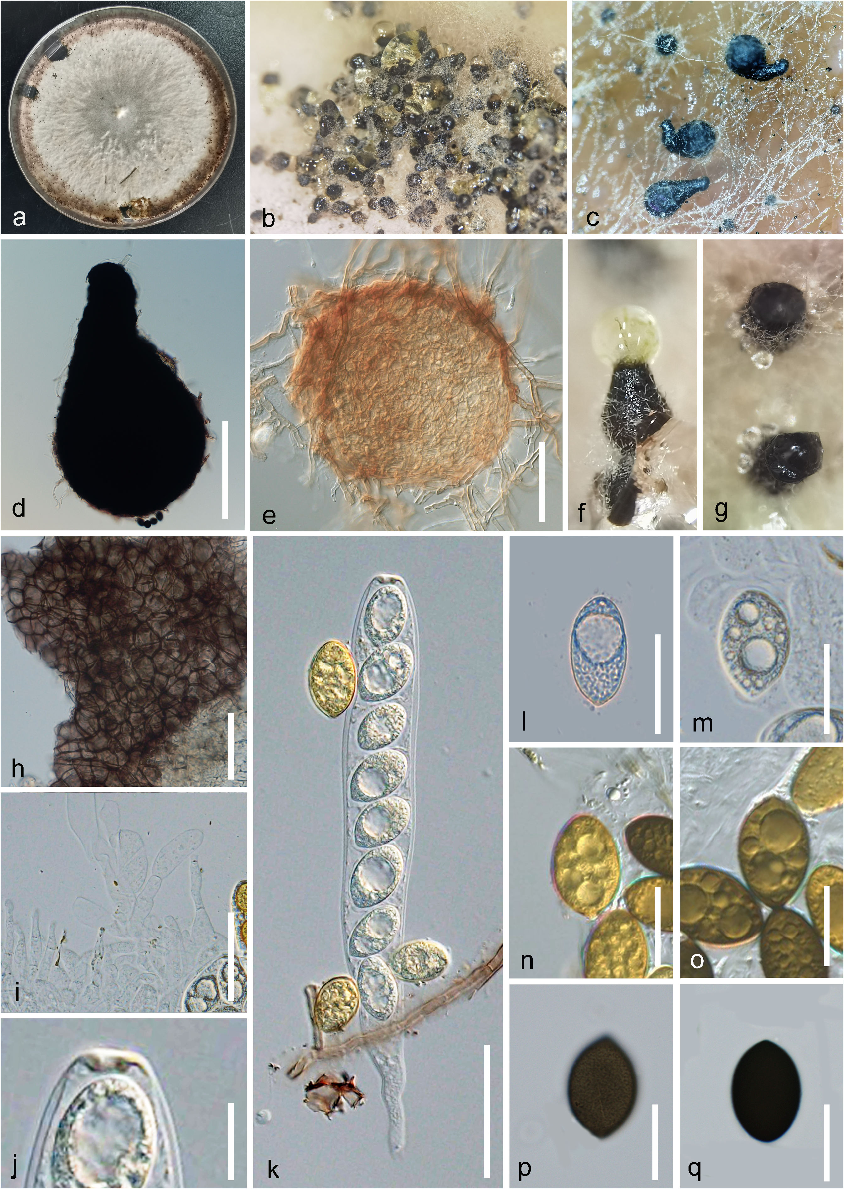
Sordaria macrospora Auersw. View in CoL View at ENA , Fungi Europaei exsiccati, Klotzschii herbarii vivi mycologici continuatio. Editio nov. Series secunda. Cent. 10: no. 954 (1866) ( Figure 5 View FIGURE 5 )
Index Fungorum number: IF 237763
Isolated from intestinal contents of dead American bullfrog larvae. Sexual morph on PDA: Ascomata 340–420 × 270–380 μm (x̅ = 330 × 380 μm, n=20), semi-immersed to superficial, perithecial, solitary or gregarious, ampulliform or pyriform, globose, coriaceous, brown to black, ostiolate. Neck 140–180 × 85–120 μm (x̅ = 160 × 99 μm, n=10) central, oblong, straight to bent, extended, blunt ends. Peridium consist of dark-brown, thick-walled cells of textura angularis. Hamathecium 6–15 wide (x̅ = 9.5 μm, n=20), numerous, wide, branched, septate, markedly constricted at the septa, hyaline, paraphyses. Asci 130–180 × 17–22 μm (̄x̅ = 155×19 μm, n=20), unitunicate, eight-spored, slenderly cylindrical, with a lobate pedicel and a prominent J- ring. Ascospores 20–30 × 15–20 μm (x̅ =25.5 × 15.5 μm, n=20), one-celled, ellipsoidal to ovoid, uniseriate, hyaline to golden to brown to black when mature, guttulate, with some prominent oil droplets, with a thin gelatinous sheath, acute to round ends, with a basal germ pore. Asexual morph: Undetermined.
Culture characteristics: Colonies growing on PDA reach around 60 mm in diameter after one week at 27 °C, forming dark brown fruiting bodies within one month in PDA. Obverse: flat, white, entire edge, peripheral fertile. Reverse: pale brown. Without pigments produced in PDA.
Known substratum: Pinus sylvestris , Fagus sylvatica ( Petrini & Fisher 1988) ; Salix fragilis , Quercus robur ( Petrini & Fisher 1990) ; Olea europaea ( Fisher et al. 1992) ; Eucalyptus nitens ( Fisher et al. 1993) ; Dactylis glomerata ( Sanchez Marquez et al. 2007) ; Herbivore dung ( Lytvynenko & Hayova 2018); Intestinal contents of dead American bullfrog larvae (this study).
Known Distribution: England (Petrini & Fisher 1998,1990); Balearic Islands ( Fisher et al. 1992); Australia ( Fisher et al. 1993); Spain ( Sanchez Marquez et al. 2007); Ukraine ( Lytvynenko & Hayova 2018); China (This study).
Material examined: China, Yunnan Province, Qujing Normal University, on intestinal contents of dead American bullfrog larvae, GPS: 103°44’35”E, 25°30’46”N, 1856.6 m, Wen-hua Lu, ER3 , (Herb. HKAS 125766 View Materials ), living culture 22-12509 GoogleMaps .
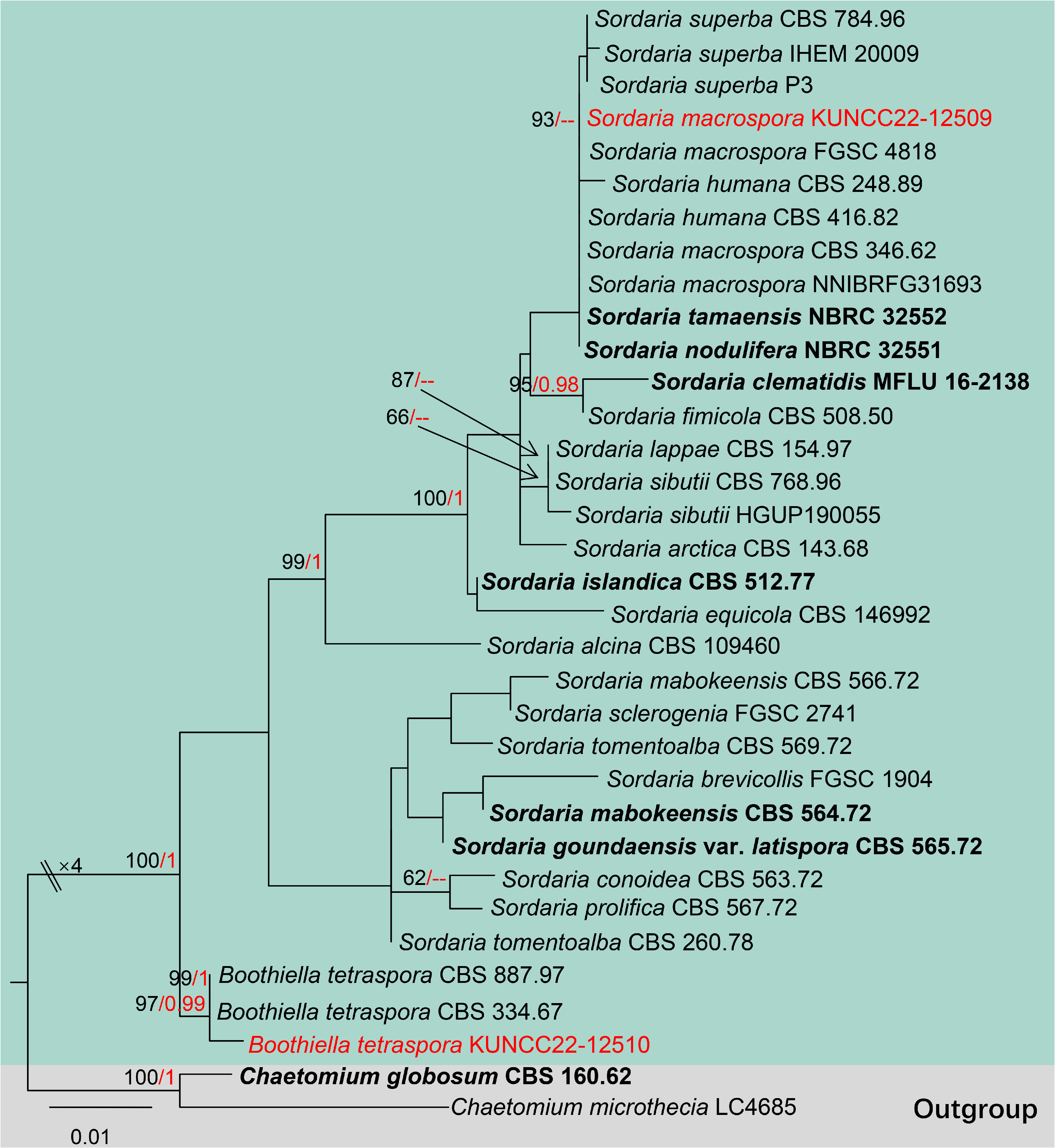
Notes: Sordaria macrospora is a coprophilic homothallic pyrenomycete, a fungal model organism in biology that was first described in 1866 by Auerswald. Our isolate fits the concept of S. macrospora by having elongated neck, oblong asci with a “J-” apical ring, enwrap eight hyaline to golden to dark brown ascospores, ascospores uniseriate, visible distinct oil droplets when its young, present verruculose surface when it is mature, without gelatinous sheath ( Lord & Read 2011). The BLASTn results of ITS, and LSU showed our isolate (KUNCC22-12509) are highly similar to Sordaria species ( Sordaria macrospora CBS :346.62, S. humana CBS :416.82, S. fimicola CBS :566.67, S. lappae CBS :154.97, S. tamaensis and S. nodulifera NBRC :32551), with 99–100% similarity. However, the tub2 gene is mostly similar to Sordaria macrospora (FGSC 4818) and S. superba (CBS 784.96) with 99% similarity, the tub2 gene of other species is not available ( Table 1). From morphology, Sordaria superba is different, as the ascospores are wrapped by a distinct gelatinous sheath with a single basal germ pore ( Yul et al. 2010), our isolate is more similar to S. macrospora (ascomata: 370–400 (500) × 250–300 µm VS. 340–420 × 270–380 μm; and asci: 160–175 ×20 VS. 20–30 × 15–20 μm) ( Ivanová et al. 2015). The phylogenetic tree indicated that our Sordaria macrospora KUNCC 22-12509 is complex with the other species that have high similarity in ITS, and LSU genes ( Figure 2 View FIGURE 2 ), and this problem may be caused by lacking tub2 genes, therefore, our isolate KUNCC22-12509 is identified based on morphological features (Table 2) coupled with BLASTn results and phylogenetic analyses as Sordaria macrospora which is a new host and country record.
No known copyright restrictions apply. See Agosti, D., Egloff, W., 2009. Taxonomic information exchange and copyright: the Plazi approach. BMC Research Notes 2009, 2:53 for further explanation.
|
Kingdom |
|
|
Phylum |
|
|
Class |
|
|
Order |
|
|
Family |
|
|
Genus |