
Guimaraesiella ( Guimaraesiella ) sphagmotica Gustafsson, 2022
|
publication ID |
https://doi.org/10.11646/zootaxa.5165.1.1 |
|
publication LSID |
lsid:zoobank.org:pub:A03F9711-19D7-4D7A-B30E-842DA141B2A0 |
|
DOI |
https://doi.org/10.5281/zenodo.6836487 |
|
persistent identifier |
https://treatment.plazi.org/id/03B15059-B37F-FFE2-FF41-F878FE6BF863 |
|
treatment provided by |
Plazi |
|
scientific name |
Guimaraesiella ( Guimaraesiella ) sphagmotica Gustafsson |
| status |
new species |
Guimaraesiella ( Guimaraesiella) sphagmotica Gustafsson View in CoL & Bush, new species
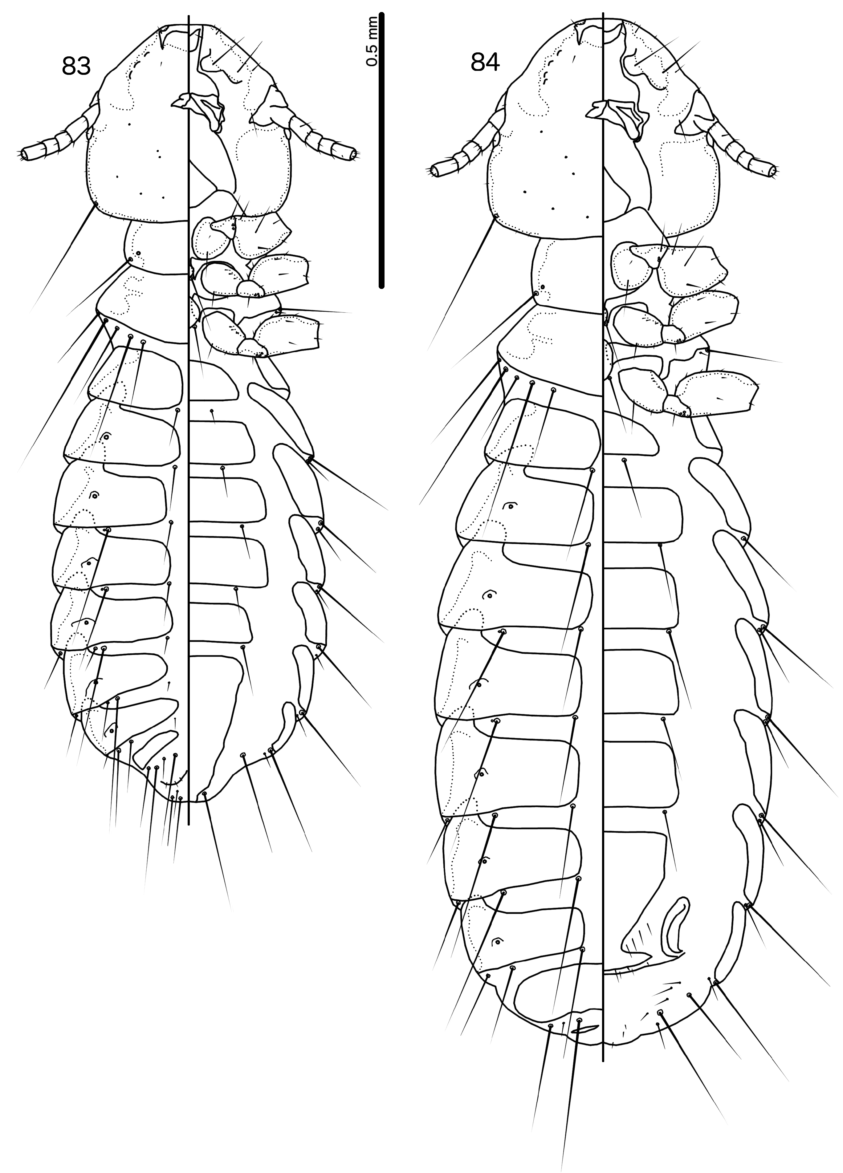
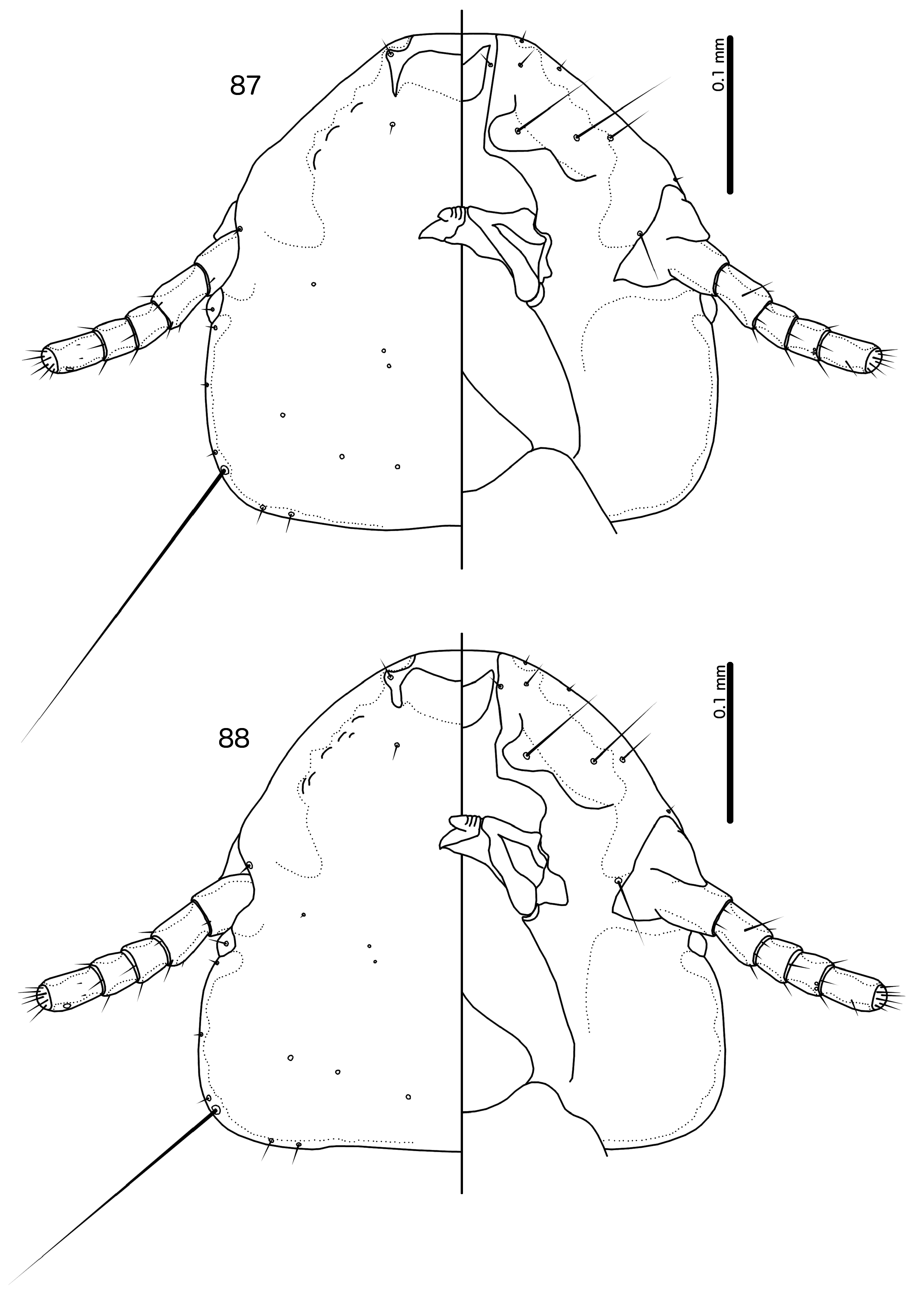
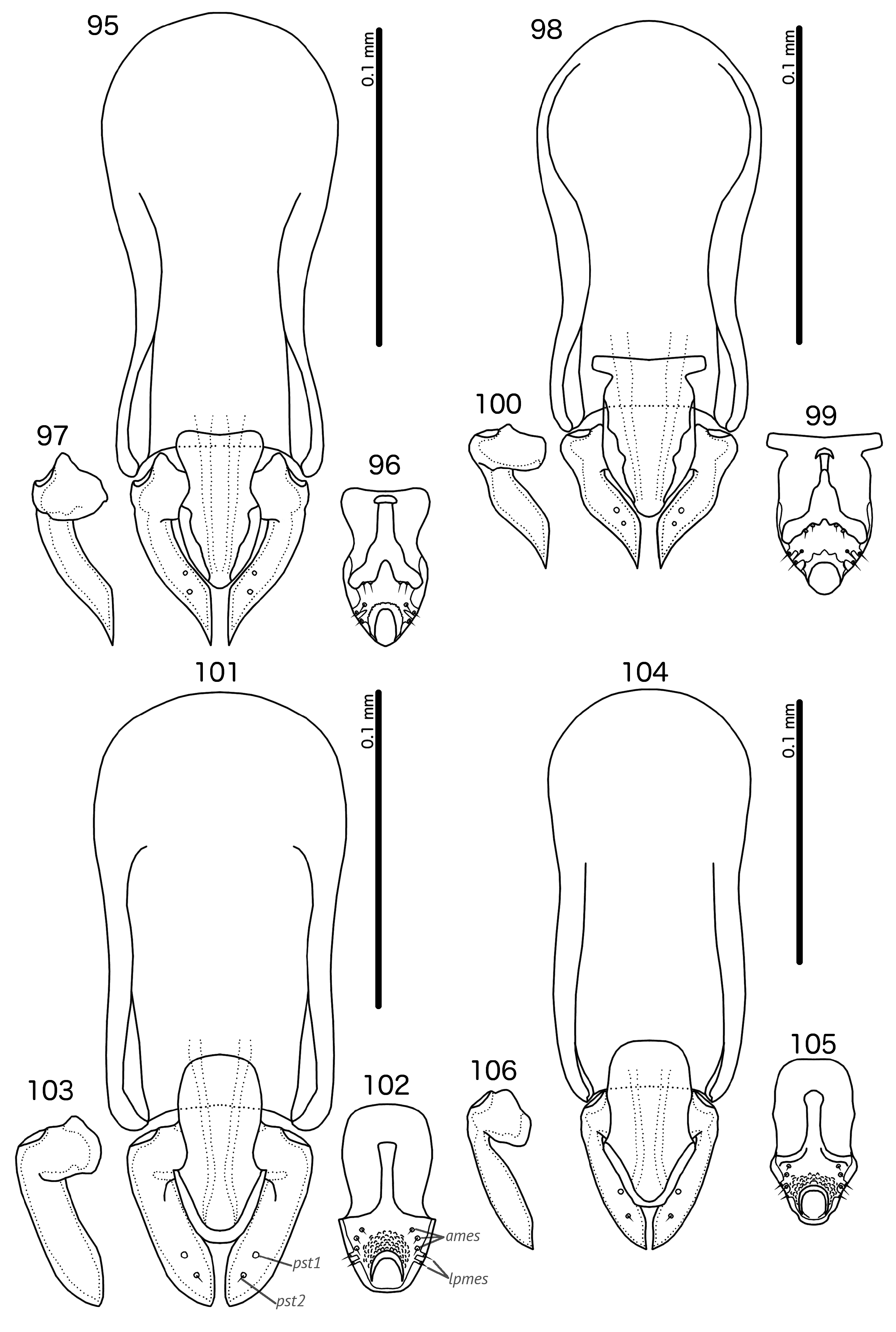
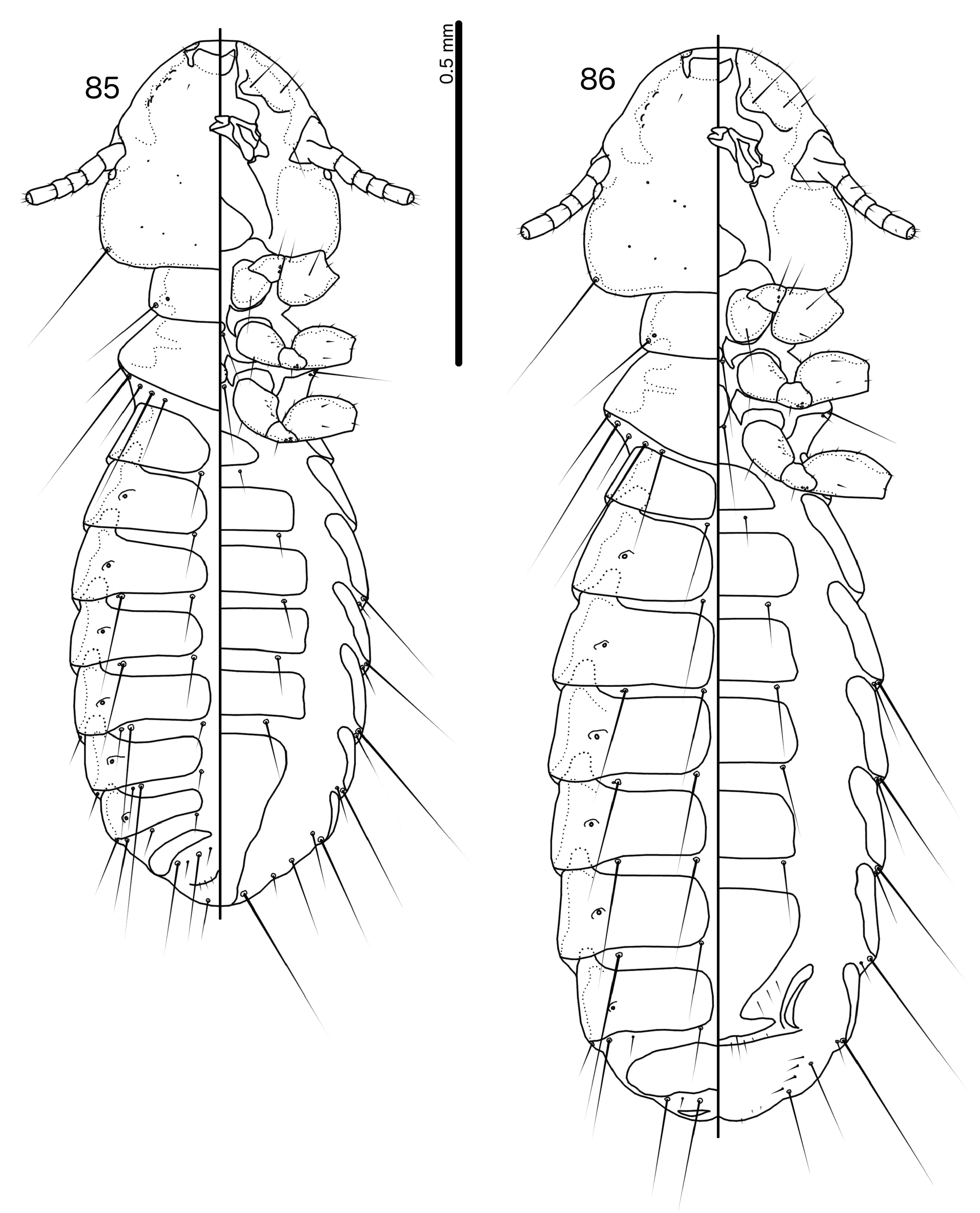
( Figs 83–84 View FIGURES 83–84 , 87 View FIGURES 87–88 , 89 View FIGURES 89–90 , 95–97 View FIGURES 95–106 )
Type host. Coracina caeruleogrisea strenua (Schlegel, 1871) – stout-billed cuckooshrike.
Type locality. Mount Bosavi , Southern Highlands Province, Papua New Guinea .
Other host. Coracina caeruleogrisea adamsoni Mayr & Rand, 1936 – stout-billed cuckooshrike.
Diagnosis. Head shape indicates that the closest relative of Gu. sphagmotica new species is Gu. nouankaoensis new species, as this differs from all known species of the “core group” of Guimaraesiella , except Guimaraesiella amsel ( Eichler, 1951) and Guimaraesiella haftorni ( Balát, 1958) . In both Gu. amsel and Gu. haftorni the dorsal preantennal suture reaches the ads, which is not the case in Gu. sphagmotica and Gu. nouankaoensis .
Guimaraesiella sphagmotica can be separated from Gu, nouankaoensis by the following characters: frons more angular and lateral margins of preantennal head straight in Gu. sphagmotica ( Fig. 87 View FIGURES 87–88 ), but frons more rounded and lateral margins of preantennal head convex in Gu. nouankaoensis ( Fig. 88 View FIGURES 87–88 ); ps present on abdominal segment III in both sexes in Gu. sphagmotica ( Figs 83–84 View FIGURES 83–84 ), but absent in Gu. nouankaoensis ( Figs 85–86 View FIGURES 85–86 ); female subgenital plate more angular in Gu. sphagmotica ( Fig. 89 View FIGURES 89–90 ) than in Gu. nouankaoensis ( Fig. 90 View FIGURES 89–90 ); proximal mesosome, ventral sclerite of mesosome, and gonopore all differ in shape between the two species ( Figs 96, 99 View FIGURES 95–106 ); parameres longer in Gu. sphagmotica ( Fig. 97 View FIGURES 95–106 ) than in Gu. nouankaoensis ( Fig. 100 View FIGURES 95–106 ).
Description. Both sexes. Head broad, flat dome-shaped ( Fig. 87 View FIGURES 87–88 ), lateral margins of preantennal area more or less straight, frons slightly rounded to flattened. Marginal carina broad, narrowing conspicuously near dsms, interrupted submedianly. Dorsal preantennal suture reaches dsms but not ads or lateral head margin, and does not separate dorsal anterior plate posteriorly. Ventral anterior plate crescent-shaped. Head chaetotaxy as in Fig. 87 View FIGURES 87–88 . Preantennal nodi very large, bulging. Preocular nodi larger than postocular nodi. Marginal temporal carina slender, of more or less even width. Gular plate with concave lateral margins converging into median point. Thoracic and abdominal segments as in Figs 83–84 View FIGURES 83–84 ; ps present on abdominal segment III in both sexes. All examined specimens stained prior to mounting, and true pigmentation unknown.
Male. Thoracic and abdominal chaetotaxy as in Fig. 83 View FIGURES 83–84 . Basal apodeme with rounded proximal end, and concave lateral margins ( Fig. 95 View FIGURES 95–106 ). Proximal mesosome rounded trapezoidal, with concave anterior margin ( Fig. 96 View FIGURES 95–106 ). Mesosomal lobes convex, convergent. Ventral sclerite as in Fig. 96 View FIGURES 95–106 , with deep median concavity on distal margin. Gonopore somewhat elongated, with rugged proximal margin. Parameres and pst1–2 as in Figs 95, 97 View FIGURES 95–106 . Measurements as in Table 1 View TABLE 1 .
Female. Thoracic and abdominal chaetotaxy as in Fig. 84 View FIGURES 83–84 . Subgenital plate angular, broad distally, with broad lateral submarginal bulges ( Fig. 89 View FIGURES 89–90 ). Vulval margin gently rounded, with 2 short, slender vms and 1–2 short, thornlike vss on each side; 4–6 short, slender vos on each side of subgenital plate; distal 1 vos median to vss. Measurements as in Table 1 View TABLE 1 .
Etymology. The species epithet is derived from “ sphagmos ”, Greek for “pulse”, referring to the rather regularly undulating median margin of the marginal carina.
Type material. Ex Coracina caeruleogrisea strenua : Holotype ♂, Mt. Bosavi, Southern Highlands Province, Papua New Guinea, 16 May 1973, 103199 ( NHML). Paratypes: 4♂, 5♀, same data as holotype ( NHML) .
Additional material examined (non-types): Ex Coracina caeruleogrisea adamsoni : 7♂, 4♀, 10 km W of Bulolo , elev. 780 m, Morobe Province, Papua New Guinea, 19 Aug. 1967, A.C. Ziegler, BBM-NG-54058 ( BPBM) ; 1♂, 3♀, 6 km NW of Wau , elev. 1070 m, Morobe Province, Papua New Guinea, 12 Sep. 1967, A.B. Mirza, BBM-NG-54489 ( BPBM) ; 2♀, Wakata Ridge , elev. 5000 ft., Wau, Morobe Province, Papua New Guinea, 8 May 1963, P.J. Shanahan, BBM-NG-27783 ( BPBM) ; 2♀, Wau Creek , elev. 3800 ft., Morobe Province, Papua New Guinea, 11 Mar. 1963, H. Clissold, BBM-NG-20397 ( BPBM) .
Remarks. Specimens from the type host are larger in all measurements than specimens from C. c. adamsoni. Otherwise, specimens from the two host subspecies are similar but, since most specimens from C. c. adamsoni are poorly preserved, examination of fresh specimens is needed to confirm that the same species of Guimaraesiella occurs on both host subspecies.

No known copyright restrictions apply. See Agosti, D., Egloff, W., 2009. Taxonomic information exchange and copyright: the Plazi approach. BMC Research Notes 2009, 2:53 for further explanation.
|
Kingdom |
|
|
Phylum |
|
|
Class |
|
|
Order |
|
|
Family |
|
|
Genus |