
Cacopsylla ( Hepatopsylla ) qiuzili Li
|
publication ID |
https://doi.org/10.5281/zenodo.213975 |
|
publication LSID |
lsid:zoobank.org:pub:2C43EA7B-94F7-4133-9070-21AC4A8AB734 |
|
DOI |
https://doi.org/10.5281/zenodo.6178510 |
|
persistent identifier |
https://treatment.plazi.org/id/03B1723D-FFFE-FF9D-FF60-FA3F517970DC |
|
treatment provided by |
Plazi |
|
scientific name |
Cacopsylla ( Hepatopsylla ) qiuzili Li |
| status |
|
Cacopsylla ( Hepatopsylla) qiuzili Li View in CoL
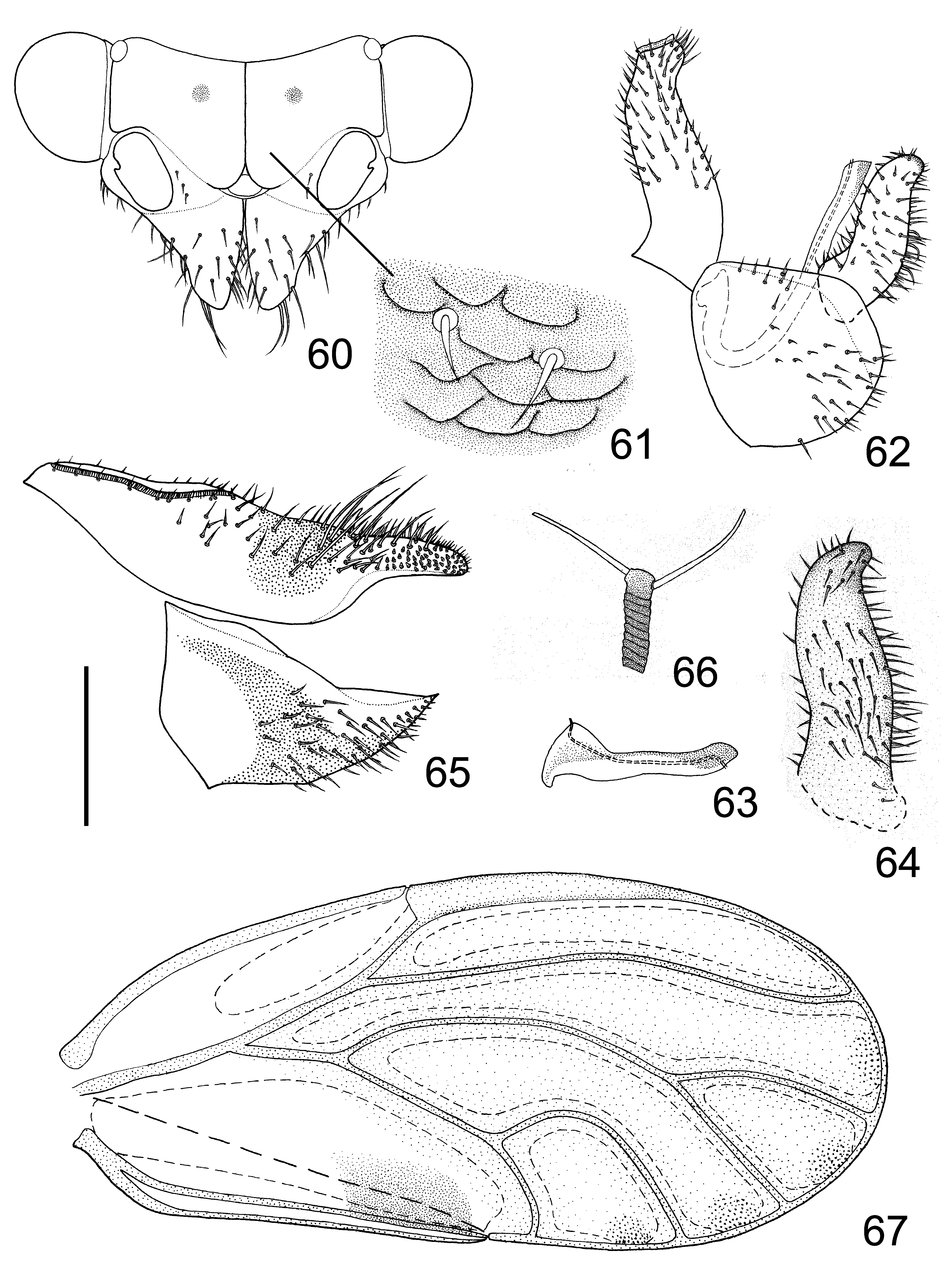
( Figs 60–67 View FIGURES 60 – 67 )
Cacopsylla qiuzili Li, 2011: 881 View in CoL .
Adult. Coloration: Body yellow. Vertex bright yellow, sometimes with orange patterns varying from irregular small cloudings in the sub margin to occupying vast major of vertex; discal foveae brown. Genal process yellow, apex sometimes orange. Antenna brown, segments III–VIII with darker apices, segments IX–X entirely black. Thorax brown in ground colour except for pronotum, metascutum and metascutellum which are yellow, with wide black stripes. Legs yellow, profemora and mesofemora more or less darkened into black. Fore wing transparent, with one dark brown marking near apex of claval suture; fields alongside veins R, R1, Rs, M+Cu, M, M1+2, M3+4, Cu, Cu1a and Cu1b yellowish, forming broad obscure bands along these veins, leaving small uncolored fields in the centre of cells r1, r2, m1, m2 and cu1, making apical 2/3 of fore wing appearing yellowish in general ( Fig. 67 View FIGURES 60 – 67 ); veins yellow. Abdomen yellow, terga of segments III–V (the first 3 visible segments) dark brown, the corresponding sterna sometimes also. Male terminalia black. Female terminalia brown, proctiger and subgenital plate with dark brown pattern as shown in Fig. 65 View FIGURES 60 – 67 .
Structures: Body glabrous. Head nearly vertical with longitudinal body axis, slightly wider than mesoscutum. Vertex ( Fig. 61 View FIGURES 60 – 67 ) finely sculptured with microscopic setae and scaly micro structures that are relatively large, smooth and more or less attached with each other. Genal processes ( Fig. 60 View FIGURES 60 – 67 ) elongate cone-shaped and moderately divergent, about as long as vertex along median suture, and covered with long setae; apex acute. Antenna long and slender, slightly squiggly; terminal setae ( Fig. 66 View FIGURES 60 – 67 ) about as long as each other, and obviously longer than antennal segment X. Metatibia with sharp basal spine, apical spurs arranged in (1+3+1). Fore wing ( Fig. 67 View FIGURES 60 – 67 ) oval, widest in about the middle; pterostigma short, ending in the middle of cell r1; cell cu1 tall and near quadrate, turning of vein Cu1a near right angle; surface spinules present in all cells, leaving narrow spinule-free stripes along veins, fields narrowing along wing margin in cells r2, m1, m2 and cu1; 4 sets of radular spinules present in cells r2, m1, m2 and cu1, not obviously reduced in r2.
Male terminalia: Proctiger ( Fig. 62 View FIGURES 60 – 67 ) slender, slightly arched, evenly covered with short setae. Paramere ( Figs 62 & 64 View FIGURES 60 – 67 ) lamellar, relatively short and robust, apex rounded and projected caudad, posterior margin thickened; short setae present in both inner and outer surface, moderately longer and denser in posterior margin than in anterior margin. Apical dilatation ( Fig. 63 View FIGURES 60 – 67 ) of aedeagus near triangular, with the sclerotised end tube of ductus ejaculatorius projecting beyond the dorsal margin and curved. Subgenital plate ( Fig. 62 View FIGURES 60 – 67 ) near spherical, with several setae that vary in length in dorsal margin; ventral surface covered with sparse short setae.
Female terminalia ( Fig. 65 View FIGURES 60 – 67 ) relatively short. Proctiger slightly sinuate dorsally, dorsal surface covered with setae that vary in length; laterally and apex of apical part covered with peg setae, the field involved completely surrounded by fields of short setae. Subgenital plate evenly covered with short setae and peg setae.
Material examined. Holotype: male, dry mounted, China, Jilin, Liudaogou, Hunjiang, 840 m, 4.viii.1983, Li Fasheng.
Paratypes: 5 male, 13 female, with same data as holotype.
Non-paratypic specimens: China, Jilin, 4 male, 5 female, Liudaogou, Hunjiang, 840 m, 4.viii.1983, Yang Chikun; 1 female, Hunjiang, 800 m, 2.viii.1983, Li Fasheng; 1 male, Songjianghe, Fusong, 710 m, 8.viii.1983, Li Fasheng; 1 male, Ji’an, 150 m, 11.viii.1983, Li Fasheng; 1 male, Tonghua, 450 m, 31.vii.1983, Li Fasheng. Liaoning, 2 male, 1 female, Qingyuan, vi.1989, Sun Lihua.
Distribution. China: Jilin, Liaoning.
Host plant. Pyrus ussuriensis .
Remarks. This species is easily diagnosed by its unique abdominal color with the terga of the first three visible segments dark brown.
adult 5th-instar nymph (n=8)
BL HW AL WL TL BL AW
C. accincta Male (n=5) 3.48±0.15 0.77±0.04 1.23±0.11 2.87±0.16 0.60±0.04
Female (n=5) 3.72±0.13 0.78±0.03 1.28±0.03 3.06±0.11 0.59±0.02
C. burckhardti Male (n=5) 3.55±0.11 0.86±0.02 1.22 2.94±0.06 0.59±0.02 2.17±0.16 0.22±0.01
Female (n=5) 3.96±0.07 0.93±0.02 1.26±0.04 3.31±0.04 0.61±0.02
Female (n=5) 3.21±0.08 0.71±0.02 1.36±0.06 2.67±0.06 0.55±0.03 C. liaoli Male (n=5) 2.58±0.06 0.66±0.03 0.94±0.01 2.01±0.03 0.50±0.03
Female (n=5) 3.07±0.16 0.69±0.02 0.95±0.01 2.40±0.06 0.49±0.01 C. maculatili Male (quotation 3.53 0.81 - 2.78 -
of Li, 2011)
Female (n=2) 3.43±0.05 0.75±0.08 1.58±0.06 2.76±0.04 0.58
C. qiuzili Male (n=5) 2.74±0.14 0.73±0.04 1.45±0.10 2.18±0.11 0.51±0.03
Female (n=5) 2.97±0.06 0.76±0.01 1.37±0.04 2.38±0.06 0.53±0.01
No known copyright restrictions apply. See Agosti, D., Egloff, W., 2009. Taxonomic information exchange and copyright: the Plazi approach. BMC Research Notes 2009, 2:53 for further explanation.
|
Kingdom |
|
|
Phylum |
|
|
Class |
|
|
Order |
|
|
SuperFamily |
Psylloidea |
|
Family |
|
|
Genus |
Cacopsylla ( Hepatopsylla ) qiuzili Li
| Luo, Xinyu, Li, Fasheng, Ma, Yanfang & Cai, Wanzhi 2012 |
Cacopsylla qiuzili
| Li 2011: 881 |