
Ophiolepididae Ljungman, 1867
|
publication ID |
https://doi.org/ 10.12782/specdiv.28.107 |
|
persistent identifier |
https://treatment.plazi.org/id/03B687DB-FF97-FFF7-FEA6-6E90FA6751A7 |
|
treatment provided by |
Felipe |
|
scientific name |
Ophiolepididae Ljungman, 1867 |
| status |
|
Family Ophiolepididae Ljungman, 1867 View in CoL View at ENA Genus Ophioteichus H. L. Clark, 1938
[New Japanese name: Joheki-kumohitode-zoku] Ophioteichus multispinum H. L. Clark, 1938 [New Japanese name: Tashin-joheki-kumohitode] ( Figs 2–6 View Fig View Fig View Fig View Fig View Fig )
Ophioteichus multispinum H. L. Clark, 1938: 360–362 View in CoL , pl. 25, fig. 5; A. M. Clark and Rowe 1971: 129; Pineda-Enriquez 2014: 242, fig. 2.
Non Ophioteichus multispinum: Starmer 2003: 557 View in CoL (= Ophiomora elegans Koehler, 1907 View in CoL ).
Material examined. NSMT E-14161: one specimen, Sabiura , Arita, Kushimoto, Wakayama Prefecture, central Japan, depth 12.6 m, SCUBA diving, collected by IH, 20 August 2018 . NSMT E-14162: one specimen, same locality as NSMT E-14161, 2.3 m, 3 October 2018 . NSMT E-14163: one specimen, same locality as NSMT E-14161, 10.2 m, 9 November 2018 .
Diagnosis (from Pineda-Enriquez 2014). Disc nearly circular in outline, higher than the arms, and covered with a coat of numerous swollen, smooth larger plates; each plate surrounded by a belt of polygonal smaller flat scales. Disc scales arranged in an identical pattern on dorsal and ventral surfaces. Radial shields smooth and flat, each pair separat- ed by 2 conspicuous tubercles. In each interradius, the disc margin is encircled by 3 or 4 large tubercles. Accessory dorsal and ventral arm plates fragmented. Minute pores present on the lateral margins of the dorsal arm plates. Infradental papilla, secondary infradental papilla, buccal scale, adoral shield spine, secondary adoral shield spines, Lyman’s ossicle on each side. Arm spines up to 11, acute, slender, and closely spaced. Each tentacle pore with 2 or 3 tentacle scales of which abradial two scales are flat and of the same size, while the third adradial one is sometimes reduced.
Description of external morphology (NSMT E-14161). Disc. Circular, 9.7 mm in diameter ( Fig. 2 View Fig ), covered by scales of two sizes. On dorsal surface, polygonal or circular larger scales, approximately 300–800 µm in length, surrounded by smaller polygonal scales, approximately 50–80 µm in length ( Fig. 3A–C View Fig ). Radial shields hexagonal, slightly longer than wide, approximately 1000 µm in length and 900 µm in width, separated by smaller scales ( Fig. 3C View Fig ). A pair of tubercles between a pair of radial shields ( Figs 2C View Fig , 3C View Fig ). A trio of larger hemispherical tubercles, approximately 1500 µm in length and 1100µm in width, on marginal edges of each interradius ( Figs 2C View Fig , 3A, D View Fig ). On ventral surface, adoral shields slightly curved parallelogram, wider than long, approximately 1000 µm in length, 250 µm in width inner side, 350 µm in width outer side ( Fig. 3E–G View Fig ). Oral plates triangular, approximately 450 µm in length, 200 µm in width inner side, 50 µm in width outer side, in contact with each other ( Fig. 3F View Fig ). Oral shields pear-shaped, slightly rounded, and convex on distal side, approximately 900 µm in length, 700 µm in width, and pointed inward ( Fig. 3F, G View Fig ). One oral shield slightly larger in size, centrally concaving, serving as madreporite ( Fig. 3E View Fig ). Adoral shield oblong, wider than long, contacting each other adradially ( Fig. 3F View Fig ). Interradial ventral disc also covered by larger scales, approximately 250–700 µm in length, surrounded by and smaller scales, approximately 40–120 µm in length, like dorsal disc ( Fig. 3F, G View Fig ). Genital slits long, almost extending to the disc edge, approximately 0.01 mm in width ( Fig. 3H View Fig ). Approximately 6 small plates arranged parallel on their adradial edge. One trapezoidal infradental papilla, 1 polygonal secondary infradental papilla, 1 trapezoidal buccal scale, 1 circular secondary adoral shield spine, 1 trapezoidal adoral shield spine and 1 triangular Lyman’s ossicle on each side of jaw ( Fig. 3F View Fig ). Of these 6 ossicles, inner one smaller and becoming larger toward the outer side ( Fig. 3F View Fig ). Teeth cylindrical, pointed, forming vertical row on dental plate ( Fig. 3F View Fig ). Second tentacle pore completely inside the mouth slit.
Arms. Five in number, 34–37 mm in length, 4.2 mm wide and 1.1 mm high in proximal portion, oblong in section. Arms tapering gradually toward the arm tip ( Fig. 2 View Fig ).
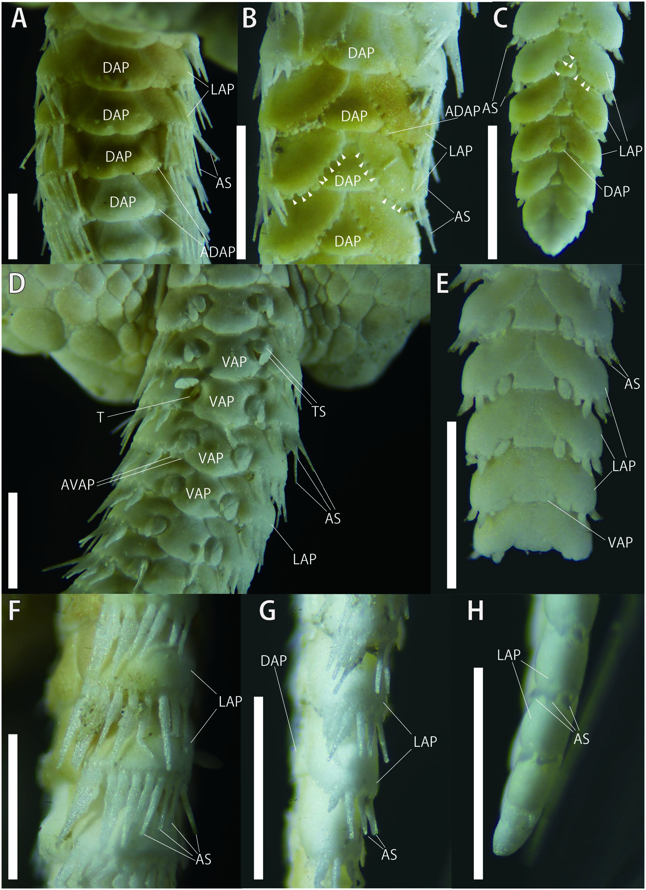
Each arm segment with a dorsal arm plate, 2 lateral arm plates and a ventral arm plate ( Fig. 4 View Fig ). On proximal portion of the arm, dorsal arm plates fan shaped with straight proximal edge, in contact with each other ( Fig. 4A View Fig ). On proximal portion of arm, each dorsal arm plate carries 1 or 2 small accessary plates which decrease in number to 1 on middle portion of arm and disappear on distal portion ( Fig. 4A–C View Fig ). No tubercles on dorsal arm plates. On middle portion of the arm, dorsal arm plates sharpen, triangular, separated and gradually decreasing in size toward the arm tip ( Fig. 4A, B View Fig ). Dorsal arm plates two times wider than long on proximal portion of arm and as wide as long on middle to distal portion ( Fig. 4A–C View Fig ). Lateral arm plates thick, protruding from arm, widely separated by dorsal and ventral arm plates on proximal portion of arm ( Fig. 4A, D View Fig ), in contact with each other on middle to distal portion of arm at proximal edge on both dorsal and ventral sides ( Fig. 4B, C, E View Fig ). On middle to distal portion of arm, dorsal side of lateral arm plates carries smaller accessory pores, 8–10 in number on middle portion, and the number decreasing toward distal portion of arm ( Fig. 4B, C View Fig ). Ventral arm plates squarish fan-shaped with slightly concave lateral edges, almost as wide as long, contiguous on proximal portion of arms and separated on subsequent segments ( Fig. 4D, E View Fig ). On proximal portion of arm, each ventral arm plate carries 1 or 2 small accessary plates which disappear on middle to distal portion ( Fig. 4D, E View Fig ). Ten long, acute arm spines, about the same length as the corresponding arm segment on dorsal-most one and about three-quarters length of the corresponding arm segment on ventral-most one ( Fig. 4F View Fig ); the number decreasing to 4 on the middle portion of arm ( Fig. 4G View Fig ); and decreasing to 3 on distal portion of arm, the length subequal, spear-shaped, about one-sixth length of the corresponding arm segment ( Fig. 4H View Fig ; see detailed descriptions of each ossicle in next section). Two oval tentacle scales at each tentacle pore on proximal portion of arm, subequal in size ( Fig. 4D View Fig ); and the number decreasing to 1 on middle to distal portion of arm ( Fig. 4E View Fig ).
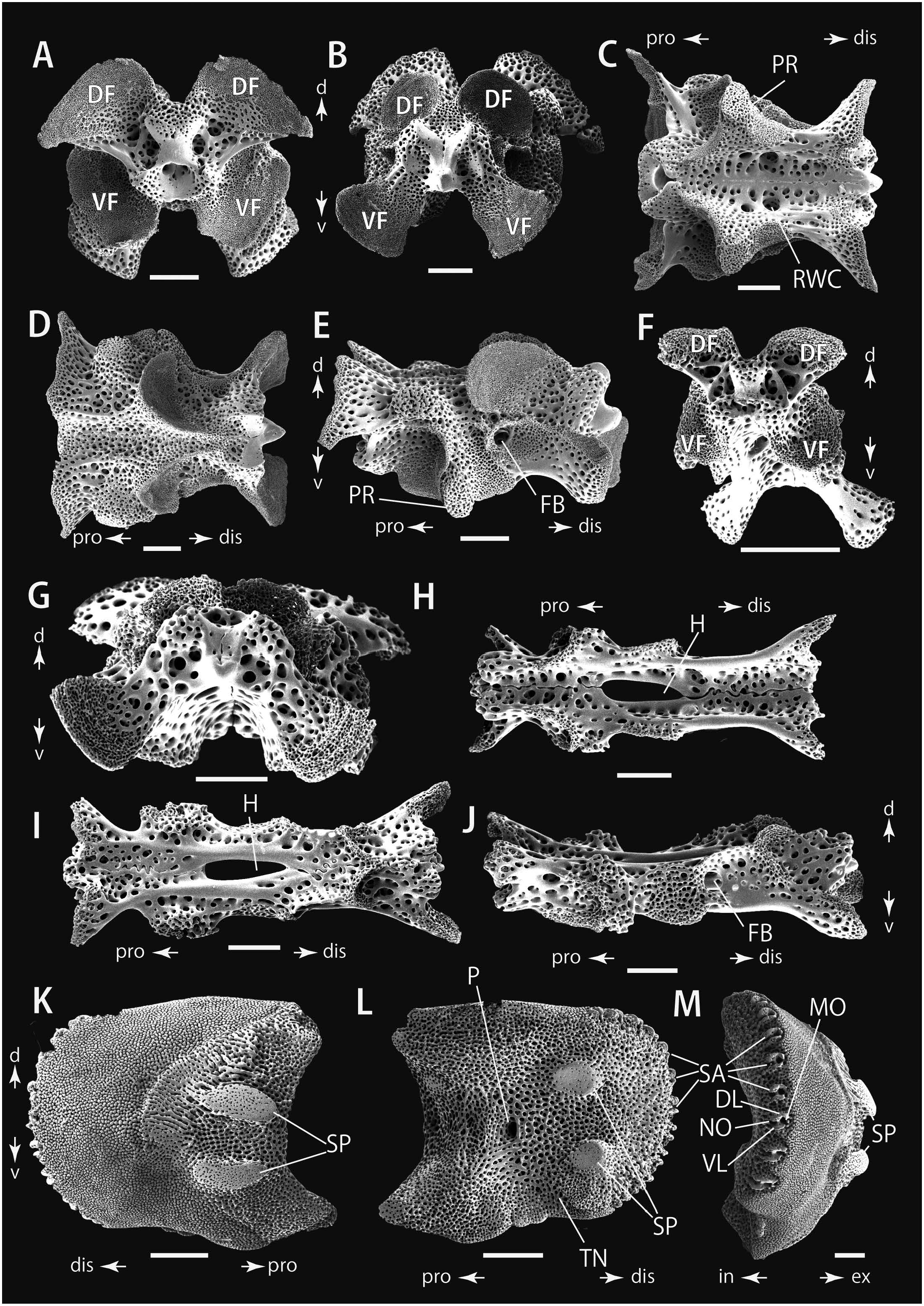
Description of ossicle morphology (NSMT E-14161). Vertebrae with zygospondylous articulation, elongate, large wing-like muscle flanges on ventral-distal side and dorsalproximal side ( Fig. 5A, B, F, G View Fig ). Longitudinal groove on both dorsal and ventral side ( Fig. 5C, D, H, I View Fig ). A pair of radial water canals opening inside ventral groove ( Fig. 5C View Fig ). Foot basins located on lateral central part of vertebra ( Fig. 5E, J View Fig ). Protrusions present on lateral middle portion ( Fig. 5C, E View Fig ). A large hole opening on central part of distal vertebrae ( Fig. 5H, I View Fig ).
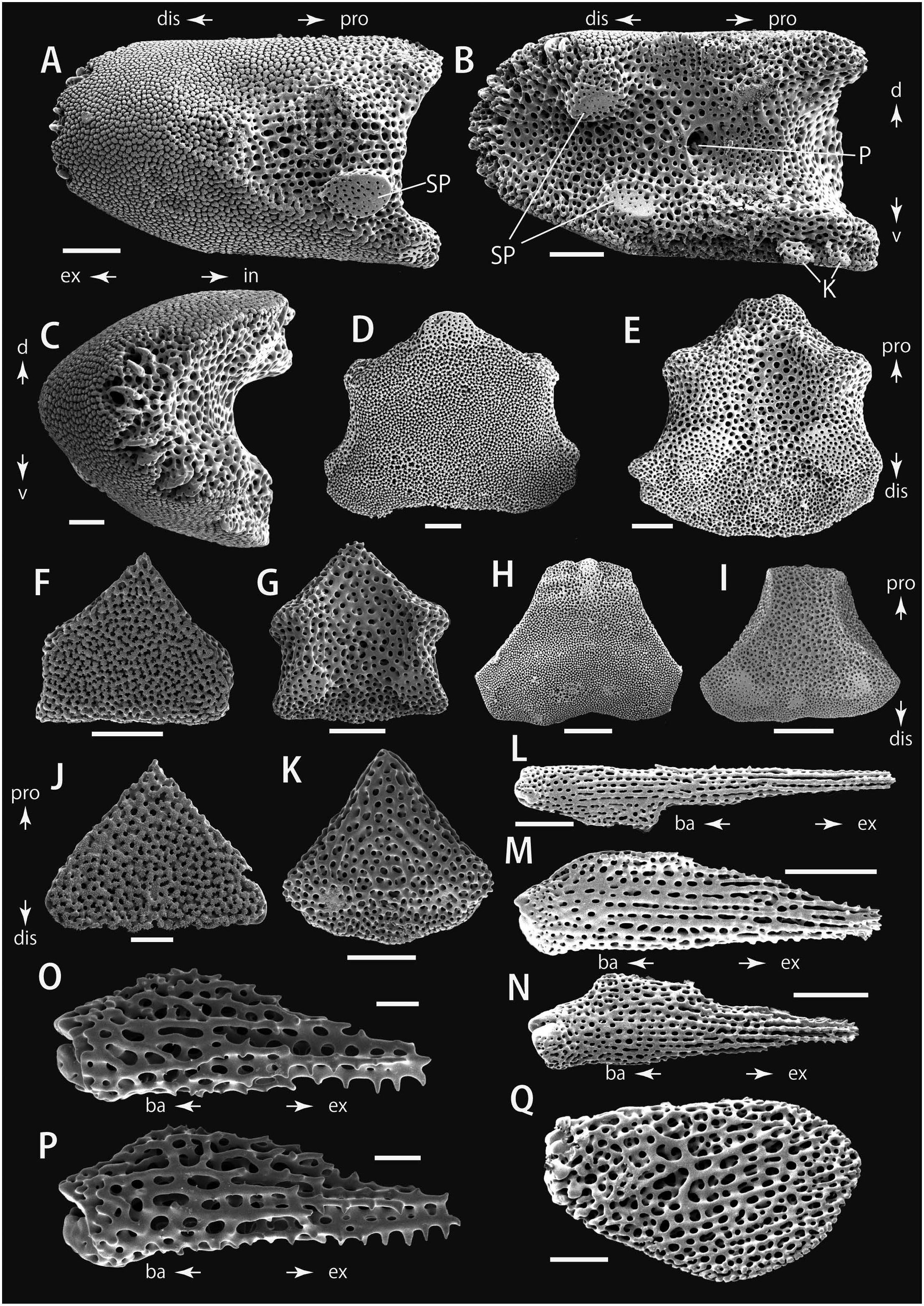
Lateral arm plates slightly longer than high, dorsal edge straight, and ventral-proximal edge slightly convex ( Fig. 5K–M View Fig ). In external view, proximal edge concave with 2 subequal (proximal portion of arm; Fig. 5K View Fig ) or 1 (distal portion of arm; Fig. 6A View Fig ) well defined, prominent, and horizontally elongated spurs composed of more densely meshed stereom than remaining proximal edge. Bands of conspicuous, fine horizontal striations almost the same length on the distal side of spurs on proximal portion of arm ( Fig. 5K View Fig ). The striations unrecognizable on distal portion of arm ( Fig. 6A View Fig ). Eight to 10 equal-sized spine articulations on distal edge on proximal portion of arm ( Fig. 5M View Fig ) and 5 on distal portion of arm ( Fig. 6C View Fig ), and the articulations composed of parallel, horizontal dorsal and ventral lobes ( Figs 5M View Fig , 6A View Fig ). Dorsal lobes larger than ventral lobes and the muscle and nerve openings almost the same size ( Figs 5M View Fig , 6C View Fig ).
On inner-distal side, 2 well-defined, round and elevat- ed spurs which are almost the same size ( Figs 5L View Fig , 6B View Fig ). On proximal portion of arm, large tentacle notch opening on ventral side and end in center of lateral arm plates, where a single perforation is opening ( Fig. 5L View Fig ). On the other hand, no such distinct tentacle notch recognizable on distal portion of arm ( Fig. 6B View Fig ). Two small knobs on proximal-ventral edge on distal portion of arm ( Fig. 6B View Fig ). Ventral arm plates basically tetragonal, notched on lateral sides ( Fig. 6D, E View Fig ). Proximal edge shaper on distal portion of arm ( Fig. 6D, E View Fig ) than on proximal portion of arm ( Fig. 6F, G View Fig ).
Dorsal arm plates triangular with straight proximal edge on proximal portion of arm and sharpened proximal edge on distal portion of arm ( Fig. 6H–K View Fig ).
Arm spines long, cylindrical with wider basal part ( Fig. 6L–P View Fig ). Longer and more slender at proximal portion of arms than at distal portion of arm ( Fig. 6L–P View Fig ). On proximal arm, dorsal arm spines longer and more slender than ventral ones ( Fig. 6L–N View Fig ), however, they subequal in size in distal portion of arm ( Fig. 6O, P View Fig ). Tentacle scales on proximal portion oval ( Fig. 6Q View Fig ), and decreasing in size toward following segments, and finally disappearing on middle to distal portion of arm.
External surfaces of lateral, ventral and dorsal arm plates with finer porous stereom than inner surfaces ( Figs 5K View Fig , 6D, F, H, J View Fig ).
Color in life. On dorsal side, central part of the disc brown and peripheral part creamy white. Marginal tubercles brown ( Fig. 2C View Fig ). Brown transverse bands on 2 to 5 arm segments alternately with creamy white ones on 2 to 3 arm segments ( Fig. 2C View Fig ). Ventral side, uniform creamy white ( Fig. 2D View Fig ).
Variations. Different from the specimen described above [NSMT E-14161: disc diameter (d.d.) = 9.7 mm, up to 10 arm spines], slightly smaller specimens (NSMT E-14162, E-14163: d.d. = 9.3 mm and 9.6 mm, respectively) have up to 11 arm spines on the proximal portion of the arm. The numbers of arm spines and of the ossicles on each side of jaw (infradental papilla, secondary infradental papilla, buccal scale, adoral shield spine, secondary adoral shield spines, Lyman’s ossicle, hereafter “oral papillae s.l.”) were 10–11 and 6, respectively, in our specimens (d.d. = 9.3–9.7 mm), and both of them were lower than those of the holotype (MCZ 5306: d.d. = 15 mm; Pineda-Enriquez et al. 2014). The difference between our examined specimens and the holotype may be due to size differences between specimens.
Ecological notes. All the collected specimens (NSMT E-14161–E-14163) of Ophioteichus multispinum were found under rocks or coral pebbles with burying of their entire body inside sand ( Fig. 2A, B View Fig ), showing sand-dwelling habitat of this species. Our video observations reveal that O. multispinum maintained its arms curled up for a while after being dug out of the sand, then slowly extended its arms ( Fig. 2 View Fig ; Supplementary Movie 1). This curled-up arm posture does not appear to be the normal condition, but even taking this into account, this species was very sluggish compared to other brittle stars. Other sand-dwelling brittle stars, such as Ophiopsila xmasilluminans Okanishi, Oba, and Fujita, 2019 dig back into the sand as soon as they are exposed ( Okanishi et al. 2019), but O. multispinum remained in place or moved only a short distance (Supplementary Movie 2). Considering this difference, we can expect that the ecology of O. multispinum is quite different from that of the other sand-dwelling taxa (e.g., Ophiopsila Forbes, 1843 and Amphiuridae ), although more periodic field observations and rearing experiments are essential to further elucidation of the ecology and behavior of O. multispinum .
Distribution. Ophioteichus multispinum is known from: Lindeman Island, Great Barrier Reef, near MacKay, Queensland, Australia (type locality: H. L. Clark 1938, depth unknown; a record of topotype: Pineda-Enriquez et al. 2014, 45.3 m); Padada Beach, Gulf of Davao, Celebes Sea, Phillippines, depth unknown ( Pineda-Enriquez et al. 2014); Sabiura, Kushimoto, Wakayama Prefecture, Japan 2.3–12.3 m depth (this study).
Japanese Name. The new Japanese name for Ophioteichus (“Joheki-kumohitode-zoku”) is formed as a compound of “joheki” (meaning “rampart”), referring to the large rampart-like marginal tubercles on their disc (H. L. Clark 1938), and “kumohitode” (meaning “brittle star”), which are translations of Ancient Greek “ teichus ”, and “ ophis ”, respectively, and “zoku” (meaning “genus”).
The new Japanese name for the specific name “ multispinum ” is formed as a compound of “Tashin” (meaning “many spines”) which is a translation of the Latin “ multispinum ”, referring to the more than 10 arm spines of this species.
Remarks. The observations of the ossicles in this study confirm that the examined specimens are properly placed within the Ophiolepididae , based on the following characteristics ( O’Hara et al. 2018): the lateral arm plate has at least 1 spur on the ventral third of the external proximal side ( Figs 5K View Fig , 6A View Fig ), arm spine articulation with muscle and nerve openings of similar size ( Figs 5M View Fig , 6C View Fig ), and finer porous stereom in external surfaces of lateral, ventral and dorsal arm plates than those of inner surfaces ( Figs 5K View Fig , 6D, F, H, J View Fig ).
The 3 specimens fall within the genus Ophioteichus by the following characters: a circular disc that is covered with a coat of numerous swollen, smooth larger plates; each plate is surrounded by a belt of polygonal smaller flat scales; the disc scales are arranged in the same pattern dorsally and ventrally; in each interradius, the disc margin is encircled by 3 or 4 large tubercles; accessory dorsal and ventral arms plates; infradental papilla, secondary infradental papilla, buccal scale, adoral shield spine, secondary adoral shield we believe that reports of such “sightings” are also valuable.


| NSMT |
National Science Museum (Natural History) |
No known copyright restrictions apply. See Agosti, D., Egloff, W., 2009. Taxonomic information exchange and copyright: the Plazi approach. BMC Research Notes 2009, 2:53 for further explanation.
|
Kingdom |
|
|
Phylum |
|
|
Class |
|
|
Order |
|
|
Family |
Ophiolepididae Ljungman, 1867
| Okanishi, Masanori, Dan, Ichinosuke & Hirabayashi, Isao 2023 |
Ophioteichus multispinum: Starmer 2003: 557
| Starmer, J. A. 2003: 557 |
Ophioteichus multispinum H. L. Clark, 1938: 360–362
| Pineda-Enriquez, T. & Solis-Marin, F. A. & Laguarda-Figueras, A. 2014: 242 |
| Clark, H. L. 1938: 362 |