
Eusparassus Simon, 1903
|
publication ID |
https://doi.org/ 10.11646/zootaxa.3675.1.1 |
|
publication LSID |
lsid:zoobank.org:pub:7F4D5550-8B85-4694-9482-8A125E9A2650 |
|
DOI |
https://doi.org/10.5281/zenodo.6422601 |
|
persistent identifier |
https://treatment.plazi.org/id/03B787E9-822B-135B-25A5-813BFA2EFC88 |
|
treatment provided by |
Felipe |
|
scientific name |
Eusparassus Simon, 1903 |
| status |
|
Genus Eusparassus Simon, 1903 View in CoL View at ENA
Type species: Eusparassus dufouri Simon, 1932 View in CoL , subsequent designation by Simon (1932). The type species was misidentified by Simon (1903) under the name “ E. argelasius ” sensu Latreille, 1818 . The females misidentified by Latreille (1818) under the name “ Micrommata argelasia ” were type specimens which are not available. Thus, the neotype was designated from Montalvão ( Portugal), re-described and illustrated by Moradmand and Jäger (2012a) [for more details on the nomenclature, see Moradmand and Jäger (2012b)].
Micrommata Latreille, 1804 View in CoL [part]. Latreille 1818: 517; Dufour 1820: 299, pl. 2 (misidentification).
Sparassus Walckenaer, 1805 [part]. Walckenaer 1830: 108, pl. 7, fig. 1; Walckenaer 1837: 584, 585; Simon 1874: 252; Simon 1880: 290; Simon 1897b: 388; Bonnet 1958: 4098; Levy 1989: 138, fig. 20 (misidentification).
Olios Walckenaer, 1837 View in CoL [part]. Pocock 1901: 489–493; Lawrence 1927:42, pls 2, 3, figs 29, 67.
Cercetius Simon, 1902: 253 (description of juvenile, holotype examined from Dibba, Persian Gulf). Simon 1903: 1020, 1023, 1026; Jäger & Kunz 2005: 170, figs 201–204 (illustration of juvenile holotype) [see the nomenclatural note in the description of Cercetius perezi Simon, 1902 , below].
Eusparassus Simon, 1903: 1020 View in CoL , 1023, 1025. Simon 1909: 31; Järvi 1912: 57, 175, fig. 49, pl. 4, figs 9, 10; 1914: 173–175; Reimoser 1919: 200; Petrunkevich 1928: 155; Gravely 1931: 238; Schenkel 1936: 9, 283; Roewer 1928: 118, pl. 2, figs 38–39; 1955a: 775; 1962: 4, figs 82–84; Caporiacco 1935: 216, pl. 6, fig. 4; 1939: 353; 1941: 109, fig. 40; Denis 1937: 1050; 1938: 388; 1945: 54; 1947: 49, pl. 2, fig. 12; 1958: 102, f. 30; Barrientos & Urones 1985: 356, figs 4, 5; Jäger 1999: 1, 4, 6; 2001: 16, 18, figs 13 a–c, ä, ö; Song et al. 1999: 467, f. 268H, K; Jäger & Yin 2001: 132; Jäger & Kunz 2005: 168, 169, figs 205–213; Urones 2006: 100, figs 1–43; Dunlop et al. 2011: 519, figs 1–3; Deltshev 2011: 28; Gabriel 2011: 9–12, figs 2, 9; Moradmand & Jäger 2012a: figs 1–23.
Eusparassus View in CoL (Doubtful usage). Strand 1906a: 630; 1907a: 437; 1907b: 671; 1908b: 19.
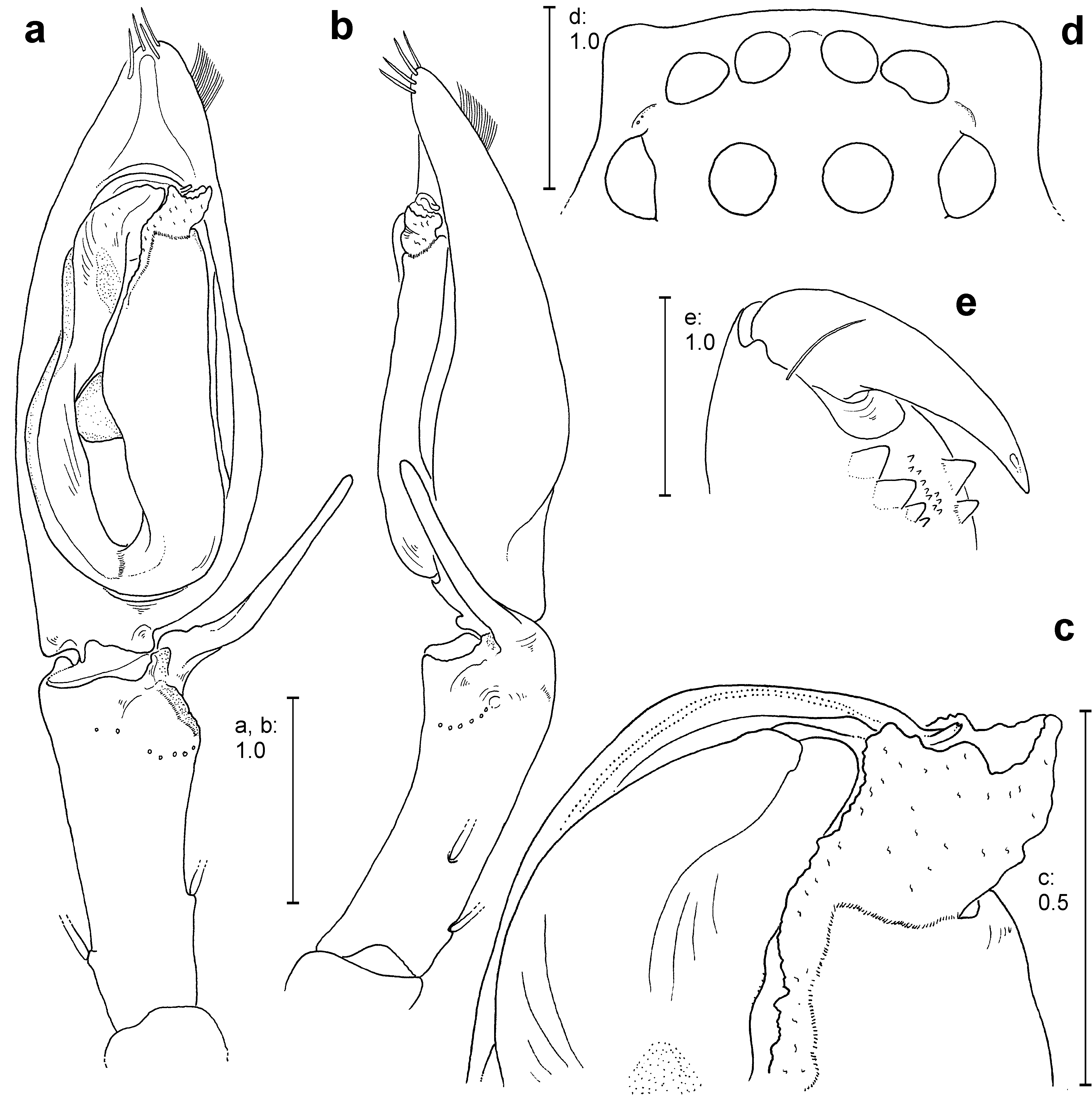
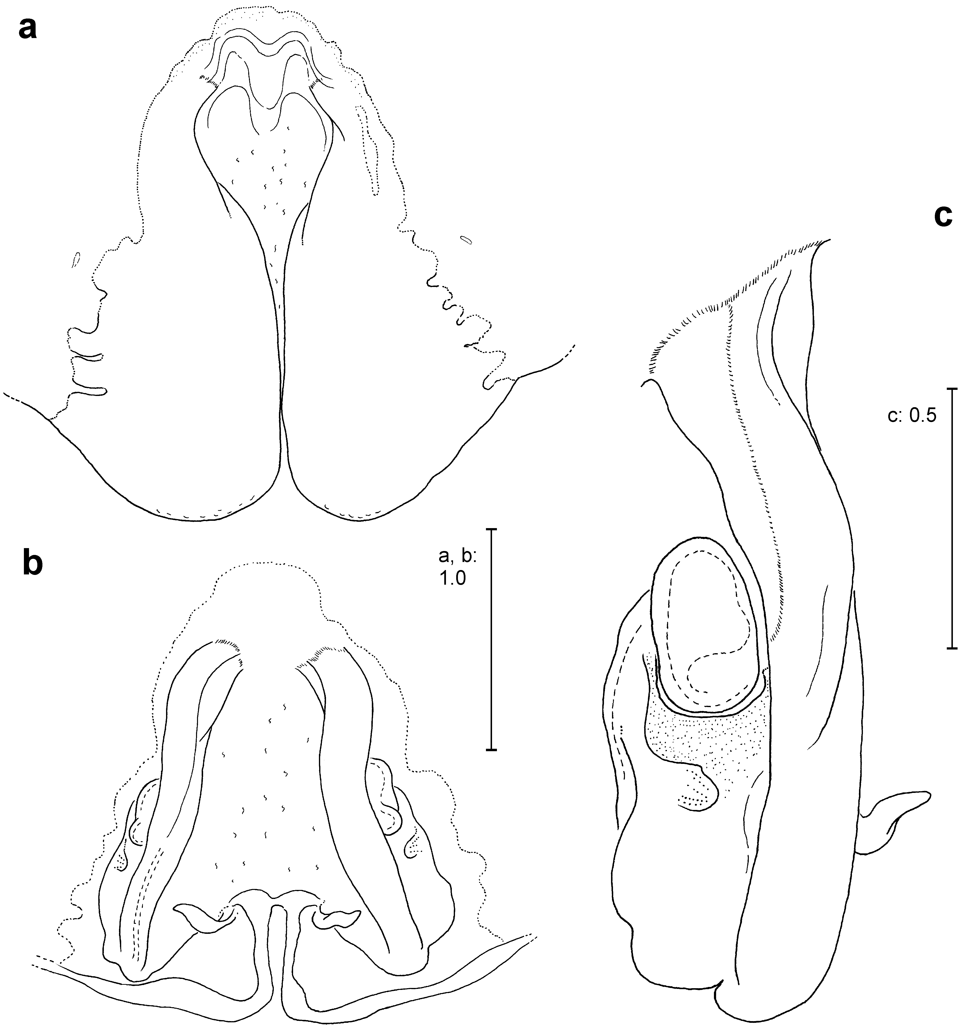
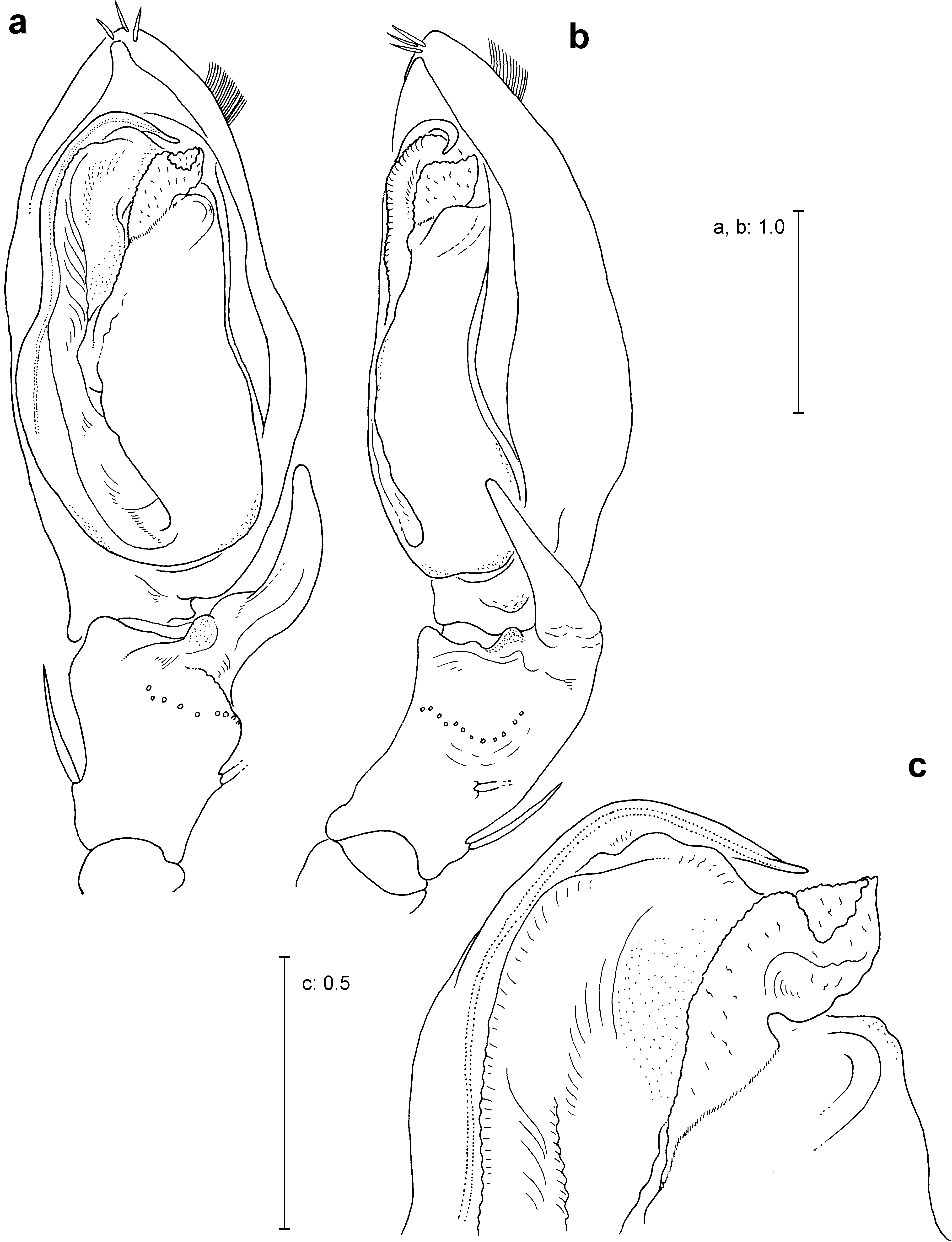
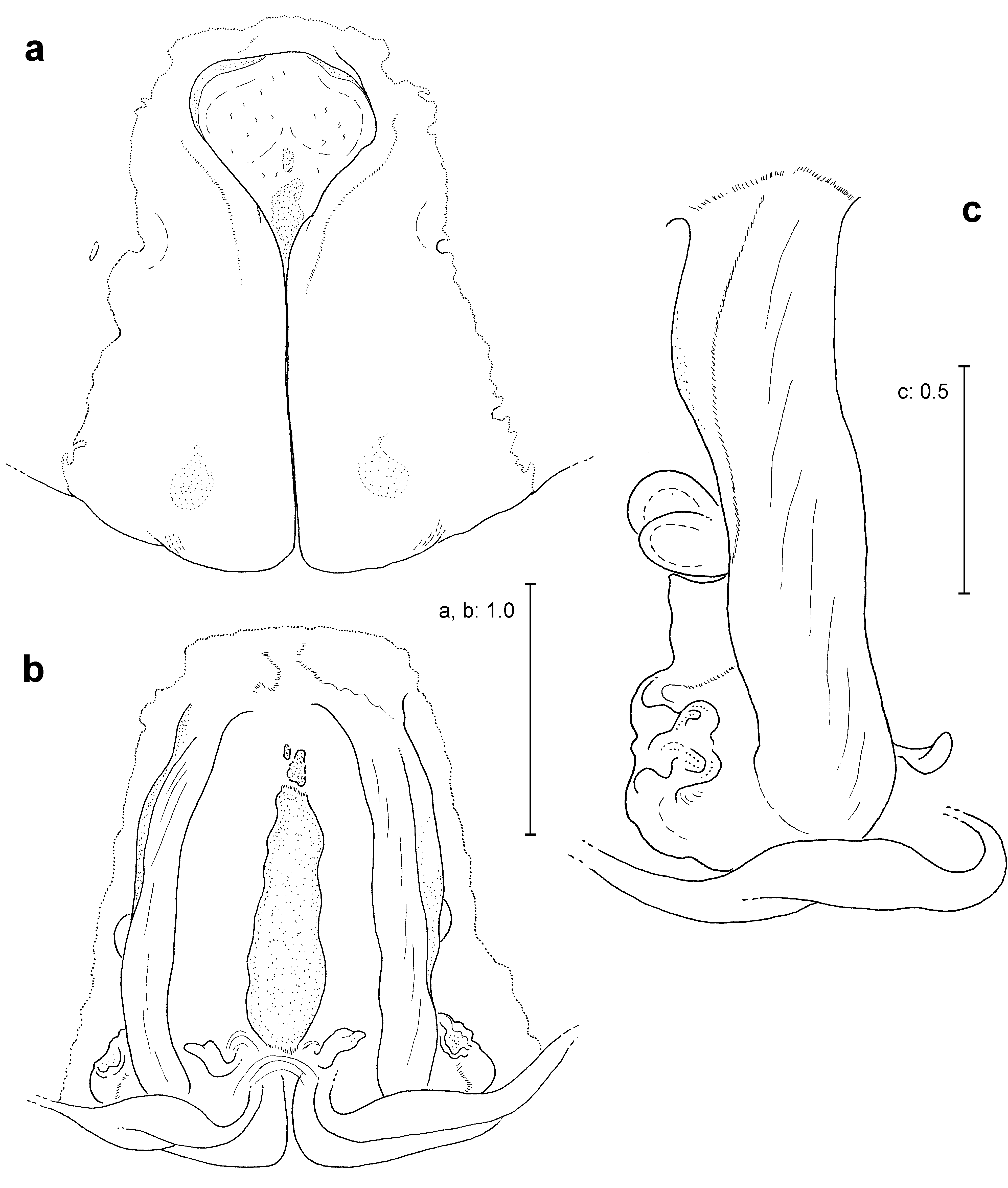
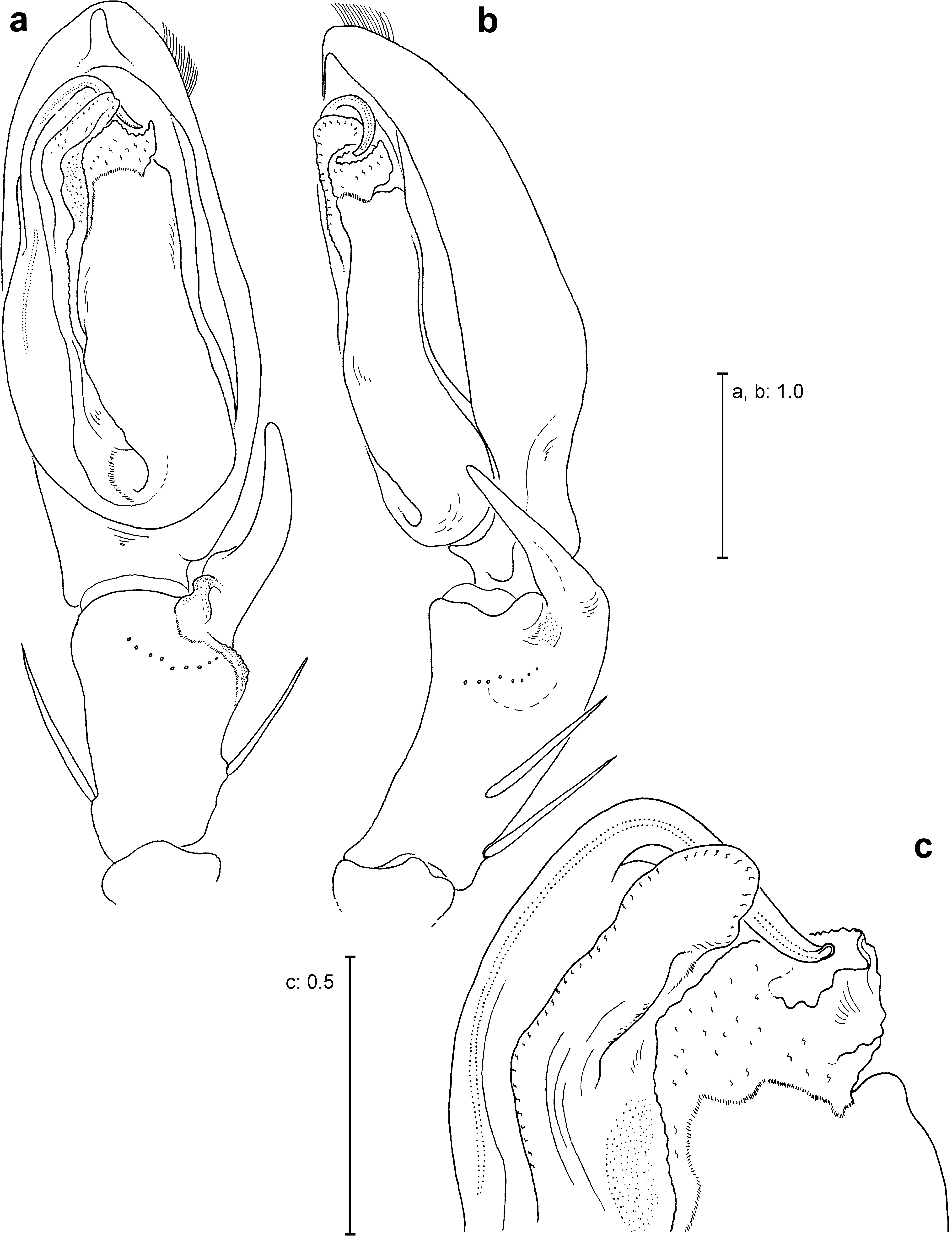
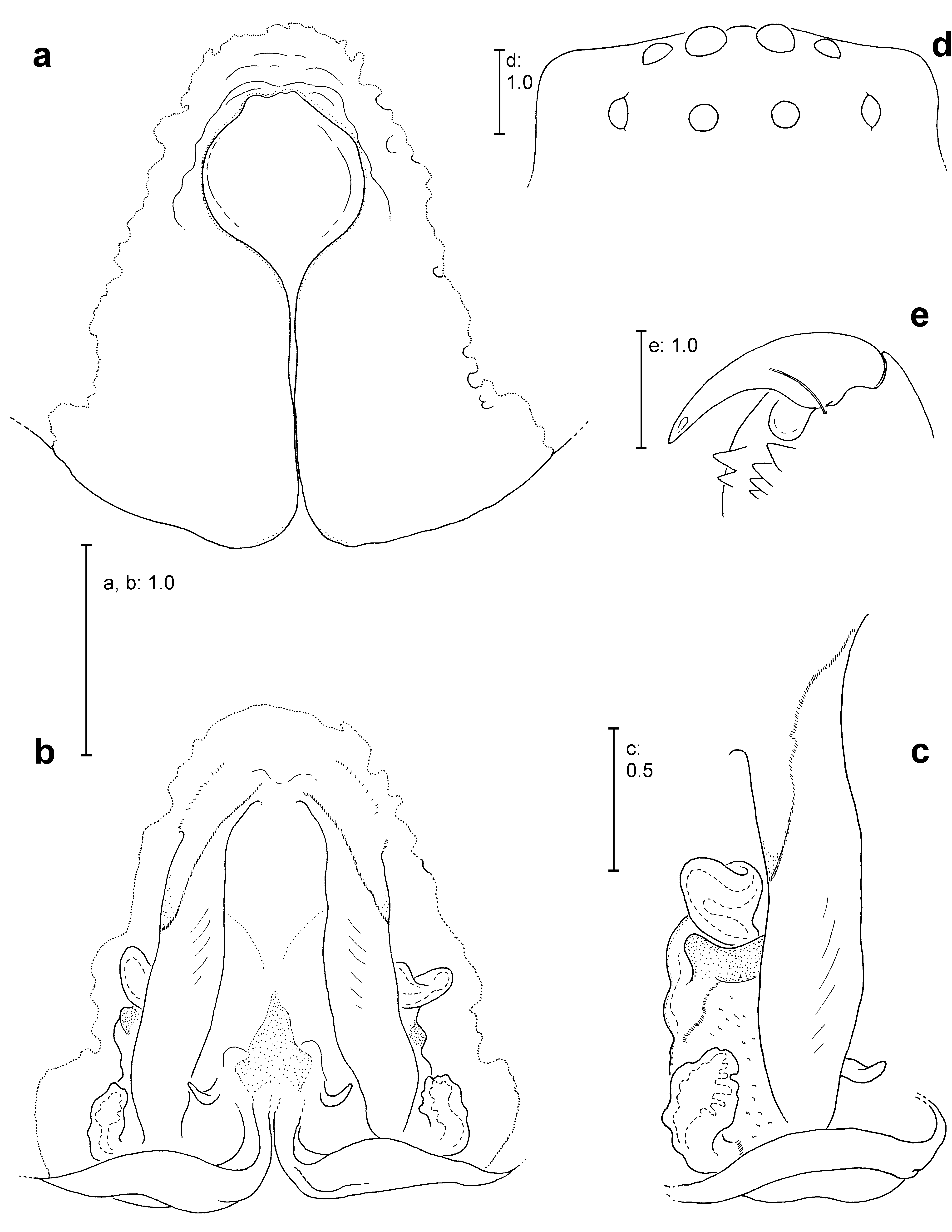
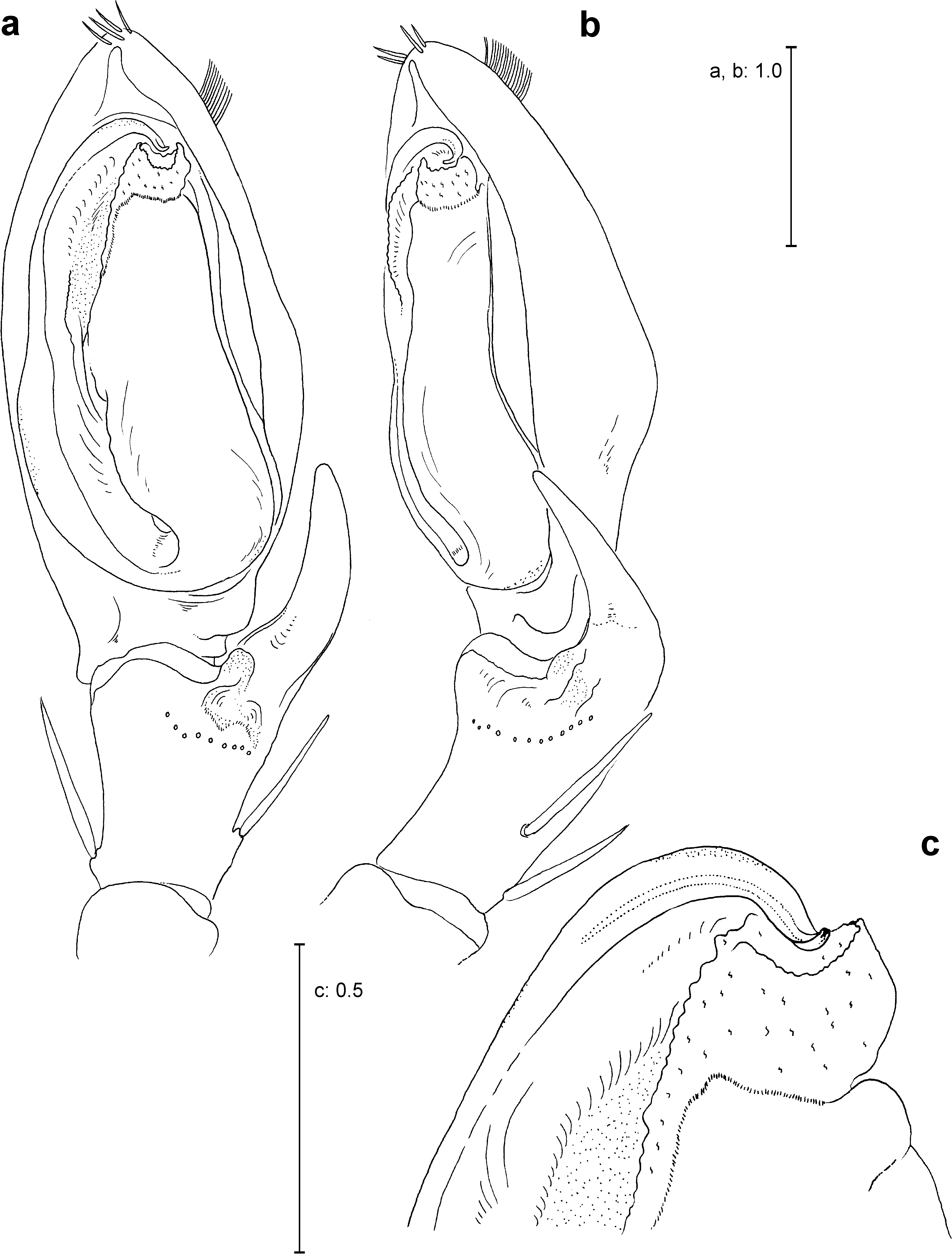
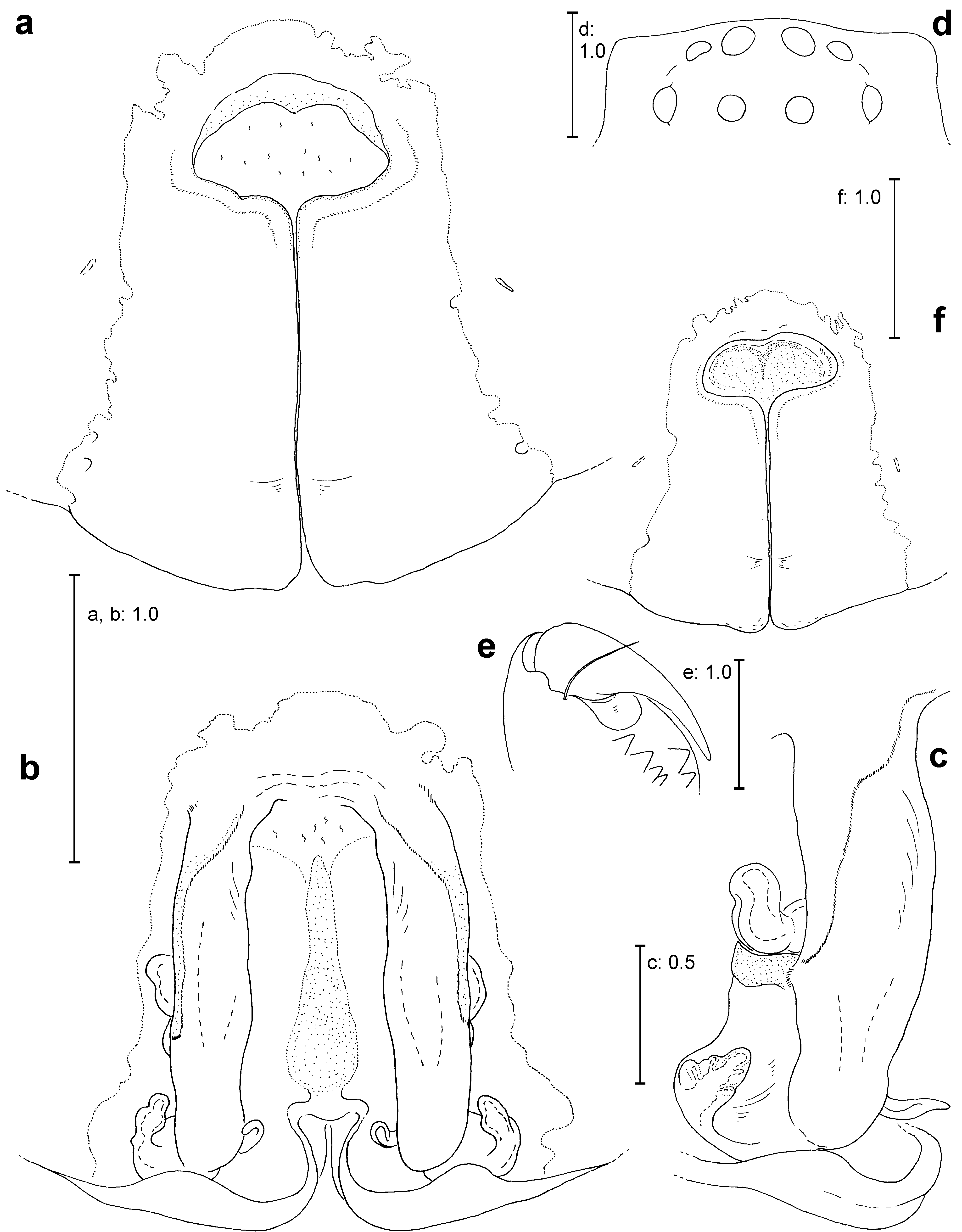
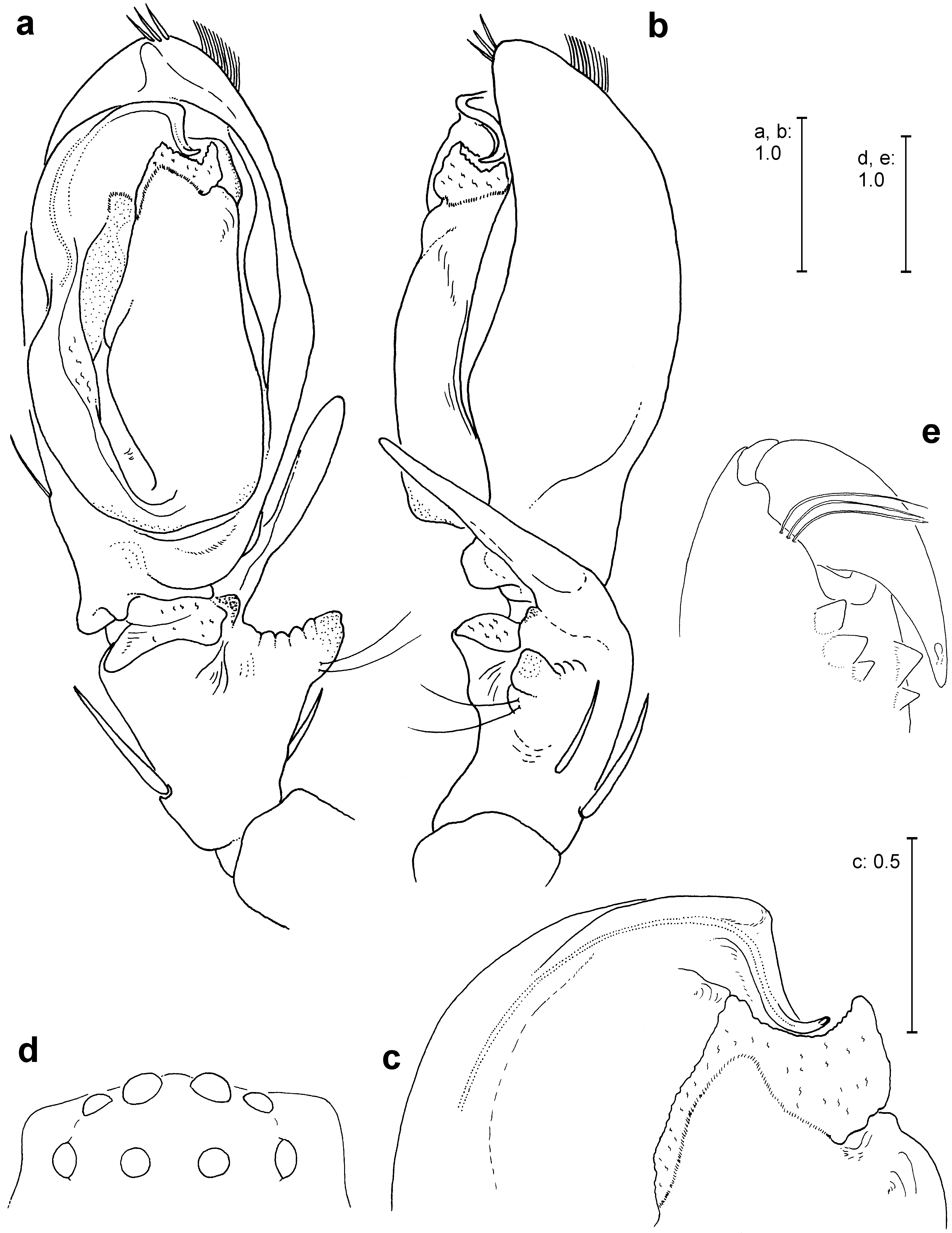
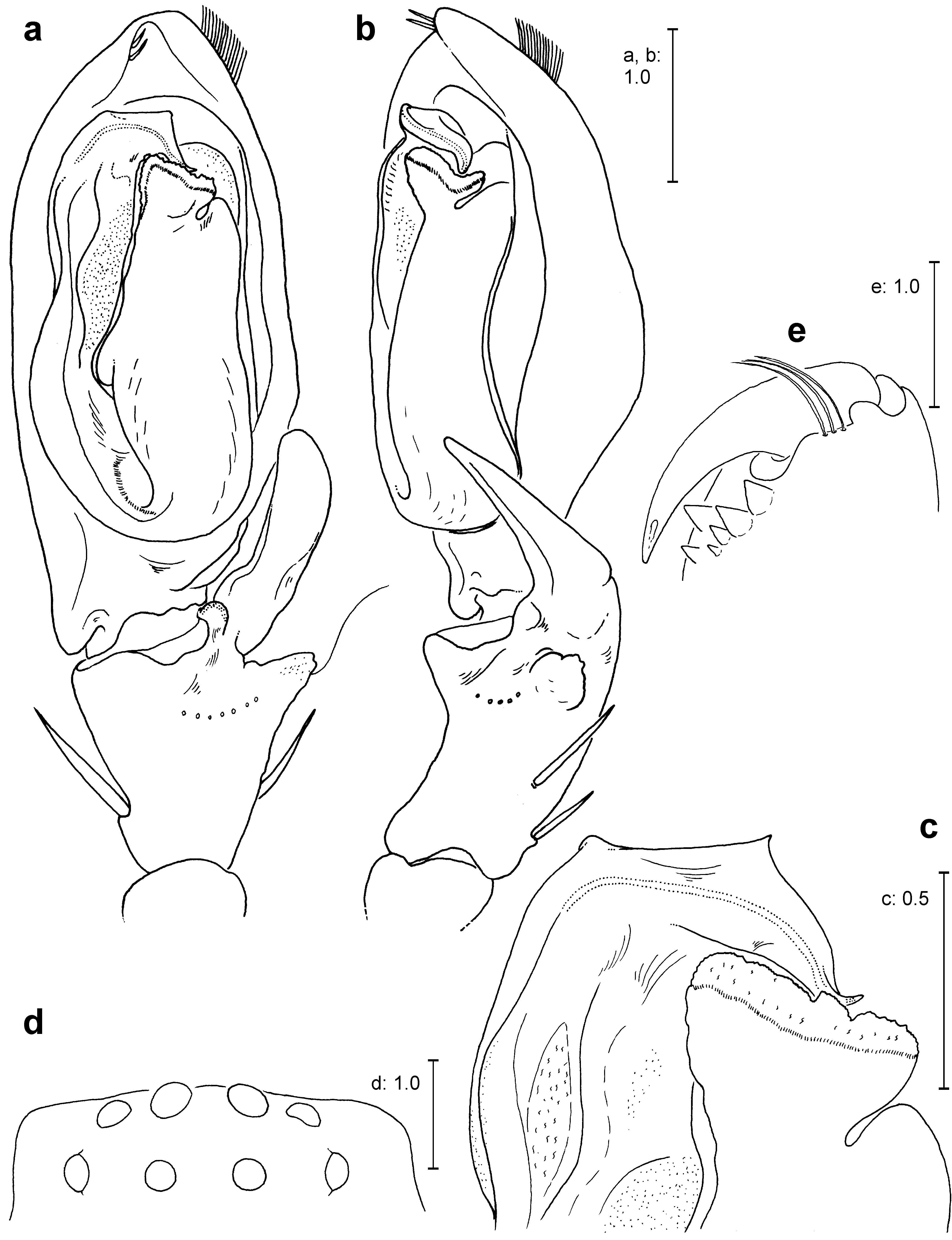
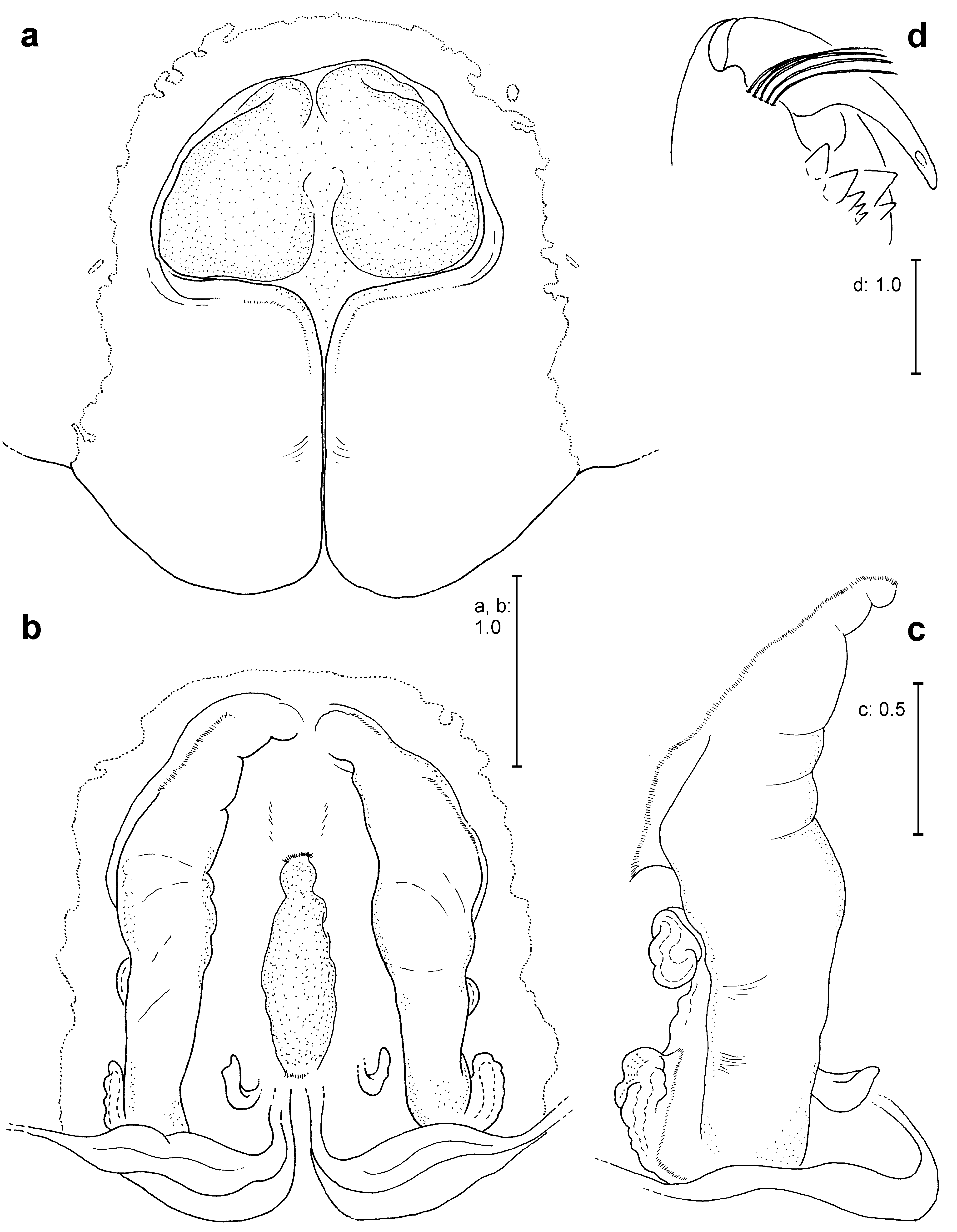
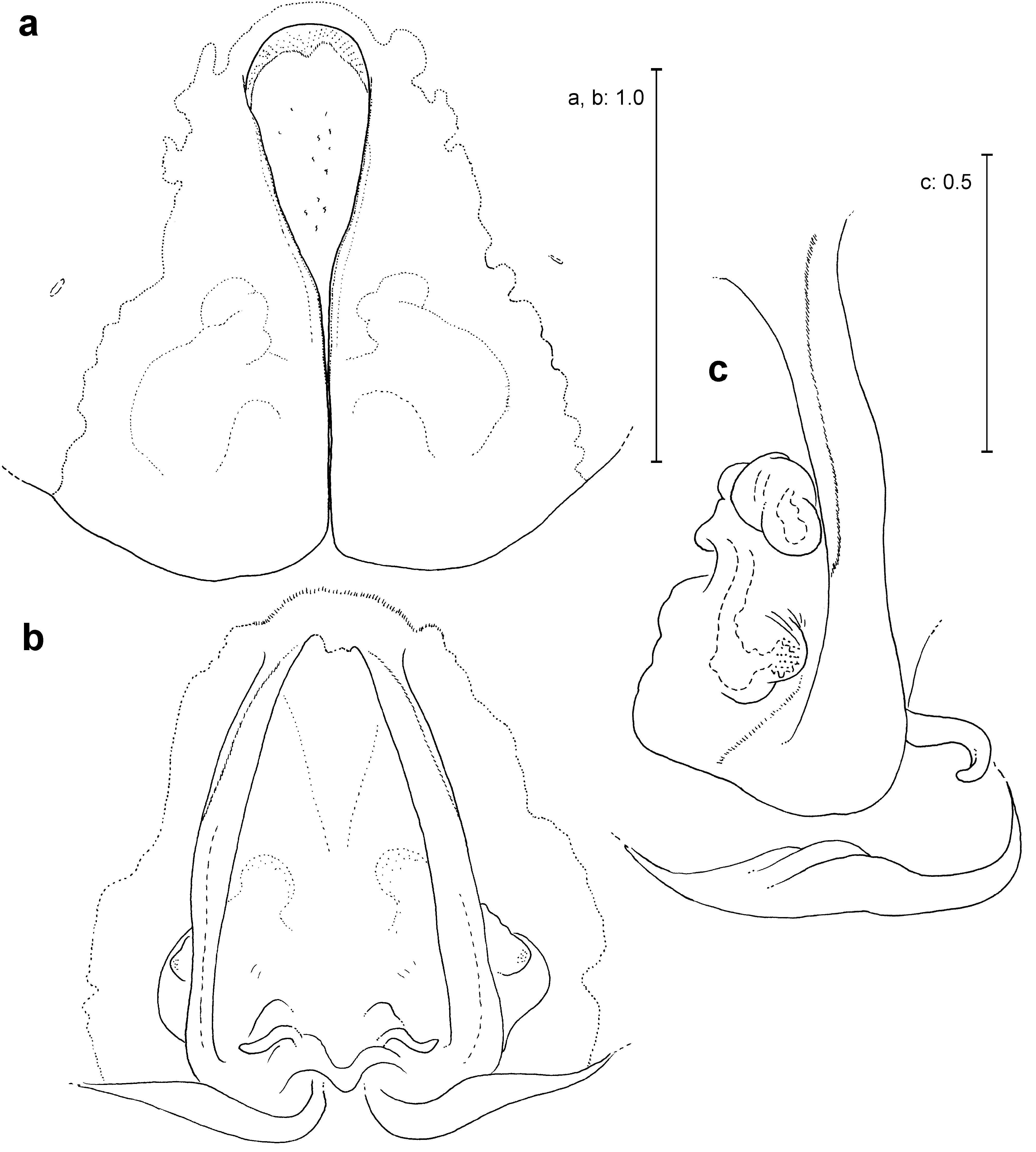
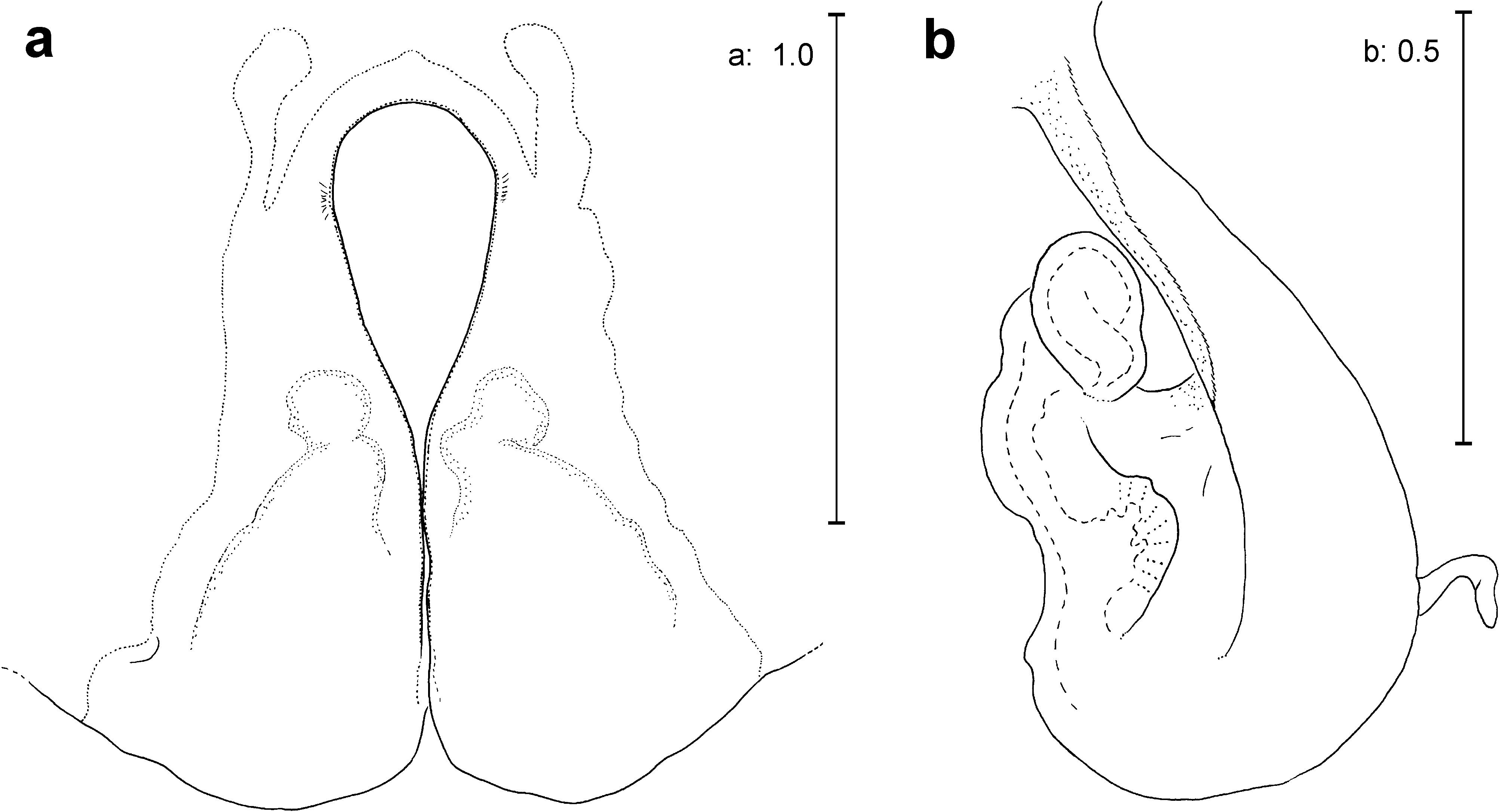
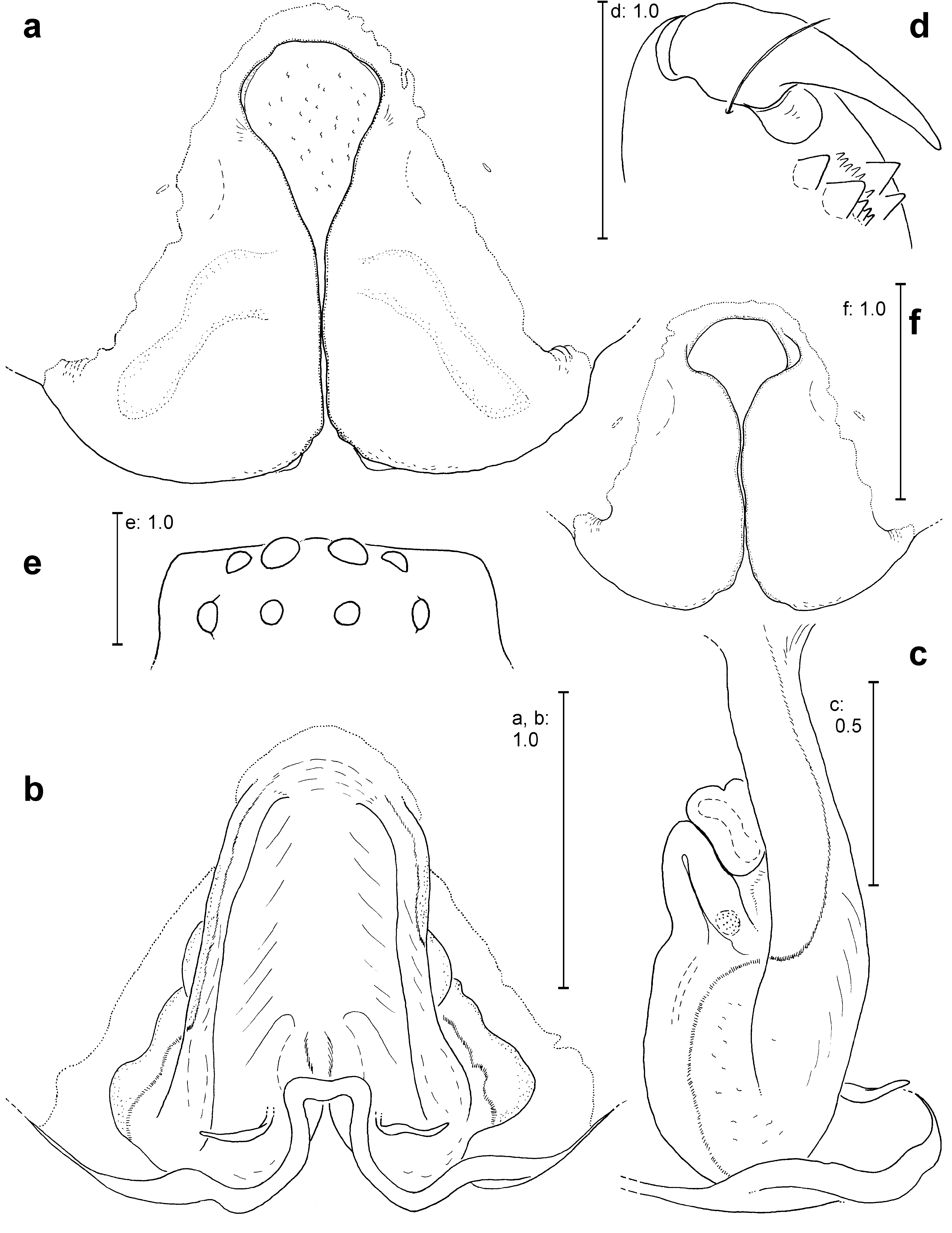
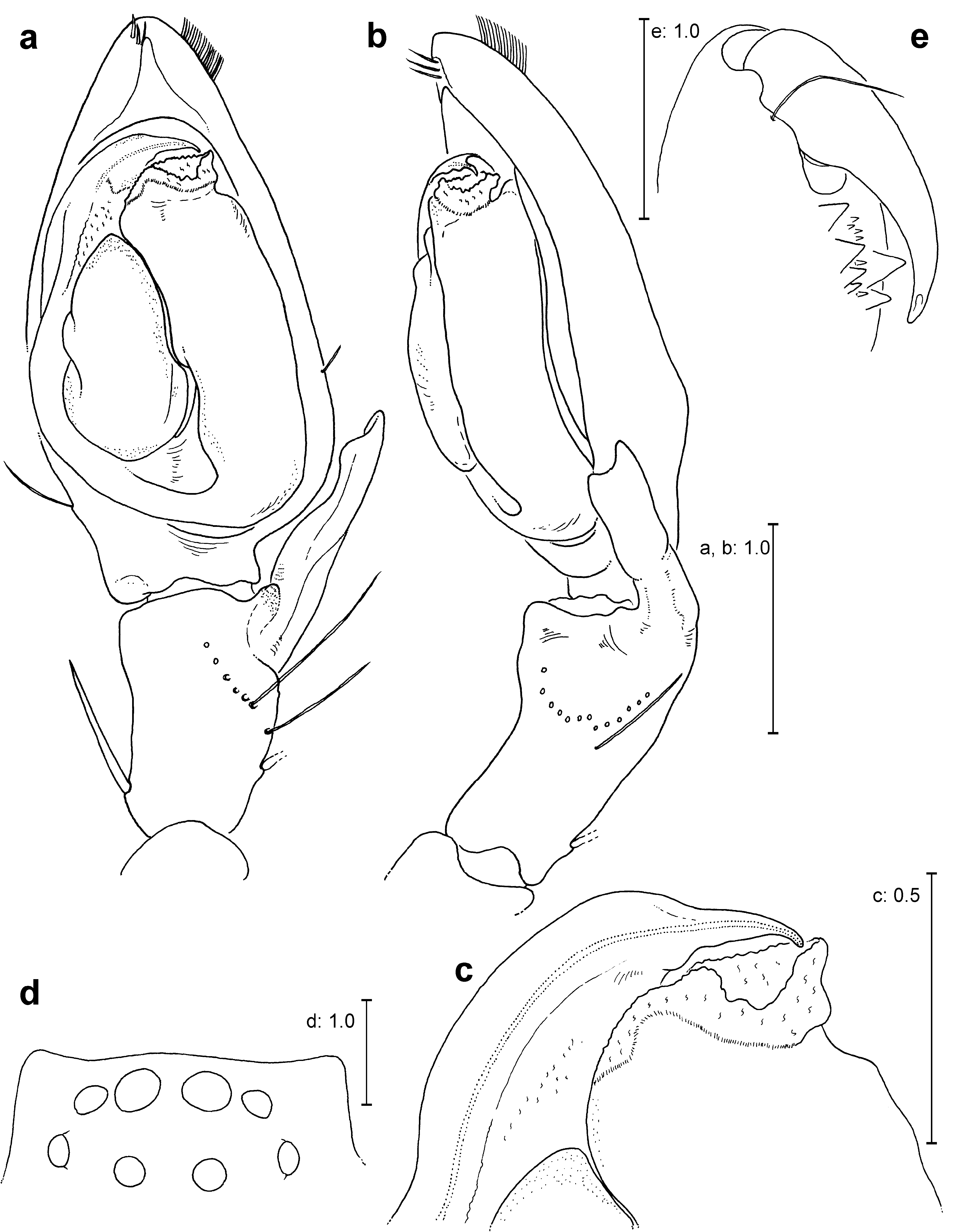
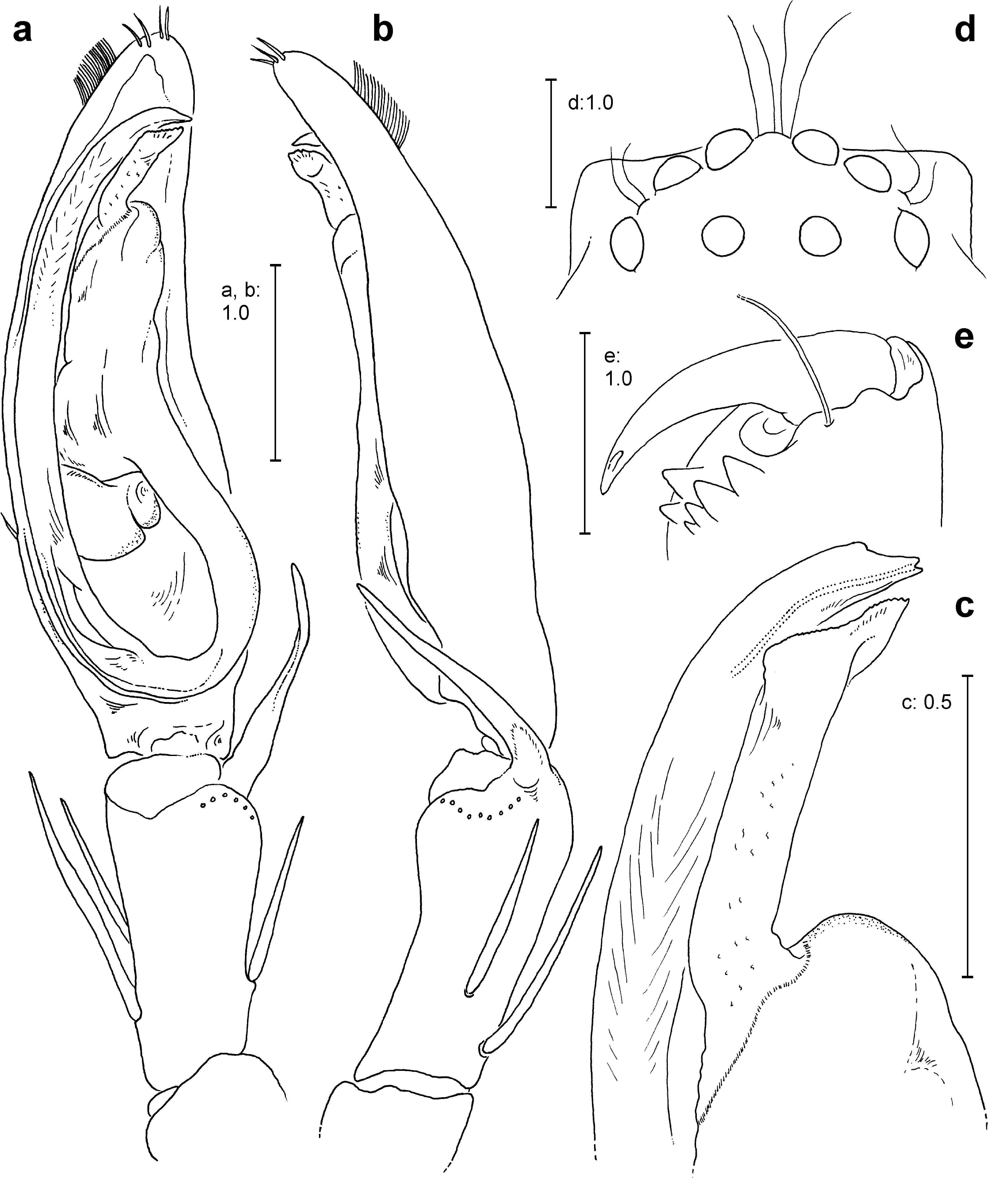
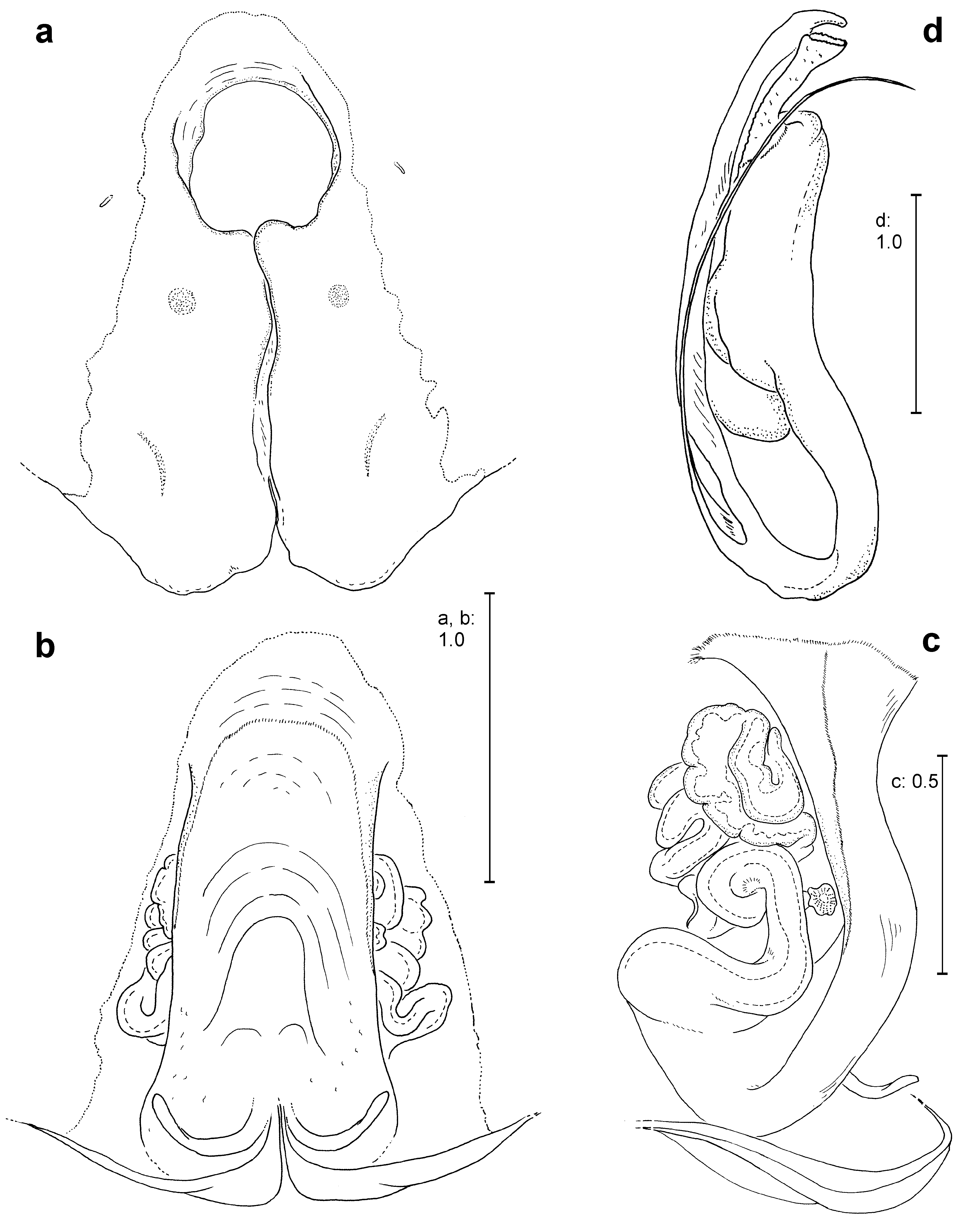
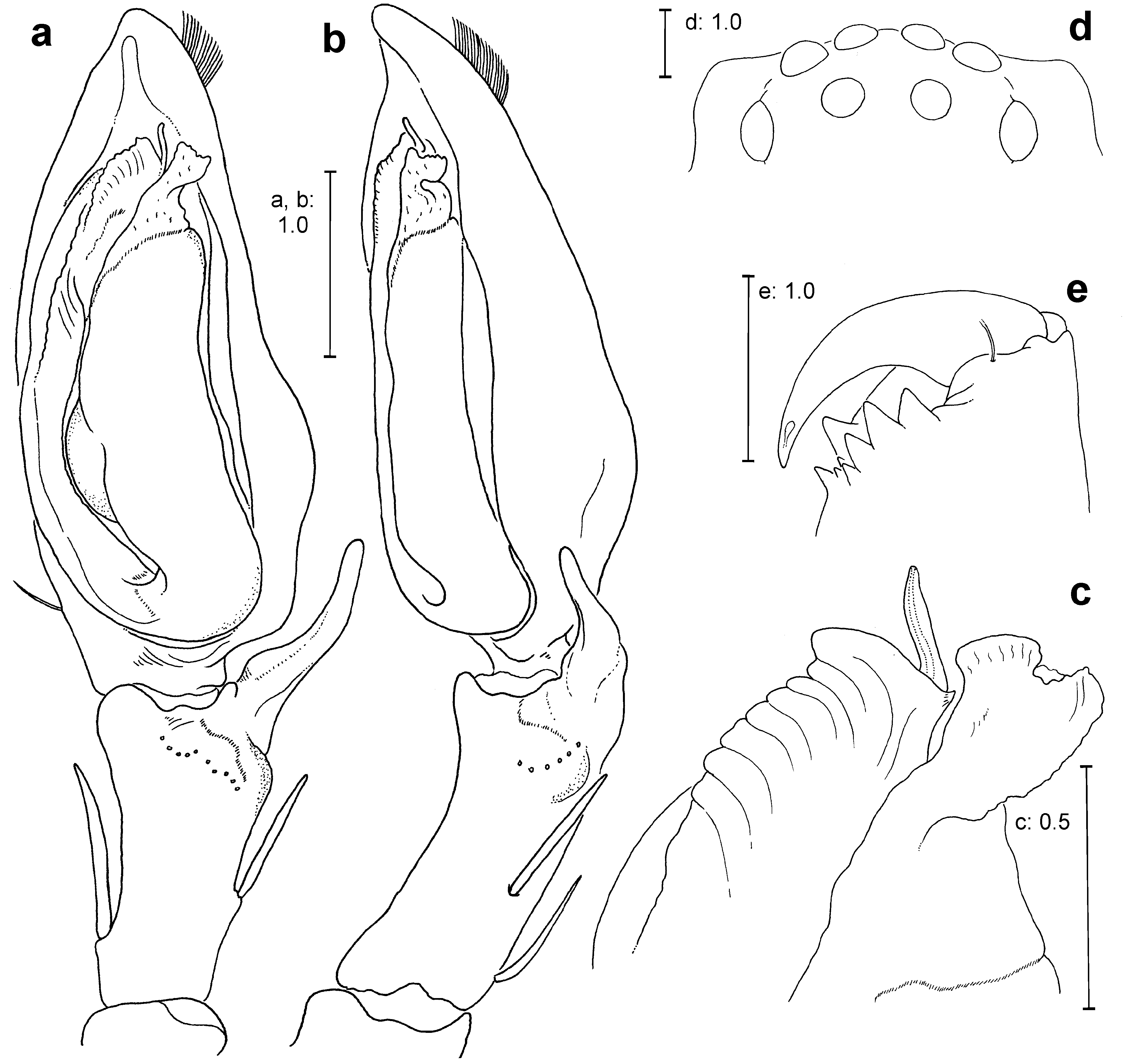
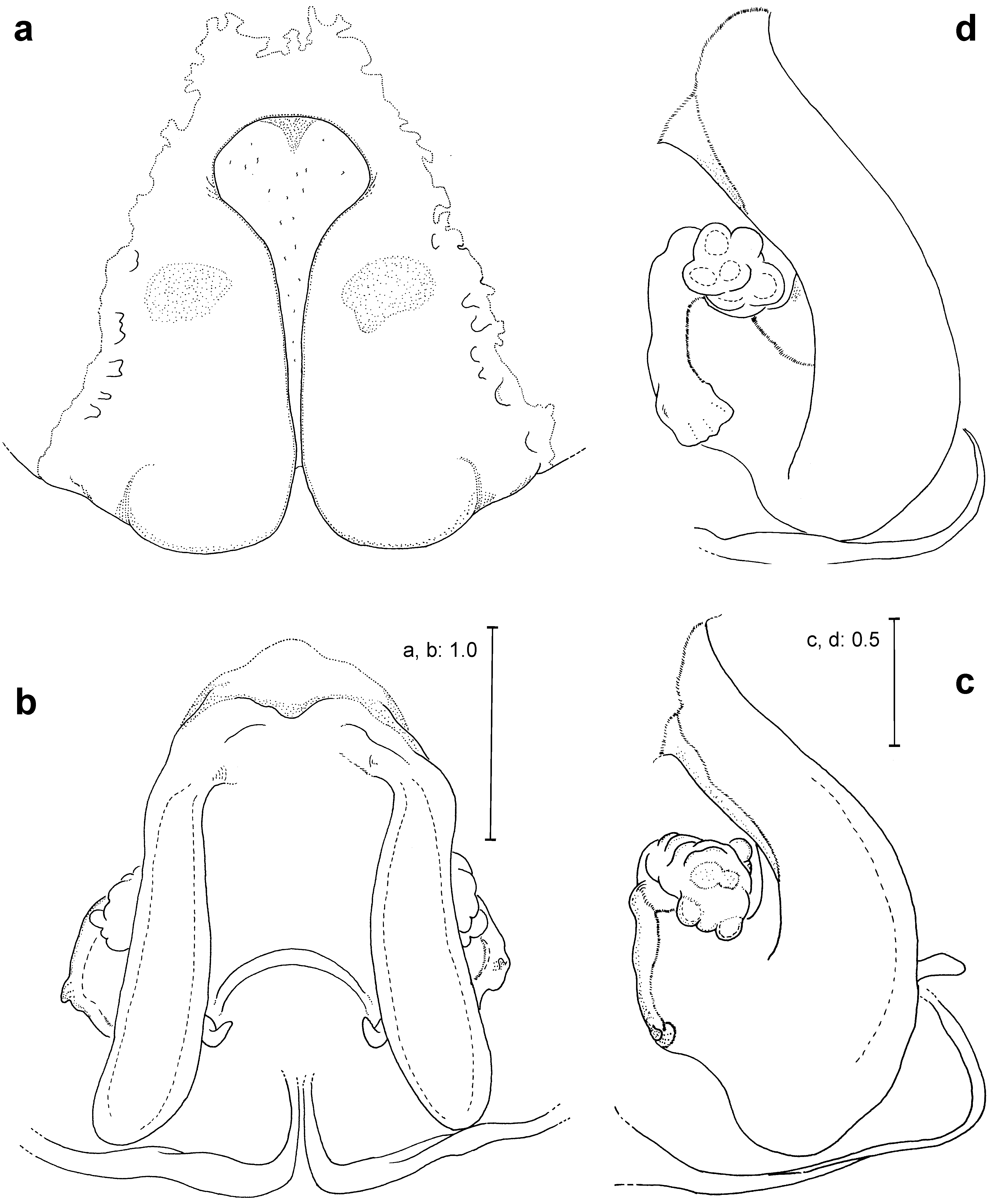
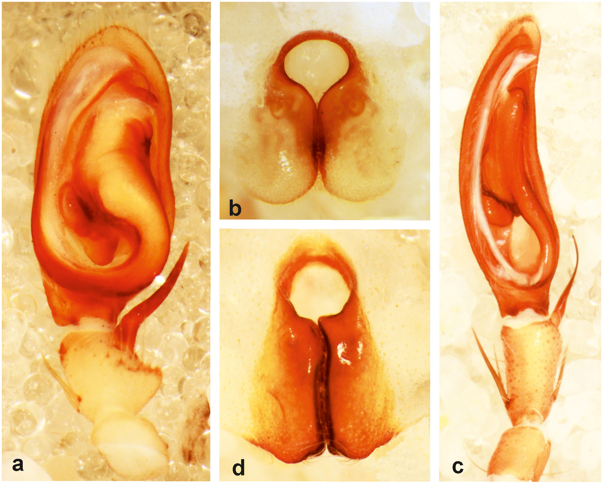
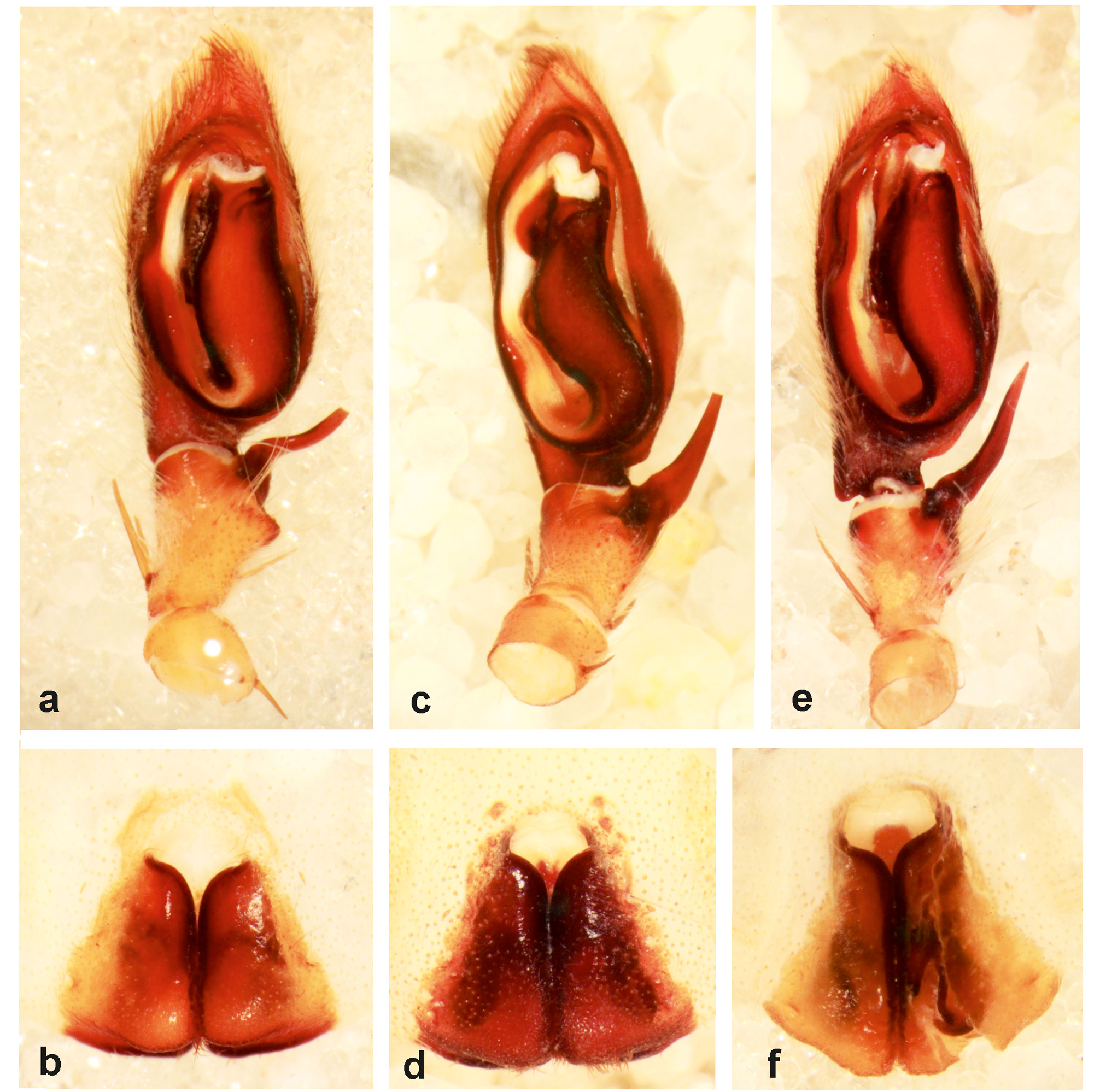
Diagnosis. Eusparassus is easily diagnosable from other members of subfamily Eusparassinae by the presence of two pairs of ventral tibial spines on legs I–IV (three pairs in Pseudomicrommata , Arandisa , Leucorchestris Lawrence, 1962 and Carparachne Lawrence, 1962 ); from Olios (subfamily Sparassinae ) by a combination of characters including the presence of intermarginal denticles in some Eusparassus spp. (absent in Olios spp. ), presence of a single bristle on the anterior margin of cheliceral basal segment below fangs but that number can reach a maximum of five (mostly> 10 in Olios spp. ). However, the best characters to distinguish between these two morphologically closely similar genera are those of the copulatory structures. In Eusparassus spp. the male palp is characterized by embolus and tegulum nearly of the same length arranged as a U-shaped structure, presence of embolus membrane (EM) [EM can be considered a well developed pars pendula, personal communication with C.A. Rheims], lack of any tegulum apophysis ( Fig. 1 View FIGURE 1 ); female epigyne shows two large lateral lobes (LL), and simple straight copulatory ducts leading to a more complex turning loop (TL) ( Fig. 2 View FIGURE 2 ).
Description. See Moradmand and Jäger (2012a).
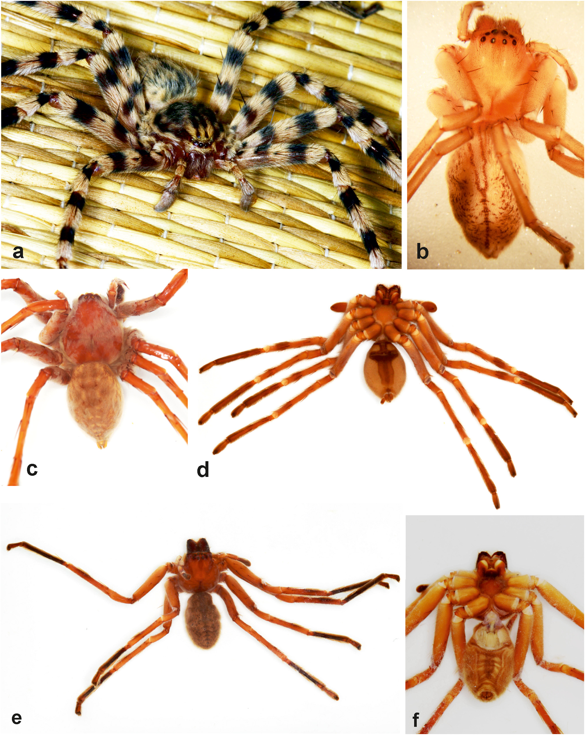
Natural history and habitat preferences. The knowledge on the biology of stone huntsman spiders is quite scanty. They produce large silken papery retreats attached to the underside of stones or in crevices of rocks. They hide during the day in these retreats and also use them to moult in. The excuvia are mostly found within the abandoned retreats (personal observation). Females construct a sealed egg-sac inside the larger retreat and guard it until the spiderlings hatch. In E. walckenaeri ( Audouin, 1826) , it took nearly one month from pre-larval stage to hatching stage ( Gabriel 2011). Like most Sparassidae , the stone huntsman spiders are nocturnal predators. They are known from semi-arid pine forest in the Atlas Mountains and the borders of the Sahara in Northern Africa to the Wahiba sand dunes and Wadis in Arabia, from the Mediterranean area to Central Asian deserts and the slopes of the Himalayas, and throughout the Eastern and Southern African Savannah to the arid borders of the Namib and Kalahari deserts. They can occur in very high elevations above sea level (e.g., E. pontii up to 3000–4000 m in Himalayas, Moradmand & Jäger 2012a). Earlier biological notes are restricted to some observations on the species E. walckenaeri by Gerhardt (1928, 1933) who documented his observations on the mating behaviour of this species (sub Sparassus sp. from Greece). Gabriel (2011) published his observations of the developments of spiderlings and some parasites and predators from Turkey.
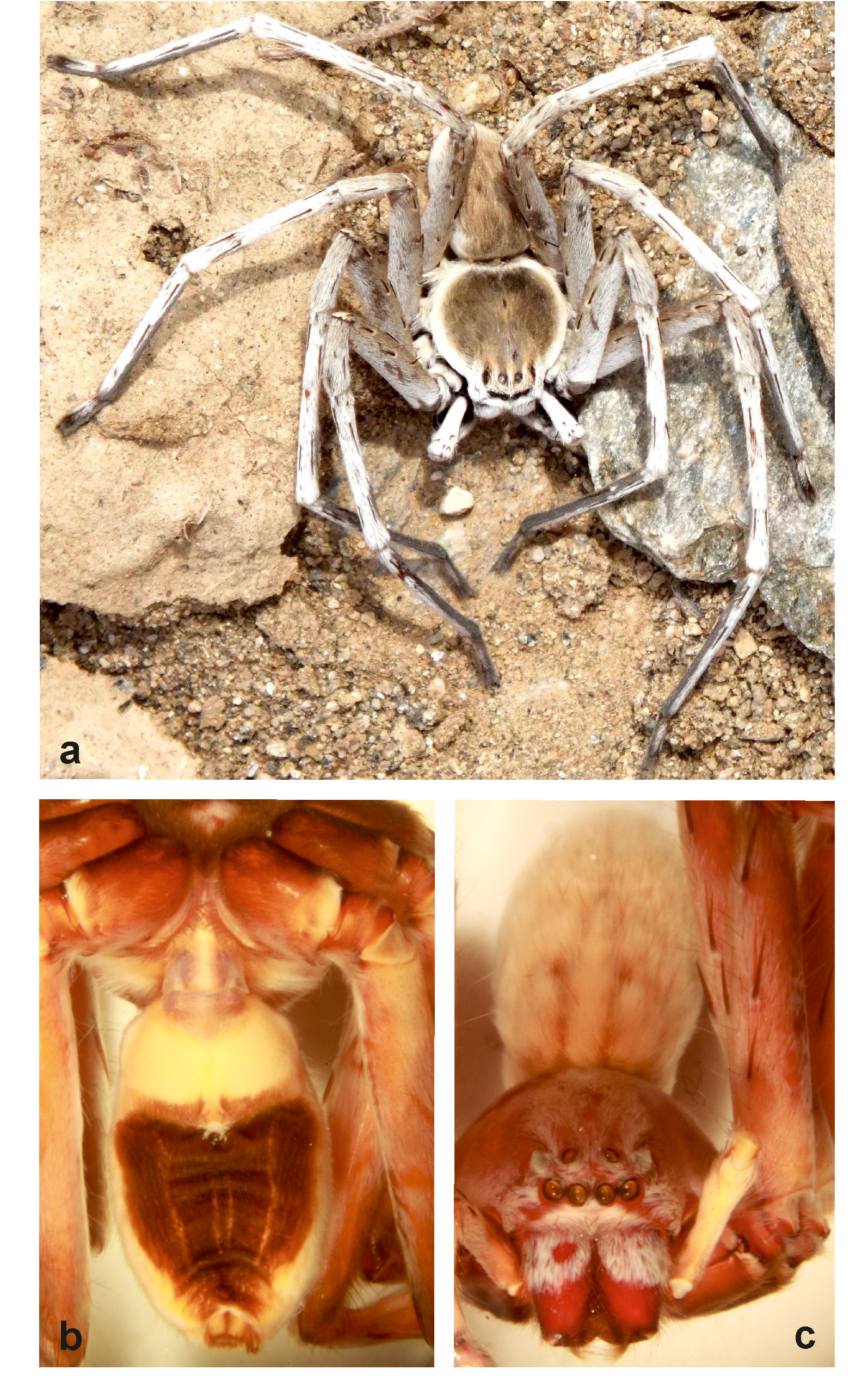
Copulation. The first photographical documentation of the copulation process of palp and epigyne in the genus Eusparassus is recorded and presented here. Combining knowledge of the morphology of the copulatory structures in Eusparassus spp. and the detailed documentation on how they function in action provide some valuable data on the functional morphology of the pedipalp and epigyne. Juvenile specimens of E. walckenaeri were collected by Dr Peter Jäger in the Negev desert (during the 26 th European congress of Arachnology) in September 2011. Specimens were reared in captivity until they reached maturity in August 2012. On the 7 th of August, the female was housed in a glass terrarium (30cm diameter x 20cm high) and one day later, the male was introduced into the terrarium. A few minutes later, the male started searching and tracing the female. Suddenly he attacked her and tried to grab her by the legs and chelicerae but the female autotomized one leg and escaped. He fed on the leg of the female and subsequently killed a cricket roaming in the terrarium but did not consume it. The male approached the female again. This time the female did not struggle and the male seized her, face to face, using both his legs and chelicerae. He gently bit the female’s pedicel area between prosoma and opisthosoma and held her with his legs ( Fig. 44a View FIGURE 44 ). They remained in this position for a few seconds until the female was totally subdued and did not move till the end of mating. The male attempted to reach the female’s epigyne, first from her right side using his left palp but without inserting his embolus ( Figs 44b–d View FIGURE 44 ). Then he shifted to the left side of the female. The process of coupling palp and epigyne was initiated by anchoring the RTA (dRTA) into the posterior margin of epigyne between the lateral lobes ( Figs 45a, d View FIGURE 45 ), the male stretched his right palp next, which suddenly expanded and the embolus was inserted into the copulatory opening ( Fig. 45b View FIGURE 45 ). This observation (inserting dRTA into posterior margin between lateral lobes of epigyne) gives some evidence about a similar structure in the vulva which was recently recognized in the species of the genus Sinopoda Jäger, 1999 . This structure was named membranous sac (Msa) and is supposed to hold the dRTA during copulation ( Jäger 2012). The Msa can be mistaken for intermediate tissue and muscles around vulva, and is usually removed during vulva preparation since its presence restricts the view on scleriotized vulva structures.. The Msa in Eusparassus species is located medially between the fertilization ducts ( Figs 11d, e View FIGURE 11 ). Another modification in the female copulatory organ might be the following: Eusparassus species with a more robust dRTA have special modifications dorsally of the median septum, from a simple hyaline structure ( Fig. 2b View FIGURE 2 ) to a sclerotized longitudinal band ( Fig. 16b View FIGURE 16 ) and even a complex folded structure ( Fig. 36b View FIGURE 36 ).
Identification key to species of Eusparassus
In the following key, a combination of the somatic and copulatory characters are used, nevertheless, species identification should be confirmed by checking the detailed diagnoses and descriptions given in the text for each species. The key should be used with special care when identifying females. Species descriptions of the doriae group as well as E. pearsoni ( Pocock, 1901) ( vestigator group), E. pontii Caporiacco, 1935 and E. xerxes ( Pocock, 1901) (both incertae sedis), E. dufouri Simon, 1932 and E. levantinus Urones, 2006 (both dufouri group) are given in Moradmand and Jäger (2012a). The character ventral opisthosoma dark marking must be used with special care as preserved specimens could have been faded. Since Cercetius perezi is regarded congeneric (retained usage until ICZN decision on case 3596), this species is included in the Eusparassus key.
1. Cheliceral furrow with intermarginal denticles (e.g. Fig. 1f View FIGURE 1 ).................................................... 2
– Cheliceral furrow without intermarginal denticles (e.g. Fig. 13e View FIGURE 13 )............................................... 16
2. Male [unknown in E. borakalalo View in CoL spec. nov.]................................................................ 3
– Female.............................................................................................. 9
3. Palp with enlarged and bulged ST (e.g. Fig. 35a View FIGURE 35 )............................................................. 4
– Palp with small and hidden ST behind T (e.g. Fig. 1a View FIGURE 1 )......................................................... 6
4. dRTA bifurcated at its tip ( Figs 35a, b View FIGURE 35 ) [ Zimbabwe]............................................ jocquei View in CoL spec. nov.
– dRTA pointed and not bifurcated at its tip.................................................................. 5
5. ET triangular and flattened proximally and pointed distally ( Fig. 31c View FIGURE 31 ) [ South Africa: Northern Cape Province]............................................................................................... schoemanae View in CoL spec. nov.
– ET slender and curved at its distal end ( Fig. 29c View FIGURE 29 ) [ South Africa].................................... jaegeri View in CoL spec. nov.
6. dRTA bent toward cymbium and pointed disto-ventrad ( Fig. 4a View FIGURE 4 ) [Horn of Africa to Arabia]..................... laevatus View in CoL
– dRTA directed distad................................................................................... 7
7. Ventral opisthosoma with large solid black marking ( Fig. 57b View FIGURE 57 ), ET directed distad ( Fig. 42c, f View FIGURE 42 ) [Arabia and Horn of Africa].......................................................................................... Cercetius perezi
– Ventral opisthosoma pale, ET directed retrolaterad (e.g. Fig. 1d View FIGURE 1 )................................................ 8
8. Palp and dRTA robust, PE and AE roughly subequal ( Figs 1a–e View FIGURE 1 ) [Eastern Mediterranean to Egypt and Algeria].. walckenaeri View in CoL
– Palp and dRTA elongated and slender, PE distinctly larger than AE ( Figs 7a–d View FIGURE 7 ) [Arabian Peninsula]..... arabicus View in CoL spec. nov.
9. Epigyne with AMLL fused together anteriorly (e.g. Fig. 32a View FIGURE 32 ).................................................. 10
– Epigyne with AMLL not fused together anteriorly (e.g. Fig. 2a View FIGURE 2 )................................................ 14
10. Epigyne with MS clearly visible posteriorly ( Fig. 36a View FIGURE 36 ) [ Zimbabwe]................................ jocquei View in CoL spec. nov.
– Epigyne with MS not visible posteriorly (LL are in contact)................................................... 11
11. Vulva composed of several bulbous parts in TL ( Figs 43c, d View FIGURE 43 ) [Horn of Africa to Arabia]................. Cercetius perezi
– Vulva different (with single large TL)..................................................................... 12
12. MS as long as wide, CD and MS partially to fully sclerotized ( Figs 30a–d View FIGURE 30 ) [ South Africa]............... jaegeri View in CoL spec. nov.
– MS longer than wide and membranous, CD hyaline......................................................... 13
13. EF longer than wide ( Figs 32a View FIGURE 32 , 33a View FIGURE 33 ) [ South Africa: Northern Cape Province].................... schoemanae View in CoL spec. nov.
– EF wider than long ( Figs 34a, f View FIGURE 34 ) [ South Africa]............................................. borakalalo View in CoL spec. nov.
14. PE distinctly larger than AE ( Fig. 7d View FIGURE 7 ), EF bridge present, ( Fig. 8a View FIGURE 8 ) [Arabian Peninsula]............... arabicus View in CoL spec. nov.
– PE and AE nearly equal, EF bridge mostly absent........................................................... 15
15. MS as wide as long, MS length ¼ EF length, vulva with Gpo situated in a depression in connection with collar form a continuous ridge ( Figs 5a–c View FIGURE 5 ) [Horn of Africa to Arabia]....................................................... laevatus View in CoL
– MS mostly longer than wide, MS length ½ of EF length, vulva with Gpo situated in a depression separated from collar part ( Figs 2a–c View FIGURE 2 ) [Eastern Mediterranean to Egypt and Algeria]............................................ walckenaeri View in CoL
16. Male [unknown in syrticus View in CoL , pearsoni View in CoL , maynardi View in CoL , pontii View in CoL ]...................................................... 17
– Female............................................................................................. 35
17. Ventral opisthosoma with distinct dark marking............................................................. 18
– Ventral opisthosoma lacking distinct dark marking.......................................................... 28
18. vRTA well developed: as long as one-third of dRTA (e.g. Fig. 25a View FIGURE 25 )............................................. 19
– vRTA not well developed: less than one-third of dRTA....................................................... 20
19. ET flat and wide with a pointed triangular process, dRTA robust and flattened dorso-ventrally ( Figs 27a–c View FIGURE 27 ) [ Burkina Faso and Nigeria]............................................................................. reverentia View in CoL spec. nov.
– ET and dRTA different ( Figs 25a–c View FIGURE 25 ) [Eastern Africa: Ethiopia, Kenya and Tanzania]......................... vestigator View in CoL
20. EM with projecting bulge covering proximal end of ET in ventral view ( Figs 20a–c View FIGURE 20 ) [Eastern Morocco]........... fritschi View in CoL
– EM without any projecting bulge........................................................................ 21
21. ET directed proximad ( Fig. 11a View FIGURE 11 )......................................................................... 22
– ET pointing in differentdirection......................................................................... 23
22. ET robust and flat, dRTA sickle-like ( Figs 11a, b View FIGURE 11 ), ventral opisthosoma with V-shaped marking ( Fig. 48b View FIGURE 48 ) [Western Iberian Peninsula]...................................................................................... dufouri View in CoL
– ET slim, dRTA more straight ( Fig. 60c View FIGURE 60 ), V-shaped marking with additional median band ( Fig. 48d View FIGURE 48 ) [Eastern Iberian Peninsula] levantinus View in CoL
23. AE larger than or subequal as PE....................................................................... 24
– PE generally larger than AE, PLE largest.................................................................. 27
24. ET directed retrolaterad ( Figs 12a, c View FIGURE 12 ) [ Morocco]...................................................... atlanticus View in CoL
– ET directed ventrad ( Fig. 22c View FIGURE 22 ), PLE subequal to PME........................................................ 25
25. ET flattened, dRTA bent toward cymbium, directed ventrad ( Figs 15a–c View FIGURE 15 ) [Northern Algeria]................... barbarus View in CoL
– ET slim, dRTA directed distad ( Figs 22a–c View FIGURE 22 )................................................................ 26
26. Small to medium Eusparassus species (16 to 18 mm) with ventral opisthosoma marking more solid in fresh samples and Vshaped in preserved ones (lines of marking are bold dark) ( Fig. 49f View FIGURE 49 ) [North-Eastern Algeria]................. letourneuxi View in CoL
– Large Eusparassus species (21 to 25 mm), a vase-like black marking on ventral opisthosoma ( Fig. 56d View FIGURE 56 ) [ Iran to Pakistan].................................................................................................... xerxes View in CoL
27. ET directed retrolaterad, vRTA pointed and triangular in ventral view ( Figs 17a–c View FIGURE 17 , 62a View FIGURE 62 ) [ Algeria to Morocco].... oraniensis View in CoL
– ET directed distad, vRTA broad and not pointed ( Figs 42a–c View FIGURE 42 , 66a, e View FIGURE 66 )................................. Cercetius perezi
28. Embolus long and ET slender (e.g. Figs 37a–c View FIGURE 37 )............................................................. 29
– ET short and robust................................................................................... 30
29. Palpal structures strongly elongated, embolus covered by slender embolus membrane ( Figs 40a–c View FIGURE 40 ) [Southern Namibia]........................................................................................... educatus View in CoL spec. nov.
– Embolus membrane projected into a folded part close to ET ( Figs 37a–c View FIGURE 37 ) [Northern Namibia, Angola]............. tuckeri View in CoL
30. AME strikingly larger (~1.5 times) than other eyes ( Fig. 58e View FIGURE 58 ) [Central Asia]................. oculatus (Kroneberg, 1875) View in CoL
– AME subequal to or <1.5 times larger than others........................................................... 31
31. ET proximad, long and robust ( Fig. 67c View FIGURE 67 ) [ Afghanistan]..................................... fuscimanus Denis, 1958 View in CoL
– ET shorter and directed in different orientations............................................................. 32
32. vRTA rounded and not well developed (e.g. Fig. 68a View FIGURE 68 )........................................................ 33
– vRTA pointed and clearly triangular (e.g. Fig. 68e View FIGURE 68 , see Moradmand & Jäger, 2012a: Fig 17C View FIGURE 17 )........................ 34
33. ET slim ( Fig. 67e View FIGURE 67 ) [ Afghanistan to Rajasthan in India]...................................... kronebergi Denis, 1958 View in CoL
– ET robust ( Fig. 68a View FIGURE 68 ) [ Iran, Iraq and Turkey]............................... mesopotamicus Moradmand & Jäger, 2012 View in CoL
34. dRTA straight and beak-like, distal end of ET pointing distad ( Fig. 68e View FIGURE 68 ) [ China: Xinjiang Uyghur]... potanini (Simon, 1895) View in CoL
– dRTA with a slight bend in proximal half, ET leaf-like, distal end of ET pointing proximad ( Fig. 67a View FIGURE 67 ) [Central Iran]............................................................................................ doriae ( Simon, 1874) View in CoL
35. Ventral opisthosoma with distinct dark marking............................................................. 36
– Ventral opisthosoma lacking distinct dark marking.......................................................... 48
36. MS widened (approximately as wide as EF), fully sclerotized and prominent (e.g. Figs 63b, d, f View FIGURE 63 ), chelicerae usually with more than two thick bristles (max. five bristles) at ventral base of fangs.............................................. 37
– MS small, hyaline to partially sclerotized, chelicerae mostly with one thick bristle (max. two bristles).................. 39
37. MS heart-shaped ( Fig. 63f View FIGURE 63 ), femur spination 323 [ India: Western Ghats].................................... pearsoni View in CoL
– MS quadrangular ( Fig. 63b View FIGURE 63 ), femur spination 424........................................................... 38
38. GP separated from CD by most of its entire length ( Figs 26b, c View FIGURE 26 ) [East Africa: Tanzania to Ethiopia]............. vestigator View in CoL
– GP attached to CD by most of its entire length ( Figs 28b, c View FIGURE 28 ) [ Burkina Faso, Nigeria]................ reverentia View in CoL spec. nov.
39. PE generally larger than AE, PLE largest..................................................................40
– AE larger than or subequal to PE........................................................................ 42
40. Vulva with several bulbous parts in the turning loop ( Figs 43b–d View FIGURE 43 ) [Arabia to Horn of Africa].............. Cercetius perezi
– Vulva different....................................................................................... 41
41. TL extending CD laterally (at least slightly) in dorsal view, GP small ( Figs 18b View FIGURE 18 , 19b View FIGURE 19 ) [ Algeria to Morocco]...... oraniensis View in CoL
– TL invisible and covered by CD in dorsal view, GP enlarged ( Figs 24b, c View FIGURE 24 ) [ Tunisia]............................ syrticus View in CoL
42. AMLL not encircling MS entirely, ventral opisthosoma with a vase-like black marking [ Iran to Pakistan]............ xerxes View in CoL
– AMLL encircling MS entirely, ventral opisthosoma with a V-shaped or solid black marking.......................... 43
43. EF quadrangular (e.g. Fig. 23a View FIGURE 23 ).......................................................................... 44
– EF rather triangular (e.g. Fig. 13a View FIGURE 13 )....................................................................... 45
44. MS semicircular ( Figs 23a, f View FIGURE 23 , 61f View FIGURE 61 ) [North-Eastern Algeria]............................................ letourneuxi View in CoL
– MS triangular ( Figs 16a View FIGURE 16 ) [North-Western Algeria]..................................................... barbarus View in CoL
45. PLE very small (PLE ~1.4 times smaller than AME) ( Fig. 21d View FIGURE 21 ); MS and EF mostly as long as wide ( Fig. 21a View FIGURE 21 ) [ Morocco].................................................................................................. fritschi View in CoL
– Eyes different; EF distinctly longer than wide.............................................................. 46
46. Lacking a sclerotized longitudinal strip on dorsal MS ( Fig. 13b View FIGURE 13 ), ventral opisthosoma with a solid dark marking ( Fig. 49b View FIGURE 49 ) [ Morocco].................................................................................... atlanticus View in CoL
– A sclerotized longitudinal strip on MS present in dorsal view (e.g. Fig. 16b View FIGURE 16 ), ventral opisthosoma with a V-shaped dark marking................................................................................................ 47
47. GP located on a continuous part distinguishable from turning loop; ventral opisthosoma with a clear V-shaped marking ( Fig. 48b View FIGURE 48 ) [Western Iberia].............................................................................. dufouri View in CoL
– GP situated on a semicircular process which is fused to entire body of vulva; V-shaped marking with an additional median band ( Figs 48d, f View FIGURE 48 ) [Eastern Iberia]................................................................ levantinus View in CoL
48. AMLL fused together and encircling MS entirely (e.g. Fig. 65b View FIGURE 65 )............................................... 49
– AMLL not fused together and not encircling MS entirely (e.g. Fig. 67d View FIGURE 67 )......................................... 50
49. Vulva ducts and TL coiled and twisted ( Fig. 41c View FIGURE 41 ) [ Namibia]..................................... educatus View in CoL spec. nov.
– Vulva ducts and TL simple, straight and spherical, respectively ( Fig. 38c View FIGURE 38 ) [ Namibia]........................... tuckeri View in CoL
50. EF bridge absent (e.g. Fig. 68d View FIGURE 68 )......................................................................... 51
– EF bridge present (e.g. Fig. 68f View FIGURE 68 )......................................................................... 52
51. EF distinctly longer than wide, AMLL strongly developed ( Fig. 66f View FIGURE 66 ), eyes subequal [ Pakistan: Baluchistan]............................................................................................... maynardi ( Pocock, 1901) View in CoL
– EF nearly as long as wide, AMLL not developed ( Fig. 68d View FIGURE 68 ), AME strikingly largest ( Fig. 58e View FIGURE 58 ) [Central Asia]...... oculatus View in CoL
52. EF bridge distinctly separated from AMLL and not bordering MS ( Fig. 68b View FIGURE 68 ) (see also Moradmand and Jäger 2012a: fig. 10A) ................................................................................................... 53
– EF bridge fused to AMLL and bordering MS............................................................... 55
53. EF as long as wide, AMLL not extended anteriorly ( Fig. 67b View FIGURE 67 ) [Central Iran]................................... doriae View in CoL
– EF longer than wide, AMLL extended anteriorly (e.g. Fig. 68b View FIGURE 68 )................................................ 54
54. EF bridge distinctly separated from AMLL, approximately as long as MS length ( Fig. 68f View FIGURE 68 ) [ China: Xinjiang Uyghur] potanini View in CoL
– EF bridge separated from AMLL, but less than MS half length ( Fig. 68b View FIGURE 68 ) [ Iran, Iraq and Turkey].......... mesopotamicus View in CoL
55. EF as wide as long or slightly wider than long ( Fig. 67d View FIGURE 67 ) [ Afghanistan].................................. fuscimanus View in CoL
– EF clearly longer than wide............................................................................ 56
56. MS as wide as long ( Fig. 66g View FIGURE 66 ) [ Pakistan: Karakoram; India: Ladakh]......................................... pontii View in CoL
– MS distinctly wider than long ( Fig. 67f View FIGURE 67 ) [ Afghanistan to Rajasthan in India]............................... kronebergi View in CoL


No known copyright restrictions apply. See Agosti, D., Egloff, W., 2009. Taxonomic information exchange and copyright: the Plazi approach. BMC Research Notes 2009, 2:53 for further explanation.
|
Kingdom |
|
|
Phylum |
|
|
Class |
|
|
Order |
|
|
Family |
Eusparassus Simon, 1903
| Moradmand, Majid 2013 |
Eusparassus
| Strand, E. 1908: 19 |
| Strand, E. 1907: 437 |
| Strand, E. 1907: 671 |
| Strand, E. 1906: 630 |
Eusparassus
| Dunlop, J. A. & Penney, D. & Daluge, N. & Jager, P. & McNeil, A. & Bradley, R. S. & Withers, P. J. & Preziosi, R. F. 2011: 519 |
| Deltshev, C. 2011: 28 |
| Gabriel, R. 2011: 9 |
| Urones, C. 2006: 100 |
| Jager, P. & Kunz, D. 2005: 168 |
| Jager, P. & Yin, C. M. 2001: 132 |
| Jager, P. 1999: 1 |
| Song, D. X. & Zhu, M. S. & Chen, J. 1999: 467 |
| Barrientos, J. A. & Urones, M. C. 1985: 356 |
| Denis, J. 1947: 49 |
| Denis, J. 1945: 54 |
| Denis, J. 1938: 388 |
| Denis, J. 1937: 1050 |
| Schenkel, E. 1936: 9 |
| Caporiacco, L. di 1935: 216 |
| Gravely, F. H. 1931: 238 |
| Roewer, C. F. 1928: 118 |
| Reimoser, E. 1919: 200 |
| Jarvi, T. H. 1912: 57 |
| Simon, E. 1909: 31 |
| Simon, E. 1903: 1020 |
Cercetius
| Jager, P. & Kunz, D. 2005: 170 |
| Simon, E. 1903: 1020 |
| Simon, E. 1902: 253 |
Olios
| Lawrence, R. F. 1927: 42 |
| Pocock, R. I. 1901: 489 |
Sparassus
| Levy, G. 1989: 138 |
| Bonnet, P. 1958: 4098 |
| Simon, E. 1897: 388 |
| Simon, E. 1880: 290 |
| Simon, E. 1874: 252 |
| Walckenaer, C. A. 1837: 584 |
| Walckenaer, C. A. 1830: 108 |
Micrommata
| Dufour, L. 1820: 299 |
| Latreille, P. A. 1818: 517 |